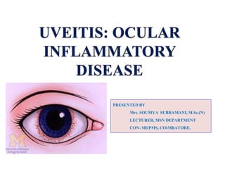
2. INTRODUCTION OF UVEITIS
• Inflammation of the uvea.
• Twentieth century referred ‘‘ophthalmia.”
• Uvea consists of middle layer of pigmented
vascular structures of the eye which includes
the iris, ciliary body, and choroid.
3. DEFINITION
•Infection of uveal tract can affect the iris, the
ciliary body or choroid.
•Inflammation of the uveal tract with associated
inflammation of the adjacent structures such as
cornea, sclera, vitreous and retina
8. Drug related side effects
Rifabutin, a derivative of Rifampin has been
shown to cause uveitis.
Quinolones especially Moxifloxacin may lead
to uveitis.
All of the widely administered vaccines have
been reported to cause uveitis.
10. Immunologic factors
Uveitis Is Driven By Th17t Cell Sub-population
That Bear T-cell Receptors Specific For Proteins Found In The
Eye.
Not Detected Centrally Whether Due To Ocular Antigen Not Being Presented
In
The Thymus.
Autoreactive T Cells Must Normally Be Held In Check By The Suppressive
Environment Produced By Microglia And Dendritic Cells In TheEye.
These Cells Produce Large Amounts Of TGF Beta And
Other Suppressive Cytokines,
Including IL-10, To Prevent Damage To The Eye By Reducing Inflammation
And Causing T Cells To Differentiate To Inducible T Reg Cells.
11. Cont….
Immune stimulation by bacteria and cellular stress is
normally suppressed by myeloid suppression while
inducible T reg cells prevention and clonal expansion of the
autoreactive Th1 and Th 17 cells that possess potential to
cause damage to the eye.
Infection or other causes, this balance can be upset and auto
reactive T cells allowed to proliferate and migrate to the eye.
Entry to the eye, these cells may be returned to an inducible
T reg state by the presence of IL-10 and TGF-beta from
microglia.
12. Genetic Factors
The cause of non-infectious uveitis is unknown.
But there are some strong genetic factors that predispose
disease onset.
Including HLA-B27 and the PTPN22genotype.
13. Infectious agents
Recent evidence has pointed to reactivation of herpes
simplex, varicella zoster and other viruses as
important causes.
Bacterial infection is another significant contributing
factor in developing uveitis.
16. Anterior uveitis
Includes iridocyclitis and iritis.
Iritis is inflammation of the anterior chamber and iris.
Iridocyclitis presents the same symptoms as iritis, but also includes
inflammation in the ciliary body.
From two-thirds to 90% of uveitis cases are anterior in location.
This condition can occur as a single episode and subside with proper
treatment.
17. Intermediate uveitis
Known as pars planitis.
Consists of vitritis -inflammation of cells in vitreous cavity.
Deposition of inflammatory material on the pars plana.
"Snowballs“,Inflammatory cells in the vitreous.
20. Clinical
– Acute uveitis: sudden symptomatic onset , last for six weeks or
more
– Chronic uveitis : insidious and asymptomatic onset ,last for
more than three
months or even years
Pathological
– Granulomatous
– Non Granulomatous
Etiological
– Infective uveitis
– Allergic uveitis
– Toxic Uveitis
– Idiopathic Uveitis
– Uveitis associated with systemic diseases
21. a) Non granulomatous
– Uveitis due to tissue invasion by leptospirae represents the
manifestation of non granulomatous uveitis
– It is acute ,occurring due to physical and toxic insult to the
tissue
– The alterations consists of dilatation and increase
permeability of vessels, breakdown of blood aqueous
barrier and infiltration of lymphocytes, plasma cells and large
macrophages of the uveal tissue
– As a consequence mobility is reduced, pupil becomes small
due to sphincter irritation and engorgement of iris vessels
Etiopathogenesis
22. b) Granulomatous:
– Chronical inflammation of proliferative nature due to irritant foreign
body, a haemorrhage or a necrotic tissue in the eye
– Characterized by infiltration with lymphocytes and proliferation of
large
mononuclear cells which aggregate into nodules
– Necrosis of adjacent structures leads to reparative process
resulting in fibrosis
and gliosis of the involved area
23. Feature Granulomatous Non granulomatous
Onset Insidious and Chronic course Acute and symptomatic
Pain Mild Marked
Photophobia Slight Marked
Ciliary congestion Minimal Marked
Keratic precipitates Mutton fat type of KP’s Fine KP’s
Aqueous flare Mild Intense flare, often with
heavy
fibrinous exudates
Iris nodules Usually present Absent
Posterior synechia Thick and broad based Thin
Fundus Nodular lesions Diffused
Area Anterior uvea and choroid
retina are equally involved
Mainly limited to anterior
uvea
24. SIGNS AND SYMPTOMS
Anterior uveitis
Burning.
Redness.
Blurred vision.
Headaches.
Irregular pupil.
Eye pain.
Photophobia or sensitivity to light.
Floaters, which are dark spots that float in the visual
field.
27. DIAGNOSIS
Diagnosis includes dilated fundus examination to rule out
posterior uveitis, which presents with white spots across
the retina along with retinitis and vasculitis.
Laboratory
underlying
testing is
diseases,
usually used to diagnose specific
including rheumatologic tests (e.g.
antinuclear antibody, rheumatoid factor) Serology for
infectious diseases (e.g. Syphilis, Toxoplasmosis,
Tuberculosis).
fig. Keratic precipitates
28. Investigations
• Routine haemogram
• Serological tests: VDRL and FTA-ABS ( for
syphilis)
• Skin tests : Mantaoux test (tuberculosis)
kviem test (sarcoidosis)
• X Ray : chest and joints
• Urine examination
30. TREATMENT
Treatment Goals:
1. Relieve pain and discomfort.
2. Prevent sight loss due to or its disease
complications.
3. Treatthe cause of the disease where
possible, that is, treat the inflammation.
The drugs usedto treat uveitis fall into 3 main
groups.
1) Steroids
2) Immunosuppressant.
3) Mydriatics.
4) NSAIDS (aspirin) (phenylbutazone ,
oxyphenylbutazone) in uvietis of rheumatoid type
31. STEROIDS
Steroids have wide ranging effects but their action
may be looked on as being anti-inflammatory and
immunosuppressant".
They are used in different forms:
• Eye drops.
• Periocular injections.
• By oral (tablets).
• Intra-venous infusion (drip).
32. Eye Drops:
Used for Anterior Uveitis.
Drops can penetrate the part of the eye in front of the lens, where anterior
uveitis occurs.
Frequency of taking the drops depending on severity of the uveitis.
Severe Cases strongest drop-every hour .
Mild inflammation weakest drop once or twice a day.
Periocular Injections:
Use of injections around the eye to deliver the steroid treatment.
In certain situations injections offer a better way than either tablets or drops.
They are used along with other forms of treatment.
Situations where injections are used include:
• Severe cases of anterior uveitis which can not be controlled by drops alone.
• Intermediate uveitis
33. Systemic Steroids:
• Oral Steroids E.g. Prednisolone Tablet.
• The use of systemic steroids is more serious than, steroid drops because in
this form there are potentially significant side effects.
• Many different situations in which oral steroids are considered.
systemic• If anterior uveitis is resistant to treatment with drops and injections then
steroids considered.
• The main use of oral steroids is to treat posterior uveitis , panuveitis.
Dosage: Prednisolone tablet 1mg and 5mg.
Intra-venous Steroids:
E.g. Methylprednisolone.
• when rapid control of inflammation is needed high dosage of steroid needs to be
delivered quickly.
34. Side Effects
Of Steroids
Nausea ,
Dyspepsia
Increased
Appetite ,
Weight Gain,
Fluid
Retention
Diabetes ,
Osteoporosis
Glaucoma ,
Cataract.
35. IMMUNOSUPPRESSANT
Steroids do suppress the immune system,but there are a
different group of drugs that may be used to treat uveitis.
These drugs tend to target the immune system more
precisely than steroids.
They are usually used in conjunction with steroids.
The main examples are:
Cyclosporine.
Azathioprine (Imuran).
Methotrexate.
Mycophenolate mofetil (cellcept).
Tacrolimus (Prograf 500).
36. MYDRIATICS
Mydriatics have 2 main aims:-
To relieve pain and light sensitivity.
To prevent sight threatening complications.
Mydriatic eye drops, Eg. Atropine and Cyclopentolate are
used.
It works by "paralyzing" the muscles of the iris and the
ciliary
body.
It taken their effect the pupils will be dilated.
because they help prevent complication which may occur
in anterior uveitis.
37. ROLE OF SOME NATURAL
PRODUCT IN UVEITIS
• TURMERIC:
• Benefits for Uveitis:
Antioxidant properties, protect and boost the functioning of the
immune system.
Turmeric help in the reduction of chronic uveitis symptoms.
Research studies which have found that turmeric can prove
beneficial for uveitis.
38. Study
study on a curcumin-phosphatidyl choline compound called
Meriva or Norflo tablets, treating chronic anterior uveitis.
given twice daily to patients with differing etiologies of this
condition.
There were 106 patients studied over a 12 month period.
They were divided into 3 groups
Autoimmune Uveitis.
Herpetic Uveitis.
Different Uveitis Etiologies.
results found that all patients well tolerated Meriva Tablet.
It reduced eye discomfort in around 80% of patients after a few
weeks.
Conclusion: curcumin based medications could benefit those
with anterior uveitis .
39. Dosage: 375mg Tablet 3 times daily.
Precautions: turmeric
Diabetes or Gall Bladder problems must avoid
supplements.
Taken in excess, it can cause Diarrhea Or Nausea.
Contraindicated in Pregnant and Breastfeeding women.
MARKETED PREPARATION:
Uvical pills.
Curcumin phytosome 500mg caps.
Turmeric curcumin 500mg caps.