![The study casts are essential diagnostic aids. They provide a three
dimensional view of the maxillary and mandibular dental arches. Model
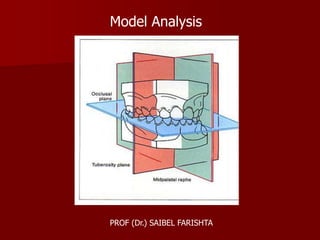
analysis involves the study of the maxillary and mandibular arches in all
the three planes of space [Sagittal, Vertical and Transverse] and is a
valuable tool in orthodontic diagnosis and treatment planning.
Ashley Howe’s Analysis
He considered tooth crowding to be due to deficiency in arch length.
He founded a relationship between the total width of the 12 anterior
teeth to the second molars and the width of the dental arch in the first
premolar region.
Determination of the Total Tooth Material [TTM]
Procedure –
The mesio-distal width of all the teeth, mesial to the second permanent
molars is measured with the help of dividers and all the values are
summed up. This value is called the total tooth material.](data:image/gif;base64,R0lGODlhAQABAIAAAAAAAP///yH5BAEAAAAALAAAAAABAAEAAAIBRAA7)
Recommended
More Related Content
Similar to Model Analysis
Similar to Model Analysis (20)
More from Saibel Farishta
More from Saibel Farishta (20)
Recently uploaded
Recently uploaded (20)
Model Analysis
- 1. Model Analysis PROF (Dr.) SAIBEL FARISHTA
- 2. The study casts are essential diagnostic aids. They provide a three dimensional view of the maxillary and mandibular dental arches. Model analysis involves the study of the maxillary and mandibular arches in all the three planes of space [Sagittal, Vertical and Transverse] and is a valuable tool in orthodontic diagnosis and treatment planning. Ashley Howe’s Analysis He considered tooth crowding to be due to deficiency in arch length. He founded a relationship between the total width of the 12 anterior teeth to the second molars and the width of the dental arch in the first premolar region. Determination of the Total Tooth Material [TTM] Procedure – The mesio-distal width of all the teeth, mesial to the second permanent molars is measured with the help of dividers and all the values are summed up. This value is called the total tooth material.
- 3. Determination of PreMolar Diameter [PMD] It refers to the arch width from the tip of the buccal cusp of one first premolar to the tip of buccal cusp of the opposite first premolar. Determination of PreMolar Basal Arch Width [PMBAW] The canine fossa is found distal to the canine eminence. The measurement of the width from canine fossa of one side to the other gives us the width of the dental arch at the apical base or the junction between the basal bone and alveolar process. If the canine fossa is not clearly distinguishable, the measurement is made from a point that is 8 mm below the crest of the inter dental papilla, distal to the canine.
- 4. Inference - Determination of the possibility of Arch expansion The PMBAW and the PMD are compared. If the PMBAW is greater than PMD, then it is an indication that arch expansion is possible. If on the other hand the PMBAW is less than PMD, then arch expansion is not possible. Calculation of PreMolar Basal Arch Width Percentage According to Ashley Howe, to achieve normal occlusion with a full complement of teeth, the basal arch width at the premolar region should be 44% the sum of mesio-distal widths of all the teeth mesial to the second permanent molar [TTM]. This ratio [expressed as %] between the apical base width at the premolar region and TTM is called the premolar basal arch width percentage. It is determined using the formula : PMBAW % = PMBAW x 100 TTM
- 5. • If PMBAW % is 37% or less, it indicates a need for extraction • If PMBAW % is 44% or more, the case can be treated without extractions • If PMBAW % is 37% - 44% the case is referred to as a borderline case
- 6. Ponts Analysis Ponts in 1909 presented a system where the measurement of the four maxillary incisors automatically established the width of the arch in Premolar and molar regions. This analysis helps in – • Determining whether the dental arch is narrow or normal • Determining the need for lateral arch expansion • Determining how much expansion is possible at Premolar & molar regions Procedure – Determination of Sum Of Incisors [SI] The mesio distal width of the 4 maxillary incisors is measured and the values summed up. This value is called sum of incisors [SI]
- 7. Determination of Measured Premolar Value [MPV] The width of the arch in the premolar region from the distal pit of one upper first premolar to the distal pit of the opposite first premolar is measured. This is called the measured premolar value. Determination of Measured Molar Value [MMV] The width of the arch in the molar region from the mesial pit of one upper first molar to the mesial pit of the opposite first molar is measured. This is called the measured molar value. Determination of Calculated Premolar Value [CPV] The calculated premolar value or the expected arch width in the premolar region is determined by the formula : SI x 100 80
- 8. Determination of Calculated Molar Value [CMV] The calculated premolar value or the expected arch width in the molar region is determined by the formula : SI x 100 64 Inference – If measured value is less than the calculated value, it indicates the need for expansion. Thus it is possible to determine how much expansion is needed in molar and premolar regions.
- 10. Linder Harth Index This analysis is quite similar to Pont’s analysis, except that a new formula has been proposed to determine the calculated premolar and molar values. The calculated premolar value is determined using the formula – SI x 100 85 The calculated molar value is determined using the formula – SI x 100 64
- 11. Korkhaus Analysis This is similar to pont’s analysis. According to this,for a given width of upper incisor,a specific value of the distance b/n midpoint of inter premolar line to point in b/n the max.incisor exist. Inference - Increase in this measurement denotes proclined upper anteriors, while reduced value indicates retroclined upper anteriors.
- 12. Carey’s Analysis Many malocclusions occur as a result of discrepancy between arch length and tooth material. This analysis helps in determining the extent of the discrepancy. It is performed on the lower cast. The same analysis on the upper cast is called Arch perimeter analysis. Procedure – Determination of Arch Length The arch length anterior to the first permanent molar is measured using a soft brass wire. The wire is placed touching the mesial surface of the first permanent molar of one side and is passed over the buccal cusps of the premolars and along the anteriors. It is continued on the opposite side in the same way upto the mesial surface of the opposite first permanent molar. In case of proclined anteriors, the wire is passed along the cingulum of anterior teeth. If the anterior teeth are retroclined, the brass wire in the anterior segment passes labial to the teeth. If the anterior teeth are well aligned, the wire passes over the incisal edge of anteriors.
- 13. Determination of Tooth Material The mesio distal width of the teeth anterior to the first molars [second premolars to second premolars] is measured and summed up. Determination of The Discrepancy It refers to the difference between the arch length and tooth material. Inference – • If the discrepancy is 0 to 2.5 mm, it indicates minimal tooth material excess. Proximal stripping can be done in these cases. • If the discrepancy is between 2.5 to 5 mm, it indicates the need to extract the second premolars • A discrepancy of more than 5 mm indicates first premolar extraction.
- 14. Carey’s Analysis
- 15. Bolton’s Analysis According to Bolton there is a ratio between the mesio distal widths of maxillary and mandibular teeth. Many malocclusions occur as a result of abnormalities in tooth size. This analysis helps in determining disproportion in size between maxillary and mandibular teeth. Procedure – Sum of Mandibular 12 The mesio distal width of all the teeth mesial to the mandibular second permanent molars is measured and summed up. Sum of Maxillary 12 The mesio distal width of all the teeth mesial to the maxillary second permanent molars is measured and summed up.
- 16. Sum of Mandibular 6 The mesio distal width of all the teeth mesial to the mandibular first premolars is measured and summed up. Sum of Maxillary 6 The mesio distal width of all the teeth mesial to the maxillary first premolars is measured and summed up. Determination of Overall Ratio According to Bolton, the sum of mesio distal widths of the mandibular teeth anterior to the second permanent molars is 91.3% the mesio distal width of the maxillary teeth mesial to the second molars. The overall ratio is determined using the formula: Overall ratio = sum of mandibular 12 x 100 Sum of maxillary 12
- 17. If the overall ratio is less than 91.3% it indicates maxillary tooth material excess. The amount of maxillary excess is determined using the formula: Sum of maxillary 12 = sum of mandibular 12 x 100 91.3 If the overall ratio is more than 91.3% it indicates mandibular tooth material excess. The amount of mandibular excess is determined using the formula: Sum of mandibular 12 = sum of maxillary 12 x 91.3 100 Determination of Anterior Ratio The sum of mesio distal width of the mandibular anteriors should be 77.2% of the mesio distal width of the maxillary anteriors. The anterior ratio is determined using the following formula: Anterior ratio = sum of mandibular 6 x 100 Sum of maxillary 6
- 18. If the anterior ratio is less than 77.2% it indicates maxillary anterior excess. The amount of maxillary anterior excess is determined by the following way: Sum of maxillary 6 - sum of mandibular 6 x 100 77.2 If the anterior ratio is more than 77.2% it indicates mandibular anterior excess. The amount of mandibular anterior excess is determined using the formula: Sum of mandibular 6 - sum of mandibular 6 x 77.2 100
- 21. Moyer’s Mixed Dentition Analysis The purpose of a mixed dentition analysis is to evaluate the amount of space available in the arch for the erupting permanent canines and premolars. In this analysis, the size of the unerupted permanent cuspids and premolars are predicted from the knowledge of the sizes of certain permanent teeth already erupted in the mouth. This analysis predicts the combined mesio distal width of 3, 4 and 5 based on the sum of the widths of the four lower permanent incisors. Procedure – The mesio distal width of the four lower incisors are measured and summed up. The amount of space available for 3, 4 and 5 after incisor alignment is determined by measuring the distance between the distal surface of lateral incisor and the mesial surface of first permanent molar. Based on the mesio distal width of the four mandibular incisors, the expected widths of the canines, first and second premolars are predicted by referring the probability chart. 75% level of probability is considered reliable.
- 22. The predicted tooth size of 3, 4 and 5 is compared with the arch length available for them so as to determine the discrepancy. If the predicted value is greater than the available arch length, crowding of teeth can be expected.
- 23. Mixed Dentition Analysis – Radiographic Method This technique makes use of a radiograph as well as a study cast to determine the width of the unerupted teeth. Radiographic measurements of unerupted teeth are by themselves unreliable due to the distortions that can occur. It is possible to determine the measurements of the unerupted teeth by studying the teeth that have already erupted in a radiograph and on a cast. The following formula is used: Y1 = X1 x Y2 X2 Y1 = width of unerupted tooth, whose measurement is to be determined Y2 = width of unerupted tooth on a radiograph X1 = width of a tooth that has erupted, measured on a cast X2 = width of a tooth that has erupted, measured on a radiograph
- 24. THANK YOU