SlideShare une entreprise Scribd logo
1  sur  49
Télécharger pour lire hors ligne
Anatomy and HA of Abdomen
2/21/2023 1
2/21/2023 3
Abdominal examination helps as to identify
abnormalities in the abdomen such as:-
Organ enlargement,
Masses,
Peritoneal irritation,
Hernias,
Fluid accumulation,
Abnormal peristaltic movement.
2/21/2023 4
Abdominal examination…Cont’d
Locating abdominal structures
• Methods of locating abdominal finding is done
by dividing abdomen in to four quadrants by
two perpendicular lines.
• One vertical pass through the umbilicus and
one horizontal line through the umbilicus.
2/21/2023 5
RUQ which contain:-
Rt adrenal gland
Liver
Gall bladder
portion of Rt kidney
head of pancreas
Duodenum
Pylorus loop of small intestine
Parts of the colon(hepatic flexure, portion of
transverse and ascending colon ).
2/21/2023 6
RLQ-which contain:-
Appendix
distended bladder
Rt ovary
Rt salpnix
Portion of ascending colon
Lower pole of Rt kidney
Cecum
Rt spermatic chord
Rt ureter
Enlarged utreus
Loop of small intestine
2/21/2023 7
LUQ- which contains:-
Lt adrenal gland
Portion of Lt kidney
Body of pancreas
Spleen
Stomach
Loop of small intestine
Portion of colon (spleenic flexure, portion of
transverse and descending colon).
2/21/2023 8
LLQ-which contain
Distended bladder
Left ovary
Lt salpnix
Lt spermatic chord
Lt ureter
Enlarged utreus
Loop of small intestine
Lower pole of Lt kidney
Sigmoid and portion of descending colon
2/21/2023 9
Palpable organs-
Often palpable
Sigmoid colon
Cecum
Iliac arteries
Distended bladder
Pregnant utreus
Pulsation of abdominal
aorta
portion of ascending,
transverse and descending
colon
2/21/2023 10
• Rt kidney and liver may palpable only in some pts.
• Much of the liver, much of the stomach and all of the
usual normal spleen are in the abdominal cavity and
extended under the rib cage to the dome of diaphragm,
therefore none palpable.
• Most of the gall bladder lies deep to the liver,
duodenum and pancreas lie deep in the upper
abdomen, hence none of them palpable.
• Techniques of examination
– The usual sequence of inspection, palpation, percussion
and auscultation is modified in the examination of the
abdomen.
– Inspection, auscultation , palpation and percussion.
2/21/2023
11
Inspection
• Observe the size and shape of the abdomen:
• it is described as flat, round, or scaphoid.
• Check for symmetry of the abdomen.
• Check for an umbilical hernia.
• A slight pulsation may be noted in the mid epigastric
area, particularly in very thin clients. This is the
pulsation of the abdominal aorta.
• In adults, observable veins may reflect major health
problems.
• Increased peristaltic waves of intestinal obstruction.
• Increased pulsation of an aortic aneurysm.
2/21/2023 13
Auscultation
• Auscultation assesses the sounds of peristalsis and vascular
abnormalities.
• Listen for bowel sounds and note their frequency and
character.
• Normal sound consists of clicks and gurgles, the frequency
which has been estimated from 5 to 34 per minute.
• Borborygmus- loud prolonged gurgles of hyper peristalsis.
• Bowel sounds may be altered in diarrhea, intestinal obstruction,
paralytic ileus, and peritonitis.
• Normal blood flow through the abdominal arteries can not be
heard.
• Blood flow through dilated or constricted blood vessels results
in a turbulence that can be detected by auscultation.
2/21/2023 14
• Recall that the bell detects low-pitched sounds. Place
the bell of stethoscope over:
– Abdominal aorta in midepigasrium above the umbilicus.
– Renal arteries to the right and left slightly above the
umbilicus.
– Hepatic and splenic arteries in the right and left
hypochondrium.
• To be considered significant, an abdominal bruit must
be heard as the client is moved in to various
positions.
• Friction rub: rough grating sound, occurs with
irritation of the peritoneal surface of an organ,
splenic infarction, primary or metastatic tumor of
liver, and peritonitis.
2/21/2023 15
• Percussion
Light percussion of the abdomen is performed to
determine:
– Enlargement of an organ
– The presence of masses
– The presence of abdominal distension or the amount
and distribution of gas in the abdomen.
• A dull percussion note will be heard over the area
of a dense abdominal organ, such as liver or spleen,
and over a solid tumor or fluid.
• Dull percussion notes detected in the suprapubic
area may be from a distended urinary bladder.
2/21/2023 16
• Tympany is the normal percussion note present
through out the abdomen except for areas over the
liver, spleen and pubic symphysis.
• With gaseous distension tympany will be elicited
throughout the abdominal area.
• The vertical span of liver dullness represents the size
of the liver in the adult; this is normally about 6-12cm
in the mid clavicular line& 4-8cm in mid sternal line.
• Vertical span is greater in men than women, tall
person than short.
• Percuss down the right anterior chest in the mid
clavicular line (MCL).
2/21/2023 17
• Resonance over the lung will be detected until
about the 5th to 7th intercostal space, where liver
dullness begins.
• Continue percussion down ward until the lower
edge of liver dullness is heard. The normal lower
level is at the costal margin.
• Ask the client to inhale, and percuss down ward
again on inspiration, there should be an increase of
about 2 to 4cm from the previous lower edge of
liver dullness, because the liver moves down ward
on inspiration.
2/21/2023 18
• The spleen lies slightly posterior to the left mid
axillaries line, b/n the 8th and 11th ICS.
• Below the 12th find dullness is called positive
spleenic percussion sign.
2/21/2023 19
Palpation
• The client should be as comfortable and relaxed as
possible.
• Light palpation-feeling the abdomen gently is
especially helpful in identifying abdominal
tenderness, muscular resistance, some superficial
organs & masses.
– If resistance is present, try to distinguish voluntary
guarding from involuntary muscular spasm.
– Feel for the relaxation of abdominal muscles that
normally accompanies exhalation.
– Ask the pt to mouth- breath with jaw dropped open,
voluntary guarding usually decreases with this
maneuvers.
– Involuntary rigidity (muscular spasm) typically persists
despite these maneuvers.
2/21/2023 20
• Visceral pain generally is a poorly localized, dull
pain, where as peritoneal pain usually is easily
localized and is described as sharp, stabbing pain.
• Abdominal pain on coughing or with light palpation
suggests peritoneal inflammation.
• Test for rebound tenderness by gently and slowly
pressing the examining hand deep in to the
abdomen away from the site of the pain, quickly
release your hand.
• the client with rebound tenderness will experience
induced pain or increased pain, immediately inquire
where the pain is felt.
2/21/2023 21
• Deep palpation is usually required to delineate
abdominal masses.
• Again using the palmar surfaces of your fingers, feel
in all four quadrants. Identify any masses and note
their location, size, shape, consistency, tenderness,
pulsations and mobility.
–Note-palpation may not be justified in some
abdominal problems such as appendicitis when
diagnosis is clear and there is risk of rapture with
manipulation.
2/21/2023 22
Liver
Place your right hand just below the lower right costal
margin with your fingers pointing toward the client's
right shoulder and parallel with the abdominal rectus
muscle.
• Push the fingers deeply in to the abdomen with a
constant pressure and simultaneously push up under
the rib cage.
• Ask the client to take a deep breath: this will cause
the liver to descend.
• On inspiration, the liver below is palpable about 4cm
below the right costal margin in the mid clavicular
line.
2/21/2023 23
• An obstructed, distended gall-bladder may form an
oval mass below the edge of the liver and merging
with it. It is dull to percussion.
• Tenderness over the liver suggests inflammation, as in
hepatitis, or congestion, as in heart failure.
•Spleen
• The spleen may be palpable in the normal infant and
young child; however, with the older child and adult,
the spleen must be considerably enlarged before it
can be palpated.
• Place your left hand under the client's left flank at the
level of the 11th and 12th ribs.
2/21/2023 24
• kidneys
– They may be felt in children and in adults with scaphoid
abdomens.
– Palpation of the right kidney:
• Although kidneys are not usually palpable, you
should learn and practice the techniques.
• Place your left hand behind the pt just below and
parallel to the 12th rib, with your fingertips just
reaching the costovertebral angle.
• Lift, trying to displace the kidney anteriorly.
• Place your right hand gently in the right upper
quadrant, lateral and parallel to the rectus muscle.
2/21/2023 25
• Ask the pt to take a deep breath.
• At the peak of inspiration, press your right hand
firmly and deeply in to the right upper quadrant,
just below the costal margin, and try to capture the
kidney between your two hands.
• Causes of kidney enlargement include
hydronephrosis, cysts, and tumors.
• Bilateral enlargement suggests poly cystic disease.
2/21/2023 26
Palpation of the left kidney
• To capture the left kidney, move to the pt’s left side.
• Use your right hand to lift from in back, and your
left hand to feel deep in the left upper quadrant.
• Instruct the client to take a deep breath while you
apply pressure with the examining hand.
2/21/2023 27
• Assessing kidney tenderness
• Use fist percussion- place the ball of your hand in
the costovrtebral angle and strike it with the ulnar
surface of your fist.
• Pain with pressure or with fist percussion in the
costovertebral angle suggests kidney infection, but
it may also have a musculoskeletal cause.
• Urinary bladder
–The bladder is non palpable unless it is
distended well above the pubic symphysis.
2/21/2023 28
• ABDOMINAL EXAMINATION: SPECIAL TECHNIQUES
1. To Assess Possible Ascites
• A protuberant abdomen with bulging flanks
suggests the possibility of ascitic fluid
• The presence and extent of ascites are assessed by
perccusing the abdomen for shifting dullness or by
detecting a fluid wave.
A. Test for shifting dullness.
• After mapping the borders of tympany and dullness, ask
the pt to turn on to one side percuss and mark the borders
again.
• In ascites, dullness shifts to the dependent side, while
tympany shifts to the top.
2/21/2023 29
B. Test for a fluid wave
• An assistant’s hand is placed along the pt’s midline
of the abdomen.
• This pressure helps to prevent the fluid wave from
being transmitted through the tissues of the
abdominal wall. The examiner places the hands
along the side of the pt’s flank, then strikes or tap
one flank sharply, feel on the opposite flank for an
impulse transmitted through the fluid.
• A fluid wave is likely to be found only when there is
a large amount of fluid present.
2/21/2023 30
2. To identify an organ or amass in an ascetic
abdomen.
• Try to ballotte the organ or mass, exemplified here
by an enlarged liver.
• Straighten and stiffen the fingers of one hand
together, place them on the abdominal surface, and
make a brief jabbing movement directly toward the
anticipated structure.
• This quick movement often displaces the fluid so
that your finger tips can briefly touch the surface of
the structure through the abdominal wall.
2/21/2023 31
3. To assess possible appendicitis
• Ask the patient to point to where the pain
began and where it is now.
• Ask the patient to cough.
• Determine whether and where pain results.
• The pain of appendicitis classically begins near
the umbilicus and then shifts to the right
lower quadrant, where coughing increases it.
2/21/2023 32
–Search for an area of local tenderness
• Localized tenderness anywhere in the right lower
quadrant may in dictate appendicitis.
• Feel for muscular rigidity
• Early voluntary guarding may be replaced by
involuntary muscular rigidity
• Check the tender area for rebound tenderness
• Rebound tenderness suggests peritoneal
inflammation, as from appendicitis.
2/21/2023 33
• Check for Rovising’s sign and for referred rebound
tenderness.
• Press deeply and evenly in the LLQ and quickly
withdraw your finger. Pain in the right lower guardant
during left- sided pressure suggests appendicitis (a
positive Rovising’s sign). Rt quadrant pain quickly
withdrawal termed as rebound tenderness.
• Look for a psoas sign. Place your hand just above the
pt’s right knee and ask the pt to raise that thigh
against your hand.
• Increased abdominal pain on this maneuver
constitutes a positive psoas sign, suggesting irritation
of the psoas muscle by an inflamed appendix.
2/21/2023 34
Images
4. To assess possible acute cholecystitis
• Cholecystitis may be detected by pressing the fingers
gently in to the right hypochondrium as the patient
takes a deep breath in, a sudden catch of breath when
the gallbladder touches your fingers indicates
inflammation (a positive Murphy’s sing).
5. To assess ventral hernias
– If you suspect this type of hernia but do not see an
umbilical or incisional hernia, ask the patient to raise both
head and shoulders off the examination coach.
– The bulge of a hernia which usually appear with this
action.
2/21/2023 38
6. To distinguish an abdominal mass from a mass in
the abdominal wall.
– Ask the patient either to raise the head and shoulders or
to strain down, thus tightening the abdominal muscles.
Feel for the mass again.
– A mass in the abdominal wall remains palpable an intra
abdominal mass is obscured by muscular contraction.
2/21/2023 39
7. Localized bulges in the abdominal wall
• Include ventral hernias and subcutaneous tumors
such as lipomas.
– The more common ventral hernias are umbilical,
incisional, and epigastric.
– Hernias usually become more evident when the patient
raises head and shoulders from a supine position.
• Lipomas, small or large, they are usually soft and
often lobulated. When your finger presses down
the edge of lipoma the tumor usually slips out from
under it.
2/21/2023 40
8. Sounds in the abdomen
a. Bowel sounds
• May be increased in clients with diarrhea or early
intestinal obstruction.
• Decreased, in paralytic ileus and peritonitis.
• High-pitched tinkling sounds suggest intestinal fluid
and air under tension in a dilated bowel.
b. Bruits
• A hepatic bruit suggests carcinoma of the liver or
alcoholic hepatitis.
• Arterial bruits with both systolic and diastolic
components suggest partial occlusion of the aorta or
large arteries.
2/21/2023 41
9. Tender abdomens
• Abdominal wall Tenderness
• Tenderness may originate in the abdominal wall.
• When the patient raises head and shoulders, this
tenderness persists; whereas tenderness from a
deeper lesion decreases.
a. Visceral tenderness: Example – Enlarged liver may
be tender to deep palpation.
b. Tenderness from disease in the chest and pelvis
2/21/2023 42
• Acute pleurisy
– Abdominal pain and tenderness may be due to acute
pleural inflammation.
• Acute salphingitis
• Frequently bilateral, the tenderness of acute
salphingitis is usually maximal just above the
inguinal ligaments.
2/21/2023 43
• Tenderness of peritoneal inflammation
• Severe than visceral tenderness.
• Muscular rigidity and rebound tenderness are
frequently present.
10. Liver enlargement
• A palpable liver does not necessarily indicate
hepatomegaly.
• Clinical estimates of liver size should be based on
both percussion and palpation.
2/21/2023 44
A. Down wards displacement of the liver by a low
diaphragm.
• Common finding in emphysema.
• The liver edge may be readily palpable well below
the costal margins.
B. Normal variations in liver shape
• In some persons, especially those with a lanky
build, the liver tends to be somewhat elongated so
that its right lobe is easily palpable as it projects
downward toward the iliac crest.
• Riedel’s lobe – represents a variation in shape, not
an increase in liver volume or size
2/21/2023 45
C. Smooth large non tender liver
• Cirrhosis may produce an enlarged liver with a
firm non tender edge.
• Many other diseases may produce similar finings.
D. Smooth large tender liver
• An enlarged liver with a smooth tender edge
suggests inflammation, as in hepatitis, venous
congestion, as in right sided heart failure.
2/21/2023 46
E. Large irregular liver
• An enlarged liver that is firm or hard and has an
irregular edge or surface suggests malignancy.
• There may be one or more nodules.
• The liver may or may not be tender.
2/21/2023 47
Reference
• Health assessment lecture notes for nursing
students by kemal ahmed 2010.
• Nitisbin medicine 1st edition-(revised).pdf
Acknowledgment
• For Wollo university college of medicine and
health sciences, department of adult health
nursing.
• For Dr. Kumar
• For my audience

Contenu connexe

Similaire à AHA.pptx

Abdomen examination.pptx
Abdomen examination.pptxAbdomen examination.pptx
Abdomen examination.pptxAkshay Shetty
 
examination of abdomen.pptx
examination of abdomen.pptxexamination of abdomen.pptx
examination of abdomen.pptxAnjanaMV4
 
abdominal x ray radiology
abdominal x ray radiologyabdominal x ray radiology
abdominal x ray radiologysarfraj Ahmad
 
Clinical Examination of Abdomen (Part II).pptx
Clinical Examination of Abdomen (Part II).pptxClinical Examination of Abdomen (Part II).pptx
Clinical Examination of Abdomen (Part II).pptxRishabhMawa1
 
Monday final abdominal examination final ppt
Monday final abdominal examination final pptMonday final abdominal examination final ppt
Monday final abdominal examination final pptroheedakhan81
 
5.Abdomen Exam.pptx
5.Abdomen Exam.pptx5.Abdomen Exam.pptx
5.Abdomen Exam.pptxmyLord3
 
Plain radiography in acute abdomen
Plain radiography in acute abdomenPlain radiography in acute abdomen
Plain radiography in acute abdomenNaik Inayat
 
Gatrointestinal assessment
Gatrointestinal assessmentGatrointestinal assessment
Gatrointestinal assessmentCHETAN RSANGATI
 
Abdominal examination.pptx
Abdominal examination.pptxAbdominal examination.pptx
Abdominal examination.pptxtsegawbiyazin
 
11. Final Abdominal Examination.ppt
11. Final Abdominal Examination.ppt11. Final Abdominal Examination.ppt
11. Final Abdominal Examination.pptSufaMengiste
 
Exam Of Abdomen.
Exam Of Abdomen.Exam Of Abdomen.
Exam Of Abdomen.Shaikhani.
 

Similaire à AHA.pptx (20)

GIT EXAMINATION.pptx
 GIT EXAMINATION.pptx GIT EXAMINATION.pptx
GIT EXAMINATION.pptx
 
Abdomen examination.pptx
Abdomen examination.pptxAbdomen examination.pptx
Abdomen examination.pptx
 
examination of abdomen.pptx
examination of abdomen.pptxexamination of abdomen.pptx
examination of abdomen.pptx
 
abdomen PE.pptx
abdomen PE.pptxabdomen PE.pptx
abdomen PE.pptx
 
abdominal x ray radiology
abdominal x ray radiologyabdominal x ray radiology
abdominal x ray radiology
 
Abdoiminal examination
Abdoiminal examinationAbdoiminal examination
Abdoiminal examination
 
Clinical Examination of Abdomen (Part II).pptx
Clinical Examination of Abdomen (Part II).pptxClinical Examination of Abdomen (Part II).pptx
Clinical Examination of Abdomen (Part II).pptx
 
ABDOMINAL EXAMINATION Presentation[1].pptx
ABDOMINAL EXAMINATION Presentation[1].pptxABDOMINAL EXAMINATION Presentation[1].pptx
ABDOMINAL EXAMINATION Presentation[1].pptx
 
Monday final abdominal examination final ppt
Monday final abdominal examination final pptMonday final abdominal examination final ppt
Monday final abdominal examination final ppt
 
5.Abdomen Exam.pptx
5.Abdomen Exam.pptx5.Abdomen Exam.pptx
5.Abdomen Exam.pptx
 
Radiological anatomy of_abdomen[1]
Radiological anatomy of_abdomen[1]Radiological anatomy of_abdomen[1]
Radiological anatomy of_abdomen[1]
 
abdominal assessment
abdominal assessmentabdominal assessment
abdominal assessment
 
Plain radiography in acute abdomen
Plain radiography in acute abdomenPlain radiography in acute abdomen
Plain radiography in acute abdomen
 
Excretionurography
Excretionurography Excretionurography
Excretionurography
 
Gatrointestinal assessment
Gatrointestinal assessmentGatrointestinal assessment
Gatrointestinal assessment
 
Abdominal examination.pptx
Abdominal examination.pptxAbdominal examination.pptx
Abdominal examination.pptx
 
11. Final Abdominal Examination.ppt
11. Final Abdominal Examination.ppt11. Final Abdominal Examination.ppt
11. Final Abdominal Examination.ppt
 
Exam Of Abdomen.
Exam Of Abdomen.Exam Of Abdomen.
Exam Of Abdomen.
 
Ppt11
Ppt11Ppt11
Ppt11
 
Story of the opposite
Story of the oppositeStory of the opposite
Story of the opposite
 

Plus de Sani42793

Leadership&managment.pptx
Leadership&managment.pptxLeadership&managment.pptx
Leadership&managment.pptxSani42793
 
8-Disease associated with pregnancy - Copy.ppt
8-Disease associated with pregnancy - Copy.ppt8-Disease associated with pregnancy - Copy.ppt
8-Disease associated with pregnancy - Copy.pptSani42793
 
AUB lecture.pptx
AUB lecture.pptxAUB lecture.pptx
AUB lecture.pptxSani42793
 
Almaz case (1).pptx
Almaz case (1).pptxAlmaz case (1).pptx
Almaz case (1).pptxSani42793
 
asthmappt-140524034207-phpapp01.pdf
asthmappt-140524034207-phpapp01.pdfasthmappt-140524034207-phpapp01.pdf
asthmappt-140524034207-phpapp01.pdfSani42793
 
5. Normal preg, Dignosis o preg,.pptx
5. Normal preg, Dignosis o preg,.pptx5. Normal preg, Dignosis o preg,.pptx
5. Normal preg, Dignosis o preg,.pptxSani42793
 
3.FP update.ppt
3.FP update.ppt3.FP update.ppt
3.FP update.pptSani42793
 
Evaluation of Psychiatric Illness.pptx
Evaluation of Psychiatric Illness.pptxEvaluation of Psychiatric Illness.pptx
Evaluation of Psychiatric Illness.pptxSani42793
 
Ethics.PPt.Bire.pptxdsgjhdf
Ethics.PPt.Bire.pptxdsgjhdfEthics.PPt.Bire.pptxdsgjhdf
Ethics.PPt.Bire.pptxdsgjhdfSani42793
 
ambulatory ward.pptx
ambulatory ward.pptxambulatory ward.pptx
ambulatory ward.pptxSani42793
 
Vaccine ppt.pptx
Vaccine ppt.pptxVaccine ppt.pptx
Vaccine ppt.pptxSani42793
 
7. Sickle Cell Disease.pptx
7. Sickle Cell Disease.pptx7. Sickle Cell Disease.pptx
7. Sickle Cell Disease.pptxSani42793
 
19 Cystic fibrosis-nnn.pptx
19 Cystic fibrosis-nnn.pptx19 Cystic fibrosis-nnn.pptx
19 Cystic fibrosis-nnn.pptxSani42793
 
13. Thyroid disorders.pptx
13. Thyroid disorders.pptx13. Thyroid disorders.pptx
13. Thyroid disorders.pptxSani42793
 
20 IBD & IBS.pptx
20 IBD & IBS.pptx20 IBD & IBS.pptx
20 IBD & IBS.pptxSani42793
 
14 Dyslipidemia.pptx
14 Dyslipidemia.pptx14 Dyslipidemia.pptx
14 Dyslipidemia.pptxSani42793
 
14. Benign prostatic hyperplasia.pptx
14. Benign prostatic hyperplasia.pptx14. Benign prostatic hyperplasia.pptx
14. Benign prostatic hyperplasia.pptxSani42793
 
13 Veneou Thromboemblism.pptx
13 Veneou Thromboemblism.pptx13 Veneou Thromboemblism.pptx
13 Veneou Thromboemblism.pptxSani42793
 

Plus de Sani42793 (20)

Leadership&managment.pptx
Leadership&managment.pptxLeadership&managment.pptx
Leadership&managment.pptx
 
4. PROM.ppt
4. PROM.ppt4. PROM.ppt
4. PROM.ppt
 
8-Disease associated with pregnancy - Copy.ppt
8-Disease associated with pregnancy - Copy.ppt8-Disease associated with pregnancy - Copy.ppt
8-Disease associated with pregnancy - Copy.ppt
 
AUB lecture.pptx
AUB lecture.pptxAUB lecture.pptx
AUB lecture.pptx
 
Almaz case (1).pptx
Almaz case (1).pptxAlmaz case (1).pptx
Almaz case (1).pptx
 
asthmappt-140524034207-phpapp01.pdf
asthmappt-140524034207-phpapp01.pdfasthmappt-140524034207-phpapp01.pdf
asthmappt-140524034207-phpapp01.pdf
 
5. Normal preg, Dignosis o preg,.pptx
5. Normal preg, Dignosis o preg,.pptx5. Normal preg, Dignosis o preg,.pptx
5. Normal preg, Dignosis o preg,.pptx
 
3.FP update.ppt
3.FP update.ppt3.FP update.ppt
3.FP update.ppt
 
COPD.pdf
COPD.pdfCOPD.pdf
COPD.pdf
 
Evaluation of Psychiatric Illness.pptx
Evaluation of Psychiatric Illness.pptxEvaluation of Psychiatric Illness.pptx
Evaluation of Psychiatric Illness.pptx
 
Ethics.PPt.Bire.pptxdsgjhdf
Ethics.PPt.Bire.pptxdsgjhdfEthics.PPt.Bire.pptxdsgjhdf
Ethics.PPt.Bire.pptxdsgjhdf
 
ambulatory ward.pptx
ambulatory ward.pptxambulatory ward.pptx
ambulatory ward.pptx
 
Vaccine ppt.pptx
Vaccine ppt.pptxVaccine ppt.pptx
Vaccine ppt.pptx
 
7. Sickle Cell Disease.pptx
7. Sickle Cell Disease.pptx7. Sickle Cell Disease.pptx
7. Sickle Cell Disease.pptx
 
19 Cystic fibrosis-nnn.pptx
19 Cystic fibrosis-nnn.pptx19 Cystic fibrosis-nnn.pptx
19 Cystic fibrosis-nnn.pptx
 
13. Thyroid disorders.pptx
13. Thyroid disorders.pptx13. Thyroid disorders.pptx
13. Thyroid disorders.pptx
 
20 IBD & IBS.pptx
20 IBD & IBS.pptx20 IBD & IBS.pptx
20 IBD & IBS.pptx
 
14 Dyslipidemia.pptx
14 Dyslipidemia.pptx14 Dyslipidemia.pptx
14 Dyslipidemia.pptx
 
14. Benign prostatic hyperplasia.pptx
14. Benign prostatic hyperplasia.pptx14. Benign prostatic hyperplasia.pptx
14. Benign prostatic hyperplasia.pptx
 
13 Veneou Thromboemblism.pptx
13 Veneou Thromboemblism.pptx13 Veneou Thromboemblism.pptx
13 Veneou Thromboemblism.pptx
 

Dernier

Study on the Impact of FOCUS-PDCA Management Model on the Disinfection Qualit...
Study on the Impact of FOCUS-PDCA Management Model on the Disinfection Qualit...Study on the Impact of FOCUS-PDCA Management Model on the Disinfection Qualit...
Study on the Impact of FOCUS-PDCA Management Model on the Disinfection Qualit...MehranMouzam
 
Informed Consent Empowering Healthcare Decision-Making.pptx
Informed Consent Empowering Healthcare Decision-Making.pptxInformed Consent Empowering Healthcare Decision-Making.pptx
Informed Consent Empowering Healthcare Decision-Making.pptxSasikiranMarri
 
Measurement of Radiation and Dosimetric Procedure.pptx
Measurement of Radiation and Dosimetric Procedure.pptxMeasurement of Radiation and Dosimetric Procedure.pptx
Measurement of Radiation and Dosimetric Procedure.pptxDr. Dheeraj Kumar
 
SYNDESMOTIC INJURY- ANATOMICAL REPAIR.pptx
SYNDESMOTIC INJURY- ANATOMICAL REPAIR.pptxSYNDESMOTIC INJURY- ANATOMICAL REPAIR.pptx
SYNDESMOTIC INJURY- ANATOMICAL REPAIR.pptxdrashraf369
 
PHYSIOTHERAPY IN HEART TRANSPLANTATION..
PHYSIOTHERAPY IN HEART TRANSPLANTATION..PHYSIOTHERAPY IN HEART TRANSPLANTATION..
PHYSIOTHERAPY IN HEART TRANSPLANTATION..AneriPatwari
 
April 2024 ONCOLOGY CARTOON by DR KANHU CHARAN PATRO
April 2024 ONCOLOGY CARTOON by  DR KANHU CHARAN PATROApril 2024 ONCOLOGY CARTOON by  DR KANHU CHARAN PATRO
April 2024 ONCOLOGY CARTOON by DR KANHU CHARAN PATROKanhu Charan
 
Tans femoral Amputee : Prosthetics Knee Joints.pptx
Tans femoral Amputee : Prosthetics Knee Joints.pptxTans femoral Amputee : Prosthetics Knee Joints.pptx
Tans femoral Amputee : Prosthetics Knee Joints.pptxKezaiah S
 
Nutrition of OCD for my Nutritional Neuroscience Class
Nutrition of OCD for my Nutritional Neuroscience ClassNutrition of OCD for my Nutritional Neuroscience Class
Nutrition of OCD for my Nutritional Neuroscience Classmanuelazg2001
 
Biomechanics- Shoulder Joint!!!!!!!!!!!!
Biomechanics- Shoulder Joint!!!!!!!!!!!!Biomechanics- Shoulder Joint!!!!!!!!!!!!
Biomechanics- Shoulder Joint!!!!!!!!!!!!ibtesaam huma
 
History and Development of Pharmacovigilence.pdf
History and Development of Pharmacovigilence.pdfHistory and Development of Pharmacovigilence.pdf
History and Development of Pharmacovigilence.pdfSasikiranMarri
 
Presentation on General Anesthetics pdf.
Presentation on General Anesthetics pdf.Presentation on General Anesthetics pdf.
Presentation on General Anesthetics pdf.Prerana Jadhav
 
MedDRA-A-Comprehensive-Guide-to-Standardized-Medical-Terminology.pdf
MedDRA-A-Comprehensive-Guide-to-Standardized-Medical-Terminology.pdfMedDRA-A-Comprehensive-Guide-to-Standardized-Medical-Terminology.pdf
MedDRA-A-Comprehensive-Guide-to-Standardized-Medical-Terminology.pdfSasikiranMarri
 
Apiculture Chapter 1. Introduction 2.ppt
Apiculture Chapter 1. Introduction 2.pptApiculture Chapter 1. Introduction 2.ppt
Apiculture Chapter 1. Introduction 2.pptkedirjemalharun
 
Radiation Dosimetry Parameters and Isodose Curves.pptx
Radiation Dosimetry Parameters and Isodose Curves.pptxRadiation Dosimetry Parameters and Isodose Curves.pptx
Radiation Dosimetry Parameters and Isodose Curves.pptxDr. Dheeraj Kumar
 
Big Data Analysis Suggests COVID Vaccination Increases Excess Mortality Of ...
Big Data Analysis Suggests COVID  Vaccination Increases Excess Mortality Of  ...Big Data Analysis Suggests COVID  Vaccination Increases Excess Mortality Of  ...
Big Data Analysis Suggests COVID Vaccination Increases Excess Mortality Of ...sdateam0
 
Statistical modeling in pharmaceutical research and development.
Statistical modeling in pharmaceutical research and development.Statistical modeling in pharmaceutical research and development.
Statistical modeling in pharmaceutical research and development.ANJALI
 
Clinical Pharmacotherapy of Scabies Disease
Clinical Pharmacotherapy of Scabies DiseaseClinical Pharmacotherapy of Scabies Disease
Clinical Pharmacotherapy of Scabies DiseaseSreenivasa Reddy Thalla
 
PERFECT BUT PAINFUL TKR -ROLE OF SYNOVECTOMY.pptx
PERFECT BUT PAINFUL TKR -ROLE OF SYNOVECTOMY.pptxPERFECT BUT PAINFUL TKR -ROLE OF SYNOVECTOMY.pptx
PERFECT BUT PAINFUL TKR -ROLE OF SYNOVECTOMY.pptxdrashraf369
 
epilepsy and status epilepticus for undergraduate.pptx
epilepsy and status epilepticus  for undergraduate.pptxepilepsy and status epilepticus  for undergraduate.pptx
epilepsy and status epilepticus for undergraduate.pptxMohamed Rizk Khodair
 
Presentació "Real-Life VR Integration for Mild Cognitive Impairment Rehabilit...
Presentació "Real-Life VR Integration for Mild Cognitive Impairment Rehabilit...Presentació "Real-Life VR Integration for Mild Cognitive Impairment Rehabilit...
Presentació "Real-Life VR Integration for Mild Cognitive Impairment Rehabilit...Badalona Serveis Assistencials
 

Dernier (20)

Study on the Impact of FOCUS-PDCA Management Model on the Disinfection Qualit...
Study on the Impact of FOCUS-PDCA Management Model on the Disinfection Qualit...Study on the Impact of FOCUS-PDCA Management Model on the Disinfection Qualit...
Study on the Impact of FOCUS-PDCA Management Model on the Disinfection Qualit...
 
Informed Consent Empowering Healthcare Decision-Making.pptx
Informed Consent Empowering Healthcare Decision-Making.pptxInformed Consent Empowering Healthcare Decision-Making.pptx
Informed Consent Empowering Healthcare Decision-Making.pptx
 
Measurement of Radiation and Dosimetric Procedure.pptx
Measurement of Radiation and Dosimetric Procedure.pptxMeasurement of Radiation and Dosimetric Procedure.pptx
Measurement of Radiation and Dosimetric Procedure.pptx
 
SYNDESMOTIC INJURY- ANATOMICAL REPAIR.pptx
SYNDESMOTIC INJURY- ANATOMICAL REPAIR.pptxSYNDESMOTIC INJURY- ANATOMICAL REPAIR.pptx
SYNDESMOTIC INJURY- ANATOMICAL REPAIR.pptx
 
PHYSIOTHERAPY IN HEART TRANSPLANTATION..
PHYSIOTHERAPY IN HEART TRANSPLANTATION..PHYSIOTHERAPY IN HEART TRANSPLANTATION..
PHYSIOTHERAPY IN HEART TRANSPLANTATION..
 
April 2024 ONCOLOGY CARTOON by DR KANHU CHARAN PATRO
April 2024 ONCOLOGY CARTOON by  DR KANHU CHARAN PATROApril 2024 ONCOLOGY CARTOON by  DR KANHU CHARAN PATRO
April 2024 ONCOLOGY CARTOON by DR KANHU CHARAN PATRO
 
Tans femoral Amputee : Prosthetics Knee Joints.pptx
Tans femoral Amputee : Prosthetics Knee Joints.pptxTans femoral Amputee : Prosthetics Knee Joints.pptx
Tans femoral Amputee : Prosthetics Knee Joints.pptx
 
Nutrition of OCD for my Nutritional Neuroscience Class
Nutrition of OCD for my Nutritional Neuroscience ClassNutrition of OCD for my Nutritional Neuroscience Class
Nutrition of OCD for my Nutritional Neuroscience Class
 
Biomechanics- Shoulder Joint!!!!!!!!!!!!
Biomechanics- Shoulder Joint!!!!!!!!!!!!Biomechanics- Shoulder Joint!!!!!!!!!!!!
Biomechanics- Shoulder Joint!!!!!!!!!!!!
 
History and Development of Pharmacovigilence.pdf
History and Development of Pharmacovigilence.pdfHistory and Development of Pharmacovigilence.pdf
History and Development of Pharmacovigilence.pdf
 
Presentation on General Anesthetics pdf.
Presentation on General Anesthetics pdf.Presentation on General Anesthetics pdf.
Presentation on General Anesthetics pdf.
 
MedDRA-A-Comprehensive-Guide-to-Standardized-Medical-Terminology.pdf
MedDRA-A-Comprehensive-Guide-to-Standardized-Medical-Terminology.pdfMedDRA-A-Comprehensive-Guide-to-Standardized-Medical-Terminology.pdf
MedDRA-A-Comprehensive-Guide-to-Standardized-Medical-Terminology.pdf
 
Apiculture Chapter 1. Introduction 2.ppt
Apiculture Chapter 1. Introduction 2.pptApiculture Chapter 1. Introduction 2.ppt
Apiculture Chapter 1. Introduction 2.ppt
 
Radiation Dosimetry Parameters and Isodose Curves.pptx
Radiation Dosimetry Parameters and Isodose Curves.pptxRadiation Dosimetry Parameters and Isodose Curves.pptx
Radiation Dosimetry Parameters and Isodose Curves.pptx
 
Big Data Analysis Suggests COVID Vaccination Increases Excess Mortality Of ...
Big Data Analysis Suggests COVID  Vaccination Increases Excess Mortality Of  ...Big Data Analysis Suggests COVID  Vaccination Increases Excess Mortality Of  ...
Big Data Analysis Suggests COVID Vaccination Increases Excess Mortality Of ...
 
Statistical modeling in pharmaceutical research and development.
Statistical modeling in pharmaceutical research and development.Statistical modeling in pharmaceutical research and development.
Statistical modeling in pharmaceutical research and development.
 
Clinical Pharmacotherapy of Scabies Disease
Clinical Pharmacotherapy of Scabies DiseaseClinical Pharmacotherapy of Scabies Disease
Clinical Pharmacotherapy of Scabies Disease
 
PERFECT BUT PAINFUL TKR -ROLE OF SYNOVECTOMY.pptx
PERFECT BUT PAINFUL TKR -ROLE OF SYNOVECTOMY.pptxPERFECT BUT PAINFUL TKR -ROLE OF SYNOVECTOMY.pptx
PERFECT BUT PAINFUL TKR -ROLE OF SYNOVECTOMY.pptx
 
epilepsy and status epilepticus for undergraduate.pptx
epilepsy and status epilepticus  for undergraduate.pptxepilepsy and status epilepticus  for undergraduate.pptx
epilepsy and status epilepticus for undergraduate.pptx
 
Presentació "Real-Life VR Integration for Mild Cognitive Impairment Rehabilit...
Presentació "Real-Life VR Integration for Mild Cognitive Impairment Rehabilit...Presentació "Real-Life VR Integration for Mild Cognitive Impairment Rehabilit...
Presentació "Real-Life VR Integration for Mild Cognitive Impairment Rehabilit...
 

AHA.pptx

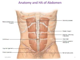
  • 1. Anatomy and HA of Abdomen 2/21/2023 1
  • 2.
  • 4. Abdominal examination helps as to identify abnormalities in the abdomen such as:- Organ enlargement, Masses, Peritoneal irritation, Hernias, Fluid accumulation, Abnormal peristaltic movement. 2/21/2023 4
  • 5. Abdominal examination…Cont’d Locating abdominal structures • Methods of locating abdominal finding is done by dividing abdomen in to four quadrants by two perpendicular lines. • One vertical pass through the umbilicus and one horizontal line through the umbilicus. 2/21/2023 5
  • 6. RUQ which contain:- Rt adrenal gland Liver Gall bladder portion of Rt kidney head of pancreas Duodenum Pylorus loop of small intestine Parts of the colon(hepatic flexure, portion of transverse and ascending colon ). 2/21/2023 6
  • 7. RLQ-which contain:- Appendix distended bladder Rt ovary Rt salpnix Portion of ascending colon Lower pole of Rt kidney Cecum Rt spermatic chord Rt ureter Enlarged utreus Loop of small intestine 2/21/2023 7
  • 8. LUQ- which contains:- Lt adrenal gland Portion of Lt kidney Body of pancreas Spleen Stomach Loop of small intestine Portion of colon (spleenic flexure, portion of transverse and descending colon). 2/21/2023 8
  • 9. LLQ-which contain Distended bladder Left ovary Lt salpnix Lt spermatic chord Lt ureter Enlarged utreus Loop of small intestine Lower pole of Lt kidney Sigmoid and portion of descending colon 2/21/2023 9
  • 10. Palpable organs- Often palpable Sigmoid colon Cecum Iliac arteries Distended bladder Pregnant utreus Pulsation of abdominal aorta portion of ascending, transverse and descending colon 2/21/2023 10
  • 11. • Rt kidney and liver may palpable only in some pts. • Much of the liver, much of the stomach and all of the usual normal spleen are in the abdominal cavity and extended under the rib cage to the dome of diaphragm, therefore none palpable. • Most of the gall bladder lies deep to the liver, duodenum and pancreas lie deep in the upper abdomen, hence none of them palpable. • Techniques of examination – The usual sequence of inspection, palpation, percussion and auscultation is modified in the examination of the abdomen. – Inspection, auscultation , palpation and percussion. 2/21/2023 11
  • 12.
  • 13. Inspection • Observe the size and shape of the abdomen: • it is described as flat, round, or scaphoid. • Check for symmetry of the abdomen. • Check for an umbilical hernia. • A slight pulsation may be noted in the mid epigastric area, particularly in very thin clients. This is the pulsation of the abdominal aorta. • In adults, observable veins may reflect major health problems. • Increased peristaltic waves of intestinal obstruction. • Increased pulsation of an aortic aneurysm. 2/21/2023 13
  • 14. Auscultation • Auscultation assesses the sounds of peristalsis and vascular abnormalities. • Listen for bowel sounds and note their frequency and character. • Normal sound consists of clicks and gurgles, the frequency which has been estimated from 5 to 34 per minute. • Borborygmus- loud prolonged gurgles of hyper peristalsis. • Bowel sounds may be altered in diarrhea, intestinal obstruction, paralytic ileus, and peritonitis. • Normal blood flow through the abdominal arteries can not be heard. • Blood flow through dilated or constricted blood vessels results in a turbulence that can be detected by auscultation. 2/21/2023 14
  • 15. • Recall that the bell detects low-pitched sounds. Place the bell of stethoscope over: – Abdominal aorta in midepigasrium above the umbilicus. – Renal arteries to the right and left slightly above the umbilicus. – Hepatic and splenic arteries in the right and left hypochondrium. • To be considered significant, an abdominal bruit must be heard as the client is moved in to various positions. • Friction rub: rough grating sound, occurs with irritation of the peritoneal surface of an organ, splenic infarction, primary or metastatic tumor of liver, and peritonitis. 2/21/2023 15
  • 16. • Percussion Light percussion of the abdomen is performed to determine: – Enlargement of an organ – The presence of masses – The presence of abdominal distension or the amount and distribution of gas in the abdomen. • A dull percussion note will be heard over the area of a dense abdominal organ, such as liver or spleen, and over a solid tumor or fluid. • Dull percussion notes detected in the suprapubic area may be from a distended urinary bladder. 2/21/2023 16
  • 17. • Tympany is the normal percussion note present through out the abdomen except for areas over the liver, spleen and pubic symphysis. • With gaseous distension tympany will be elicited throughout the abdominal area. • The vertical span of liver dullness represents the size of the liver in the adult; this is normally about 6-12cm in the mid clavicular line& 4-8cm in mid sternal line. • Vertical span is greater in men than women, tall person than short. • Percuss down the right anterior chest in the mid clavicular line (MCL). 2/21/2023 17
  • 18. • Resonance over the lung will be detected until about the 5th to 7th intercostal space, where liver dullness begins. • Continue percussion down ward until the lower edge of liver dullness is heard. The normal lower level is at the costal margin. • Ask the client to inhale, and percuss down ward again on inspiration, there should be an increase of about 2 to 4cm from the previous lower edge of liver dullness, because the liver moves down ward on inspiration. 2/21/2023 18
  • 19. • The spleen lies slightly posterior to the left mid axillaries line, b/n the 8th and 11th ICS. • Below the 12th find dullness is called positive spleenic percussion sign. 2/21/2023 19
  • 20. Palpation • The client should be as comfortable and relaxed as possible. • Light palpation-feeling the abdomen gently is especially helpful in identifying abdominal tenderness, muscular resistance, some superficial organs & masses. – If resistance is present, try to distinguish voluntary guarding from involuntary muscular spasm. – Feel for the relaxation of abdominal muscles that normally accompanies exhalation. – Ask the pt to mouth- breath with jaw dropped open, voluntary guarding usually decreases with this maneuvers. – Involuntary rigidity (muscular spasm) typically persists despite these maneuvers. 2/21/2023 20
  • 21. • Visceral pain generally is a poorly localized, dull pain, where as peritoneal pain usually is easily localized and is described as sharp, stabbing pain. • Abdominal pain on coughing or with light palpation suggests peritoneal inflammation. • Test for rebound tenderness by gently and slowly pressing the examining hand deep in to the abdomen away from the site of the pain, quickly release your hand. • the client with rebound tenderness will experience induced pain or increased pain, immediately inquire where the pain is felt. 2/21/2023 21
  • 22. • Deep palpation is usually required to delineate abdominal masses. • Again using the palmar surfaces of your fingers, feel in all four quadrants. Identify any masses and note their location, size, shape, consistency, tenderness, pulsations and mobility. –Note-palpation may not be justified in some abdominal problems such as appendicitis when diagnosis is clear and there is risk of rapture with manipulation. 2/21/2023 22
  • 23. Liver Place your right hand just below the lower right costal margin with your fingers pointing toward the client's right shoulder and parallel with the abdominal rectus muscle. • Push the fingers deeply in to the abdomen with a constant pressure and simultaneously push up under the rib cage. • Ask the client to take a deep breath: this will cause the liver to descend. • On inspiration, the liver below is palpable about 4cm below the right costal margin in the mid clavicular line. 2/21/2023 23
  • 24. • An obstructed, distended gall-bladder may form an oval mass below the edge of the liver and merging with it. It is dull to percussion. • Tenderness over the liver suggests inflammation, as in hepatitis, or congestion, as in heart failure. •Spleen • The spleen may be palpable in the normal infant and young child; however, with the older child and adult, the spleen must be considerably enlarged before it can be palpated. • Place your left hand under the client's left flank at the level of the 11th and 12th ribs. 2/21/2023 24
  • 25. • kidneys – They may be felt in children and in adults with scaphoid abdomens. – Palpation of the right kidney: • Although kidneys are not usually palpable, you should learn and practice the techniques. • Place your left hand behind the pt just below and parallel to the 12th rib, with your fingertips just reaching the costovertebral angle. • Lift, trying to displace the kidney anteriorly. • Place your right hand gently in the right upper quadrant, lateral and parallel to the rectus muscle. 2/21/2023 25
  • 26. • Ask the pt to take a deep breath. • At the peak of inspiration, press your right hand firmly and deeply in to the right upper quadrant, just below the costal margin, and try to capture the kidney between your two hands. • Causes of kidney enlargement include hydronephrosis, cysts, and tumors. • Bilateral enlargement suggests poly cystic disease. 2/21/2023 26
  • 27. Palpation of the left kidney • To capture the left kidney, move to the pt’s left side. • Use your right hand to lift from in back, and your left hand to feel deep in the left upper quadrant. • Instruct the client to take a deep breath while you apply pressure with the examining hand. 2/21/2023 27
  • 28. • Assessing kidney tenderness • Use fist percussion- place the ball of your hand in the costovrtebral angle and strike it with the ulnar surface of your fist. • Pain with pressure or with fist percussion in the costovertebral angle suggests kidney infection, but it may also have a musculoskeletal cause. • Urinary bladder –The bladder is non palpable unless it is distended well above the pubic symphysis. 2/21/2023 28
  • 29. • ABDOMINAL EXAMINATION: SPECIAL TECHNIQUES 1. To Assess Possible Ascites • A protuberant abdomen with bulging flanks suggests the possibility of ascitic fluid • The presence and extent of ascites are assessed by perccusing the abdomen for shifting dullness or by detecting a fluid wave. A. Test for shifting dullness. • After mapping the borders of tympany and dullness, ask the pt to turn on to one side percuss and mark the borders again. • In ascites, dullness shifts to the dependent side, while tympany shifts to the top. 2/21/2023 29
  • 30. B. Test for a fluid wave • An assistant’s hand is placed along the pt’s midline of the abdomen. • This pressure helps to prevent the fluid wave from being transmitted through the tissues of the abdominal wall. The examiner places the hands along the side of the pt’s flank, then strikes or tap one flank sharply, feel on the opposite flank for an impulse transmitted through the fluid. • A fluid wave is likely to be found only when there is a large amount of fluid present. 2/21/2023 30
  • 31. 2. To identify an organ or amass in an ascetic abdomen. • Try to ballotte the organ or mass, exemplified here by an enlarged liver. • Straighten and stiffen the fingers of one hand together, place them on the abdominal surface, and make a brief jabbing movement directly toward the anticipated structure. • This quick movement often displaces the fluid so that your finger tips can briefly touch the surface of the structure through the abdominal wall. 2/21/2023 31
  • 32. 3. To assess possible appendicitis • Ask the patient to point to where the pain began and where it is now. • Ask the patient to cough. • Determine whether and where pain results. • The pain of appendicitis classically begins near the umbilicus and then shifts to the right lower quadrant, where coughing increases it. 2/21/2023 32
  • 33. –Search for an area of local tenderness • Localized tenderness anywhere in the right lower quadrant may in dictate appendicitis. • Feel for muscular rigidity • Early voluntary guarding may be replaced by involuntary muscular rigidity • Check the tender area for rebound tenderness • Rebound tenderness suggests peritoneal inflammation, as from appendicitis. 2/21/2023 33
  • 34. • Check for Rovising’s sign and for referred rebound tenderness. • Press deeply and evenly in the LLQ and quickly withdraw your finger. Pain in the right lower guardant during left- sided pressure suggests appendicitis (a positive Rovising’s sign). Rt quadrant pain quickly withdrawal termed as rebound tenderness. • Look for a psoas sign. Place your hand just above the pt’s right knee and ask the pt to raise that thigh against your hand. • Increased abdominal pain on this maneuver constitutes a positive psoas sign, suggesting irritation of the psoas muscle by an inflamed appendix. 2/21/2023 34
  • 36.
  • 37.
  • 38. 4. To assess possible acute cholecystitis • Cholecystitis may be detected by pressing the fingers gently in to the right hypochondrium as the patient takes a deep breath in, a sudden catch of breath when the gallbladder touches your fingers indicates inflammation (a positive Murphy’s sing). 5. To assess ventral hernias – If you suspect this type of hernia but do not see an umbilical or incisional hernia, ask the patient to raise both head and shoulders off the examination coach. – The bulge of a hernia which usually appear with this action. 2/21/2023 38
  • 39. 6. To distinguish an abdominal mass from a mass in the abdominal wall. – Ask the patient either to raise the head and shoulders or to strain down, thus tightening the abdominal muscles. Feel for the mass again. – A mass in the abdominal wall remains palpable an intra abdominal mass is obscured by muscular contraction. 2/21/2023 39
  • 40. 7. Localized bulges in the abdominal wall • Include ventral hernias and subcutaneous tumors such as lipomas. – The more common ventral hernias are umbilical, incisional, and epigastric. – Hernias usually become more evident when the patient raises head and shoulders from a supine position. • Lipomas, small or large, they are usually soft and often lobulated. When your finger presses down the edge of lipoma the tumor usually slips out from under it. 2/21/2023 40
  • 41. 8. Sounds in the abdomen a. Bowel sounds • May be increased in clients with diarrhea or early intestinal obstruction. • Decreased, in paralytic ileus and peritonitis. • High-pitched tinkling sounds suggest intestinal fluid and air under tension in a dilated bowel. b. Bruits • A hepatic bruit suggests carcinoma of the liver or alcoholic hepatitis. • Arterial bruits with both systolic and diastolic components suggest partial occlusion of the aorta or large arteries. 2/21/2023 41
  • 42. 9. Tender abdomens • Abdominal wall Tenderness • Tenderness may originate in the abdominal wall. • When the patient raises head and shoulders, this tenderness persists; whereas tenderness from a deeper lesion decreases. a. Visceral tenderness: Example – Enlarged liver may be tender to deep palpation. b. Tenderness from disease in the chest and pelvis 2/21/2023 42
  • 43. • Acute pleurisy – Abdominal pain and tenderness may be due to acute pleural inflammation. • Acute salphingitis • Frequently bilateral, the tenderness of acute salphingitis is usually maximal just above the inguinal ligaments. 2/21/2023 43
  • 44. • Tenderness of peritoneal inflammation • Severe than visceral tenderness. • Muscular rigidity and rebound tenderness are frequently present. 10. Liver enlargement • A palpable liver does not necessarily indicate hepatomegaly. • Clinical estimates of liver size should be based on both percussion and palpation. 2/21/2023 44
  • 45. A. Down wards displacement of the liver by a low diaphragm. • Common finding in emphysema. • The liver edge may be readily palpable well below the costal margins. B. Normal variations in liver shape • In some persons, especially those with a lanky build, the liver tends to be somewhat elongated so that its right lobe is easily palpable as it projects downward toward the iliac crest. • Riedel’s lobe – represents a variation in shape, not an increase in liver volume or size 2/21/2023 45
  • 46. C. Smooth large non tender liver • Cirrhosis may produce an enlarged liver with a firm non tender edge. • Many other diseases may produce similar finings. D. Smooth large tender liver • An enlarged liver with a smooth tender edge suggests inflammation, as in hepatitis, venous congestion, as in right sided heart failure. 2/21/2023 46
  • 47. E. Large irregular liver • An enlarged liver that is firm or hard and has an irregular edge or surface suggests malignancy. • There may be one or more nodules. • The liver may or may not be tender. 2/21/2023 47
  • 48. Reference • Health assessment lecture notes for nursing students by kemal ahmed 2010. • Nitisbin medicine 1st edition-(revised).pdf
  • 49. Acknowledgment • For Wollo university college of medicine and health sciences, department of adult health nursing. • For Dr. Kumar • For my audience

Notes de l'éditeur

  1. scaph·oid ab·do·men. a condition in which the anterior abdominal wall is sunken and presents a concave rather than a convex contour. Synonym(s): navicular abdomen