SlideShare une entreprise Scribd logo
1  sur  62
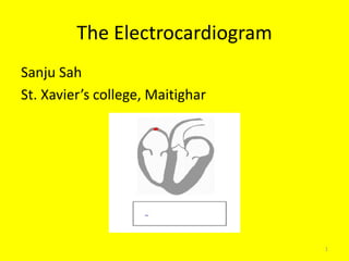
The Electrocardiogram
Sanju Sah
St. Xavier’s college, Maitighar
1
Electrocardiogram
• An electrocardiogram is a tracing of the
heart’s electrical activity
• An electrocardiograph is the machine that
produces it
Electrocardiograph
• Also called the ECG
machine, it detects
heart’s electrical activity
through electrodes
positioned on patient’s
skin
• Lead wires transfer
electrical activity back to
ECG machine where it is
displayed or printed onto
graph paper.
Lead wire and electrodes
• Lead wires and cables
transfer the ECG signal
detected through the
electrodes to the ECG
machine
– There may be 3, 4, or 5
lead wires for monitoring
purposes and up to 10 lead
wires for 12-lead ECGs
– Each lead wire has a
labeled clip, snap, or pin-
type connector on the
distal end which attaches
to the electrode
ECG Leads
• Are a combination of electrodes that form an
imaginary line in the body along which the
electrical signals, detectable during the time
course of the heartbeat, are measured
• Each lead provides a different view of the heart:
– Electrodes are placed on chest, arms and legs
– Sites vary depending on which view of the heart's
electrical activity is being assessed
• ECG leads are either bipolar or unipolar
Bipolar Leads
• Record the flow of
the electrical
impulse between
two (one is positive,
the other is
negative) selected
electrodes
• Includes I, II and III
Unipolar Leads
• Use only one
positive electrode
and a reference
point calculated by
the ECG machine
• Includes leads aVR,
aVL, aVF, and V1
through V6
• Electrodes are placed
on the extremities and
chest wall to view the
heart’s electrical
activity from the
frontal and horizontal
planes
– Provides a cross-
sectional view of the
heart
Graphical representation of Einthoven's triangle
» RA (Right Arm) -
Anywhere between the
right shoulder and right
elbow
» RL (Right Leg) - Anywhere
below the right torso and
above the right ankle
» LA(Left Arm) - Anywhere
between the left shoulder
and the left elbow
» LL (Left Leg) - Anywhere
below the left torso and
above the left ankle
• » V1 - Fourth intercostal
space on the right
sternum
» V2 - Fourth intercostal
space at the left sternum
» V3 - Midway between
placement of V2 and V4
» V4 - Fifth intercostal
space at the
midclavicular line
» V5 - Anterior axillary
line on the same
horizontal level as V4
» V6 - Mid-axillary line
on the same horizontal
level as V4 and V5
Effects of artifact on ECG recording
• Interference from the power line
• Shifting of baseline
• Muscle Tremor
Type of ECG recorders
• Single/ Three/ Six/ Twelve channel ECG
recorder
• channel ECG recorder
• ECG system for Stress Testing
• Holter recording
ELECTROENCEPHALOGRAPHY
(EEG)
Introduction
• Electroencephalography is a technique that
records the electrical activity of the brain
• The recording of the brains spontaneous
electrical activity is done over a short period
of time, usually 20–40 minutes, as recorded
from multiple electrodes placed on the scalp.
• Electroencephalograph is an instrument used for
recording of electrical activity of brain.
• The activity measured by EEG are electrical
potential created by the post-synaptic currents.
• Its an effective method for diagnosing many
neurological disorder such as epilepsy,
tumour,etc.
Principle
• The brains electrical charge is maintained by
billions of neurons.
• Neurons pass signals via action potential created
by exchange between sodium and potassium ions
in and out of the cell - Volume conduction.
• When the wave of ions reaches the electrodes on
the scalp, they can push or pull electrons on the
metal on the electrodes, the difference in push,
or voltage, between any two electrodes can be
measured by a voltmeter. Recording these
voltages over time gives us the EEG.
• Scalp EEG activity shows oscillations at a variety
of frequencies. Several of these oscillations have
characteristic frequency ranges, spatial
distributions and are associated with different
states of brain functioning.
Brain Wave Classification
• Brain patterns form wave shapes that are commonly
sinusoidal.
• Measured from peak to peak and normally range from
0.5 to 100 μV in amplitude.
• Signal is derived by means of Fourier transform power
spectrum from the raw.
• Brain waves have been categorized into four basic
groups;
- Beta (>13 hz)
- Alpha (8-13 hz)
- Theta (4-8 hz)
-Delta (0.5-4 Hz)
Applications
• Monitor alertness, coma and brain death
• Locate areas of damage following head injury,
stroke, tumor, etc.
• Test afferent pathways (by evoked potentials)
• Control anesthesia depth
• Investigate epilepsy and locate seizure origin
• Test epilepsy drug effects
Methodology
• Non-invasive and painless
• Major components;
– 1. Electrodes with conductive media
– 2. Amplifiers with filters
– 3. A/D converter
– 4. Recording device
• Electrodes read the signal from the head surface,
amplifiers bring the microvolt signals into the range
where they can be digitalized accurately, converter
changes signals from analog to digital form and
computer stores and displays obtained data.
Recording electrodes
• Types of electrodes:
1. Disposable (gel-less, and pre-gelled types)
2. Reusable disc electrodes (gold, silver, s.s. or tin)
3. Headbands and electrode caps
4. Saline-based electrodes
5. Needle electrodes
• Electrode caps are preferred, with certain number of
electrodes installed on its surface.
• Commonly used scalp electrodes consist of Ag-AgCl disks, 1 to
3 mm in diameter, with long flexible leads that can be plugged
into an amplifier.
• Needle electrodes are used for long recordings and are
invasively inserted under the scalp.
• Electrode locations and names are specified by the
International 10–20 system for most clinical and research
application
• Label 10-20 designates proportional distance in percents
between ears and nose where points for electrodes are
chosen.
• Electrode placements are labeled according adjacent brain
areas: F (frontal), C (central), T (temporal), P (posterior),
and O (occipital).
• The letters are accompanied by odd numbers at the left
side of the head and with even numbers on the right side.
• In general 25 electrodes are used in general EEG test but no
of electrode may vary as per the EEG requirement and area
of investigation In 25 electrode EEG system
– 23 electrode are active electrodes
– 1 is ground electrode
– 1 is ref electrode
Labels for points according to 10-20
electrode placement system
• Display of the EEG may be set up in one of
several ways. The representation of the EEG
channels is referred to as a montage.
1. Bipolar montage: Each channel (i.e., waveform)
represents the difference between two adjacent
electrodes. The entire montage consists of a series of
these channels.
2. Referential montage: Each channel represents the
difference between a certain electrode and a
designated reference electrode.
3. Average reference montage: The outputs of all of
the amplifiers are summed and averaged, and this
averaged signal is used as the common reference for
each channel.
4. Laplacian montage: Each channel represents the
difference between an electrode and a weighted
average of the surrounding electrodes.
Amplifiers and filters
• The input signal to the amplifier consists of five
components:
1.Desired biopotential
2.Undesired biopotential
3.A power line interference signal of 50/60 Hz and its harmonics
4.Interference signals generated by the tissue/electrode interface
5.Noise
• The A/D converter is interfaced to a computer system so
that channels of analog signal are converted into a digital
representation.
• Analog low-pass filters prevent distortion of the signal by
interference effects with sampling rate, called aliasing,
which would occur if frequencies greater than one half of
the sampling rate survive.
Artifacts
• Among basic evaluation of the EEG traces belongs
scanning for signal distortions called Artefacts.
• The Artefact in the recorded EEG may be either patient-
related or technical.
• Patient related: Technical:
– Any minor body movements - 50/60 hz
– EMG - Impedance fluctuation
– ECG (pulse, pace-maker) - Cable movements
– Eye movements - Broken wire contacts
– Sweating - Too much electrode paste/jelly
- Low battery
Evoked potentials
• Evoked potentials are used to measure the electrical
activity in certain areas of the brain and spinal cord.
Electrical activity is produced by stimulation of specific
sensory nerve pathways.
• Types of evoked potentials
– Visual Evoked Potentials (VEP): the patient sits before a
screen in which alternating patterns are displayed.
– Auditory Evoked Potentials (AEP): the patient listens to a
series of clicks in each ear.
– Sensory Evoked Potentials (SEP): short electrical impulses
are administered on the arm or leg.
– Motor Evoked Potentials: these can detect disruption on a
motor pathway of the brain or spinal cord.
ELECTROMYOGRAM
INTRODUCTION
• Electromyogram (EMG) is a technique for
evaluating and recording the electrical activation
signal of muscles.
• EMG is performed by an electromyograph, which
records an electromyogram.
• Electromyograph detects the electrical potential
generated by muscle cells when these cells contract
and relax.
EMG Apparatus Muscle Structure/EMG
EMG PROCEDURE
• Clean the site of application
of electrode;
• Insert needle/place surface
electrodes at muscle belly;
• Record muscle activity at rest;
• Record muscle activity upon
voluntary contraction of the
muscle.
EMG Contd.
• Muscle Signals are
Analog in nature.
• EMG signals are also
collected over a
specific period of
time.
Analog Signal
EMG Contd.
EMG processing:
Amplification
& Filtering
Signal pick up
Conversion of Analog
signals to Digital signals
Computer
Types of EMG
• Electrode Categories
– Inserted
• Fine-wire (Intra-muscular)
• Needle
– Surface
EMG Electrodes
Fine wire
Surface
Electrodes
Needle
electrode
Fine-wire Electrodes
• Advantages
– Extremely sensitive
– Record single muscle activity
– Access to deep musculature
– Little cross-talk concern
• Disadvantages
– Extremely sensitive
– Requires medical personnel, certification
– Repositioning nearly impossible
– Detection area may not be representative of entire muscle
Surface Electrodes
• Advantages
– Quick, easy to apply
– No medical supervision, required certification
– Minimal discomfort
• Disadvantages
– Generally used only for superficial muscles
– Cross-talk concerns
– No standard electrode placement
– May affect movement patterns of subject
– Limitations with recording dynamic muscle activity
General Concerns
• Signal-to-noise ratio
– Ratio of energy of EMG signal divided by energy of
noise signal
• Distortion of the signal
– EMG signal should be altered as minimally as
possible for accurate representation
Characteristics of EMG Signal
• Amplitude range: 0–10
mV (+5 to -5) prior to
amplification
• Useable energy: Range
of 0 - 500 Hz
• Dominant energy: 50 –
150 Hz
Characteristics of Electrical Noise
• Inherent noise in electronics equipment
• Ambient noise
• Motion artifact
• Inherent instability of signal
Inherent Noise in Electronics
Equipment
• Generated by all electronics equipment
• Frequency range: 0 – several thousand Hz
• Cannot be eliminated
• Reduced by using high quality components
Ambient Noise
• Electromagnetic radiation sources
– Radio transmission
– Electrical wires
– Fluorescent lights
• Essentially impossible to avoid
• Dominant frequency: 60 Hz
• Amplitude: 1 – 3x EMG signal
Motion Artifact
• Two main sources
– Electrode/skin interface
– Electrode cable
• Reducible by proper circuitry and set-up
• Frequency range: 0 – 20 Hz
Inherent Instability of Signal
• Amplitude is somewhat random in nature
• Frequency range of 0 – 20 Hz is especially
unstable
• Therefore, removal of this range is
recommended
Solutions for Signal Interruption Related to
Electrode and Amplifier Design
• Differential amplification
– Reduces electromagnetic radiation noise
– Dual electrodes
• Electrode stability
– Time for chemical reaction to stabilize
– Important factors: electrode movement, perspiration,
humidity changes
• Improved quality of electrodes
– Less need for skin abrasion, hair removal
Differential Amplification
• Ambient
(electromagnetic) noise
is constant
• System subtracts two
signals
• Resultant difference is
amplified
• Double differential
technique
Electrode Placement
• Away from tendon
– Fewer, thinner muscle fibers
– Closer to other muscle origins, insertions
• More susceptible to cross-talk
• Away from outer edge of muscle
– Closer to other musculature
• Orientation parallel to muscle fibers
– More accurate conduction velocity
– Increased probability of detecting same signal
EMG Electrode Placement
Surface Electrode Placement
Reference Electrode Placement
(Ground)
• As far away as possible from recording
electrodes
• Electrically neutral tissue
– Bony prominence
APPLICATION OF EMG
• EMG can be used for diagnosis of Neurogenic
or Myogenic Diseases.
Electrocardiogram pptx

Contenu connexe

Tendances

EEG - Montages, Equipment and Basic Physics
EEG - Montages, Equipment and Basic PhysicsEEG - Montages, Equipment and Basic Physics
EEG - Montages, Equipment and Basic PhysicsRahul Kumar
 
Electroencephalogram(EEG)
Electroencephalogram(EEG)Electroencephalogram(EEG)
Electroencephalogram(EEG)ashikh
 
A review of eeg recording techniques
A review of eeg recording techniquesA review of eeg recording techniques
A review of eeg recording techniquesiaemedu
 
Bioelectric potentials
Bioelectric potentialsBioelectric potentials
Bioelectric potentialssrirenga
 
various type of artifacts in ECG signal & how it's remove
various type of artifacts in ECG signal & how it's removevarious type of artifacts in ECG signal & how it's remove
various type of artifacts in ECG signal & how it's removeManish Kumar
 
Application of eeg wave
Application  of  eeg  waveApplication  of  eeg  wave
Application of eeg waveRohit vijay
 
Electroencephalogram (eeg)
Electroencephalogram (eeg)Electroencephalogram (eeg)
Electroencephalogram (eeg)divyabms
 
Electroencephalogram (EEG)
Electroencephalogram (EEG)Electroencephalogram (EEG)
Electroencephalogram (EEG)med zar
 
Introduction to EEG: Instrument and Acquisition
Introduction to EEG: Instrument and AcquisitionIntroduction to EEG: Instrument and Acquisition
Introduction to EEG: Instrument and Acquisitionkj_jantzen
 
Removal of artifacts in EEG by averaging and
Removal of artifacts in EEG by averaging andRemoval of artifacts in EEG by averaging and
Removal of artifacts in EEG by averaging andNamratha Dcruz
 

Tendances (20)

EEG - Montages, Equipment and Basic Physics
EEG - Montages, Equipment and Basic PhysicsEEG - Montages, Equipment and Basic Physics
EEG - Montages, Equipment and Basic Physics
 
Eeg from a to z
Eeg from a to zEeg from a to z
Eeg from a to z
 
EEG
EEGEEG
EEG
 
Eeg
EegEeg
Eeg
 
Eeg presentation
Eeg presentationEeg presentation
Eeg presentation
 
Electroencephalogram(EEG)
Electroencephalogram(EEG)Electroencephalogram(EEG)
Electroencephalogram(EEG)
 
A review of eeg recording techniques
A review of eeg recording techniquesA review of eeg recording techniques
A review of eeg recording techniques
 
Eeg by prc
Eeg by prcEeg by prc
Eeg by prc
 
Bioelectric potentials
Bioelectric potentialsBioelectric potentials
Bioelectric potentials
 
various type of artifacts in ECG signal & how it's remove
various type of artifacts in ECG signal & how it's removevarious type of artifacts in ECG signal & how it's remove
various type of artifacts in ECG signal & how it's remove
 
Application of eeg wave
Application  of  eeg  waveApplication  of  eeg  wave
Application of eeg wave
 
EEG
EEGEEG
EEG
 
Electroencephalogram (eeg)
Electroencephalogram (eeg)Electroencephalogram (eeg)
Electroencephalogram (eeg)
 
Electroencephalogram (EEG)
Electroencephalogram (EEG)Electroencephalogram (EEG)
Electroencephalogram (EEG)
 
Introduction to EEG: Instrument and Acquisition
Introduction to EEG: Instrument and AcquisitionIntroduction to EEG: Instrument and Acquisition
Introduction to EEG: Instrument and Acquisition
 
Removal of artifacts in EEG by averaging and
Removal of artifacts in EEG by averaging andRemoval of artifacts in EEG by averaging and
Removal of artifacts in EEG by averaging and
 
EEG INTERPRETATION
EEG INTERPRETATIONEEG INTERPRETATION
EEG INTERPRETATION
 
EEG
EEG EEG
EEG
 
EEG artifacts
EEG artifactsEEG artifacts
EEG artifacts
 
EEG
EEGEEG
EEG
 

Similaire à Electrocardiogram pptx (20)

unit i
 unit i unit i
unit i
 
ELECTROCARDIOGRAM. pptx
ELECTROCARDIOGRAM. pptxELECTROCARDIOGRAM. pptx
ELECTROCARDIOGRAM. pptx
 
ECG Fundamentals.pptx
ECG Fundamentals.pptxECG Fundamentals.pptx
ECG Fundamentals.pptx
 
Basics of EEG
Basics of EEGBasics of EEG
Basics of EEG
 
ELECTROCARDIOGRAM
ELECTROCARDIOGRAMELECTROCARDIOGRAM
ELECTROCARDIOGRAM
 
Biomedical Signals
Biomedical SignalsBiomedical Signals
Biomedical Signals
 
Lecture 3 - taking -ecg.pptx
Lecture 3 - taking -ecg.pptxLecture 3 - taking -ecg.pptx
Lecture 3 - taking -ecg.pptx
 
Cardiac monitoring.pdf
Cardiac monitoring.pdfCardiac monitoring.pdf
Cardiac monitoring.pdf
 
SUMSEM-2021-22_ECE6007_ETH_VL2021220701295_Reference_Material_I_04-07-2022_EE...
SUMSEM-2021-22_ECE6007_ETH_VL2021220701295_Reference_Material_I_04-07-2022_EE...SUMSEM-2021-22_ECE6007_ETH_VL2021220701295_Reference_Material_I_04-07-2022_EE...
SUMSEM-2021-22_ECE6007_ETH_VL2021220701295_Reference_Material_I_04-07-2022_EE...
 
Components of Pacemaker and ICDs - understanding the hardware
Components of Pacemaker and ICDs - understanding the hardwareComponents of Pacemaker and ICDs - understanding the hardware
Components of Pacemaker and ICDs - understanding the hardware
 
Eeg examples
Eeg examplesEeg examples
Eeg examples
 
ECG.pptx
ECG.pptxECG.pptx
ECG.pptx
 
ECG-Electrocardiography
ECG-ElectrocardiographyECG-Electrocardiography
ECG-Electrocardiography
 
Hawzhin
HawzhinHawzhin
Hawzhin
 
ELECTROMYOGRAPHY.pptx
ELECTROMYOGRAPHY.pptxELECTROMYOGRAPHY.pptx
ELECTROMYOGRAPHY.pptx
 
Eeg artifacts
Eeg artifactsEeg artifacts
Eeg artifacts
 
ECG Machine
ECG MachineECG Machine
ECG Machine
 
Ecg123 (1)
Ecg123 (1)Ecg123 (1)
Ecg123 (1)
 
Eeg
EegEeg
Eeg
 
8101222.ppt
8101222.ppt8101222.ppt
8101222.ppt
 

Plus de St. Xavier's college, maitighar,Kathmandu

Plus de St. Xavier's college, maitighar,Kathmandu (20)

Orders of class osteichthyes
Orders of class osteichthyesOrders of class osteichthyes
Orders of class osteichthyes
 
Mammals stomach and dentition
Mammals stomach and dentitionMammals stomach and dentition
Mammals stomach and dentition
 
Origin and Evolution of Mammals
Origin and Evolution of MammalsOrigin and Evolution of Mammals
Origin and Evolution of Mammals
 
Draco volans
Draco volansDraco volans
Draco volans
 
Digestive system of vertebrates
Digestive system of vertebratesDigestive system of vertebrates
Digestive system of vertebrates
 
Common peafowl
Common peafowlCommon peafowl
Common peafowl
 
Brain of vertebrates
Brain of vertebratesBrain of vertebrates
Brain of vertebrates
 
Integument
IntegumentIntegument
Integument
 
Water
WaterWater
Water
 
Translation
TranslationTranslation
Translation
 
The structure of dna and genome ogranization
The structure of dna and genome ogranization The structure of dna and genome ogranization
The structure of dna and genome ogranization
 
Plasmodium
PlasmodiumPlasmodium
Plasmodium
 
Comparative study of vertebrates
Comparative  study of vertebratesComparative  study of vertebrates
Comparative study of vertebrates
 
The nucleic acids
The nucleic acidsThe nucleic acids
The nucleic acids
 
Honey bee
Honey beeHoney bee
Honey bee
 
Genetic engineering of humans
Genetic engineering of humansGenetic engineering of humans
Genetic engineering of humans
 
Hazards of genetic engineering
Hazards of genetic engineeringHazards of genetic engineering
Hazards of genetic engineering
 
Azotobacter spp.
Azotobacter spp.Azotobacter spp.
Azotobacter spp.
 
Biological membrane
Biological membraneBiological membrane
Biological membrane
 
Atomic radius
Atomic radiusAtomic radius
Atomic radius
 

Dernier

METHODS OF ACQUIRING KNOWLEDGE IN NURSING.pptx by navdeep kaur
METHODS OF ACQUIRING KNOWLEDGE IN NURSING.pptx by navdeep kaurMETHODS OF ACQUIRING KNOWLEDGE IN NURSING.pptx by navdeep kaur
METHODS OF ACQUIRING KNOWLEDGE IN NURSING.pptx by navdeep kaurNavdeep Kaur
 
Primary headache and facial pain. (2024)
Primary headache and facial pain. (2024)Primary headache and facial pain. (2024)
Primary headache and facial pain. (2024)Mohamed Rizk Khodair
 
PERFECT BUT PAINFUL TKR -ROLE OF SYNOVECTOMY.pptx
PERFECT BUT PAINFUL TKR -ROLE OF SYNOVECTOMY.pptxPERFECT BUT PAINFUL TKR -ROLE OF SYNOVECTOMY.pptx
PERFECT BUT PAINFUL TKR -ROLE OF SYNOVECTOMY.pptxdrashraf369
 
Big Data Analysis Suggests COVID Vaccination Increases Excess Mortality Of ...
Big Data Analysis Suggests COVID  Vaccination Increases Excess Mortality Of  ...Big Data Analysis Suggests COVID  Vaccination Increases Excess Mortality Of  ...
Big Data Analysis Suggests COVID Vaccination Increases Excess Mortality Of ...sdateam0
 
The next social challenge to public health: the information environment.pptx
The next social challenge to public health:  the information environment.pptxThe next social challenge to public health:  the information environment.pptx
The next social challenge to public health: the information environment.pptxTina Purnat
 
Presentation on General Anesthetics pdf.
Presentation on General Anesthetics pdf.Presentation on General Anesthetics pdf.
Presentation on General Anesthetics pdf.Prerana Jadhav
 
97111 47426 Call Girls In Delhi MUNIRKAA
97111 47426 Call Girls In Delhi MUNIRKAA97111 47426 Call Girls In Delhi MUNIRKAA
97111 47426 Call Girls In Delhi MUNIRKAAjennyeacort
 
April 2024 ONCOLOGY CARTOON by DR KANHU CHARAN PATRO
April 2024 ONCOLOGY CARTOON by  DR KANHU CHARAN PATROApril 2024 ONCOLOGY CARTOON by  DR KANHU CHARAN PATRO
April 2024 ONCOLOGY CARTOON by DR KANHU CHARAN PATROKanhu Charan
 
Culture and Health Disorders Social change.pptx
Culture and Health Disorders Social change.pptxCulture and Health Disorders Social change.pptx
Culture and Health Disorders Social change.pptxDr. Dheeraj Kumar
 
Lippincott Microcards_ Microbiology Flash Cards-LWW (2015).pdf
Lippincott Microcards_ Microbiology Flash Cards-LWW (2015).pdfLippincott Microcards_ Microbiology Flash Cards-LWW (2015).pdf
Lippincott Microcards_ Microbiology Flash Cards-LWW (2015).pdfSreeja Cherukuru
 
Clinical Pharmacotherapy of Scabies Disease
Clinical Pharmacotherapy of Scabies DiseaseClinical Pharmacotherapy of Scabies Disease
Clinical Pharmacotherapy of Scabies DiseaseSreenivasa Reddy Thalla
 
Study on the Impact of FOCUS-PDCA Management Model on the Disinfection Qualit...
Study on the Impact of FOCUS-PDCA Management Model on the Disinfection Qualit...Study on the Impact of FOCUS-PDCA Management Model on the Disinfection Qualit...
Study on the Impact of FOCUS-PDCA Management Model on the Disinfection Qualit...MehranMouzam
 
History and Development of Pharmacovigilence.pdf
History and Development of Pharmacovigilence.pdfHistory and Development of Pharmacovigilence.pdf
History and Development of Pharmacovigilence.pdfSasikiranMarri
 
SYNDESMOTIC INJURY- ANATOMICAL REPAIR.pptx
SYNDESMOTIC INJURY- ANATOMICAL REPAIR.pptxSYNDESMOTIC INJURY- ANATOMICAL REPAIR.pptx
SYNDESMOTIC INJURY- ANATOMICAL REPAIR.pptxdrashraf369
 
Music Therapy's Impact in Palliative Care| IAPCON2024| Dr. Tara Rajendran
Music Therapy's Impact in Palliative Care| IAPCON2024| Dr. Tara RajendranMusic Therapy's Impact in Palliative Care| IAPCON2024| Dr. Tara Rajendran
Music Therapy's Impact in Palliative Care| IAPCON2024| Dr. Tara RajendranTara Rajendran
 
maternal mortality and its causes and how to reduce maternal mortality
maternal mortality and its causes and how to reduce maternal mortalitymaternal mortality and its causes and how to reduce maternal mortality
maternal mortality and its causes and how to reduce maternal mortalityhardikdabas3
 
CEHPALOSPORINS.pptx By Harshvardhan Dev Bhoomi Uttarakhand University
CEHPALOSPORINS.pptx By Harshvardhan Dev Bhoomi Uttarakhand UniversityCEHPALOSPORINS.pptx By Harshvardhan Dev Bhoomi Uttarakhand University
CEHPALOSPORINS.pptx By Harshvardhan Dev Bhoomi Uttarakhand UniversityHarshChauhan475104
 
Radiation Dosimetry Parameters and Isodose Curves.pptx
Radiation Dosimetry Parameters and Isodose Curves.pptxRadiation Dosimetry Parameters and Isodose Curves.pptx
Radiation Dosimetry Parameters and Isodose Curves.pptxDr. Dheeraj Kumar
 
VarSeq 2.6.0: Advancing Pharmacogenomics and Genomic Analysis
VarSeq 2.6.0: Advancing Pharmacogenomics and Genomic AnalysisVarSeq 2.6.0: Advancing Pharmacogenomics and Genomic Analysis
VarSeq 2.6.0: Advancing Pharmacogenomics and Genomic AnalysisGolden Helix
 

Dernier (20)

METHODS OF ACQUIRING KNOWLEDGE IN NURSING.pptx by navdeep kaur
METHODS OF ACQUIRING KNOWLEDGE IN NURSING.pptx by navdeep kaurMETHODS OF ACQUIRING KNOWLEDGE IN NURSING.pptx by navdeep kaur
METHODS OF ACQUIRING KNOWLEDGE IN NURSING.pptx by navdeep kaur
 
Primary headache and facial pain. (2024)
Primary headache and facial pain. (2024)Primary headache and facial pain. (2024)
Primary headache and facial pain. (2024)
 
PERFECT BUT PAINFUL TKR -ROLE OF SYNOVECTOMY.pptx
PERFECT BUT PAINFUL TKR -ROLE OF SYNOVECTOMY.pptxPERFECT BUT PAINFUL TKR -ROLE OF SYNOVECTOMY.pptx
PERFECT BUT PAINFUL TKR -ROLE OF SYNOVECTOMY.pptx
 
Big Data Analysis Suggests COVID Vaccination Increases Excess Mortality Of ...
Big Data Analysis Suggests COVID  Vaccination Increases Excess Mortality Of  ...Big Data Analysis Suggests COVID  Vaccination Increases Excess Mortality Of  ...
Big Data Analysis Suggests COVID Vaccination Increases Excess Mortality Of ...
 
The next social challenge to public health: the information environment.pptx
The next social challenge to public health:  the information environment.pptxThe next social challenge to public health:  the information environment.pptx
The next social challenge to public health: the information environment.pptx
 
Presentation on General Anesthetics pdf.
Presentation on General Anesthetics pdf.Presentation on General Anesthetics pdf.
Presentation on General Anesthetics pdf.
 
97111 47426 Call Girls In Delhi MUNIRKAA
97111 47426 Call Girls In Delhi MUNIRKAA97111 47426 Call Girls In Delhi MUNIRKAA
97111 47426 Call Girls In Delhi MUNIRKAA
 
April 2024 ONCOLOGY CARTOON by DR KANHU CHARAN PATRO
April 2024 ONCOLOGY CARTOON by  DR KANHU CHARAN PATROApril 2024 ONCOLOGY CARTOON by  DR KANHU CHARAN PATRO
April 2024 ONCOLOGY CARTOON by DR KANHU CHARAN PATRO
 
Culture and Health Disorders Social change.pptx
Culture and Health Disorders Social change.pptxCulture and Health Disorders Social change.pptx
Culture and Health Disorders Social change.pptx
 
Lippincott Microcards_ Microbiology Flash Cards-LWW (2015).pdf
Lippincott Microcards_ Microbiology Flash Cards-LWW (2015).pdfLippincott Microcards_ Microbiology Flash Cards-LWW (2015).pdf
Lippincott Microcards_ Microbiology Flash Cards-LWW (2015).pdf
 
Clinical Pharmacotherapy of Scabies Disease
Clinical Pharmacotherapy of Scabies DiseaseClinical Pharmacotherapy of Scabies Disease
Clinical Pharmacotherapy of Scabies Disease
 
Study on the Impact of FOCUS-PDCA Management Model on the Disinfection Qualit...
Study on the Impact of FOCUS-PDCA Management Model on the Disinfection Qualit...Study on the Impact of FOCUS-PDCA Management Model on the Disinfection Qualit...
Study on the Impact of FOCUS-PDCA Management Model on the Disinfection Qualit...
 
History and Development of Pharmacovigilence.pdf
History and Development of Pharmacovigilence.pdfHistory and Development of Pharmacovigilence.pdf
History and Development of Pharmacovigilence.pdf
 
SYNDESMOTIC INJURY- ANATOMICAL REPAIR.pptx
SYNDESMOTIC INJURY- ANATOMICAL REPAIR.pptxSYNDESMOTIC INJURY- ANATOMICAL REPAIR.pptx
SYNDESMOTIC INJURY- ANATOMICAL REPAIR.pptx
 
Music Therapy's Impact in Palliative Care| IAPCON2024| Dr. Tara Rajendran
Music Therapy's Impact in Palliative Care| IAPCON2024| Dr. Tara RajendranMusic Therapy's Impact in Palliative Care| IAPCON2024| Dr. Tara Rajendran
Music Therapy's Impact in Palliative Care| IAPCON2024| Dr. Tara Rajendran
 
maternal mortality and its causes and how to reduce maternal mortality
maternal mortality and its causes and how to reduce maternal mortalitymaternal mortality and its causes and how to reduce maternal mortality
maternal mortality and its causes and how to reduce maternal mortality
 
CEHPALOSPORINS.pptx By Harshvardhan Dev Bhoomi Uttarakhand University
CEHPALOSPORINS.pptx By Harshvardhan Dev Bhoomi Uttarakhand UniversityCEHPALOSPORINS.pptx By Harshvardhan Dev Bhoomi Uttarakhand University
CEHPALOSPORINS.pptx By Harshvardhan Dev Bhoomi Uttarakhand University
 
Radiation Dosimetry Parameters and Isodose Curves.pptx
Radiation Dosimetry Parameters and Isodose Curves.pptxRadiation Dosimetry Parameters and Isodose Curves.pptx
Radiation Dosimetry Parameters and Isodose Curves.pptx
 
VarSeq 2.6.0: Advancing Pharmacogenomics and Genomic Analysis
VarSeq 2.6.0: Advancing Pharmacogenomics and Genomic AnalysisVarSeq 2.6.0: Advancing Pharmacogenomics and Genomic Analysis
VarSeq 2.6.0: Advancing Pharmacogenomics and Genomic Analysis
 
Epilepsy
EpilepsyEpilepsy
Epilepsy
 

Electrocardiogram pptx

  • 1. The Electrocardiogram Sanju Sah St. Xavier’s college, Maitighar 1
  • 2. Electrocardiogram • An electrocardiogram is a tracing of the heart’s electrical activity • An electrocardiograph is the machine that produces it
  • 3. Electrocardiograph • Also called the ECG machine, it detects heart’s electrical activity through electrodes positioned on patient’s skin • Lead wires transfer electrical activity back to ECG machine where it is displayed or printed onto graph paper.
  • 4.
  • 5.
  • 6. Lead wire and electrodes • Lead wires and cables transfer the ECG signal detected through the electrodes to the ECG machine – There may be 3, 4, or 5 lead wires for monitoring purposes and up to 10 lead wires for 12-lead ECGs – Each lead wire has a labeled clip, snap, or pin- type connector on the distal end which attaches to the electrode
  • 7. ECG Leads • Are a combination of electrodes that form an imaginary line in the body along which the electrical signals, detectable during the time course of the heartbeat, are measured • Each lead provides a different view of the heart: – Electrodes are placed on chest, arms and legs – Sites vary depending on which view of the heart's electrical activity is being assessed • ECG leads are either bipolar or unipolar
  • 8. Bipolar Leads • Record the flow of the electrical impulse between two (one is positive, the other is negative) selected electrodes • Includes I, II and III
  • 9.
  • 10. Unipolar Leads • Use only one positive electrode and a reference point calculated by the ECG machine • Includes leads aVR, aVL, aVF, and V1 through V6
  • 11. • Electrodes are placed on the extremities and chest wall to view the heart’s electrical activity from the frontal and horizontal planes – Provides a cross- sectional view of the heart
  • 12.
  • 13.
  • 14. Graphical representation of Einthoven's triangle
  • 15. » RA (Right Arm) - Anywhere between the right shoulder and right elbow » RL (Right Leg) - Anywhere below the right torso and above the right ankle » LA(Left Arm) - Anywhere between the left shoulder and the left elbow » LL (Left Leg) - Anywhere below the left torso and above the left ankle
  • 16. • » V1 - Fourth intercostal space on the right sternum » V2 - Fourth intercostal space at the left sternum » V3 - Midway between placement of V2 and V4 » V4 - Fifth intercostal space at the midclavicular line » V5 - Anterior axillary line on the same horizontal level as V4 » V6 - Mid-axillary line on the same horizontal level as V4 and V5
  • 17. Effects of artifact on ECG recording • Interference from the power line • Shifting of baseline • Muscle Tremor
  • 18.
  • 19. Type of ECG recorders • Single/ Three/ Six/ Twelve channel ECG recorder • channel ECG recorder • ECG system for Stress Testing • Holter recording
  • 20.
  • 22. Introduction • Electroencephalography is a technique that records the electrical activity of the brain • The recording of the brains spontaneous electrical activity is done over a short period of time, usually 20–40 minutes, as recorded from multiple electrodes placed on the scalp.
  • 23. • Electroencephalograph is an instrument used for recording of electrical activity of brain. • The activity measured by EEG are electrical potential created by the post-synaptic currents. • Its an effective method for diagnosing many neurological disorder such as epilepsy, tumour,etc.
  • 24. Principle • The brains electrical charge is maintained by billions of neurons. • Neurons pass signals via action potential created by exchange between sodium and potassium ions in and out of the cell - Volume conduction. • When the wave of ions reaches the electrodes on the scalp, they can push or pull electrons on the metal on the electrodes, the difference in push, or voltage, between any two electrodes can be measured by a voltmeter. Recording these voltages over time gives us the EEG.
  • 25. • Scalp EEG activity shows oscillations at a variety of frequencies. Several of these oscillations have characteristic frequency ranges, spatial distributions and are associated with different states of brain functioning.
  • 26. Brain Wave Classification • Brain patterns form wave shapes that are commonly sinusoidal. • Measured from peak to peak and normally range from 0.5 to 100 μV in amplitude. • Signal is derived by means of Fourier transform power spectrum from the raw. • Brain waves have been categorized into four basic groups; - Beta (>13 hz) - Alpha (8-13 hz) - Theta (4-8 hz) -Delta (0.5-4 Hz)
  • 27. Applications • Monitor alertness, coma and brain death • Locate areas of damage following head injury, stroke, tumor, etc. • Test afferent pathways (by evoked potentials) • Control anesthesia depth • Investigate epilepsy and locate seizure origin • Test epilepsy drug effects
  • 28. Methodology • Non-invasive and painless • Major components; – 1. Electrodes with conductive media – 2. Amplifiers with filters – 3. A/D converter – 4. Recording device • Electrodes read the signal from the head surface, amplifiers bring the microvolt signals into the range where they can be digitalized accurately, converter changes signals from analog to digital form and computer stores and displays obtained data.
  • 29. Recording electrodes • Types of electrodes: 1. Disposable (gel-less, and pre-gelled types) 2. Reusable disc electrodes (gold, silver, s.s. or tin) 3. Headbands and electrode caps 4. Saline-based electrodes 5. Needle electrodes • Electrode caps are preferred, with certain number of electrodes installed on its surface. • Commonly used scalp electrodes consist of Ag-AgCl disks, 1 to 3 mm in diameter, with long flexible leads that can be plugged into an amplifier. • Needle electrodes are used for long recordings and are invasively inserted under the scalp.
  • 30. • Electrode locations and names are specified by the International 10–20 system for most clinical and research application • Label 10-20 designates proportional distance in percents between ears and nose where points for electrodes are chosen. • Electrode placements are labeled according adjacent brain areas: F (frontal), C (central), T (temporal), P (posterior), and O (occipital). • The letters are accompanied by odd numbers at the left side of the head and with even numbers on the right side. • In general 25 electrodes are used in general EEG test but no of electrode may vary as per the EEG requirement and area of investigation In 25 electrode EEG system – 23 electrode are active electrodes – 1 is ground electrode – 1 is ref electrode
  • 31. Labels for points according to 10-20 electrode placement system
  • 32.
  • 33. • Display of the EEG may be set up in one of several ways. The representation of the EEG channels is referred to as a montage. 1. Bipolar montage: Each channel (i.e., waveform) represents the difference between two adjacent electrodes. The entire montage consists of a series of these channels. 2. Referential montage: Each channel represents the difference between a certain electrode and a designated reference electrode. 3. Average reference montage: The outputs of all of the amplifiers are summed and averaged, and this averaged signal is used as the common reference for each channel. 4. Laplacian montage: Each channel represents the difference between an electrode and a weighted average of the surrounding electrodes.
  • 34. Amplifiers and filters • The input signal to the amplifier consists of five components: 1.Desired biopotential 2.Undesired biopotential 3.A power line interference signal of 50/60 Hz and its harmonics 4.Interference signals generated by the tissue/electrode interface 5.Noise • The A/D converter is interfaced to a computer system so that channels of analog signal are converted into a digital representation. • Analog low-pass filters prevent distortion of the signal by interference effects with sampling rate, called aliasing, which would occur if frequencies greater than one half of the sampling rate survive.
  • 35. Artifacts • Among basic evaluation of the EEG traces belongs scanning for signal distortions called Artefacts. • The Artefact in the recorded EEG may be either patient- related or technical. • Patient related: Technical: – Any minor body movements - 50/60 hz – EMG - Impedance fluctuation – ECG (pulse, pace-maker) - Cable movements – Eye movements - Broken wire contacts – Sweating - Too much electrode paste/jelly - Low battery
  • 36. Evoked potentials • Evoked potentials are used to measure the electrical activity in certain areas of the brain and spinal cord. Electrical activity is produced by stimulation of specific sensory nerve pathways. • Types of evoked potentials – Visual Evoked Potentials (VEP): the patient sits before a screen in which alternating patterns are displayed. – Auditory Evoked Potentials (AEP): the patient listens to a series of clicks in each ear. – Sensory Evoked Potentials (SEP): short electrical impulses are administered on the arm or leg. – Motor Evoked Potentials: these can detect disruption on a motor pathway of the brain or spinal cord.
  • 37.
  • 39. INTRODUCTION • Electromyogram (EMG) is a technique for evaluating and recording the electrical activation signal of muscles. • EMG is performed by an electromyograph, which records an electromyogram. • Electromyograph detects the electrical potential generated by muscle cells when these cells contract and relax.
  • 40. EMG Apparatus Muscle Structure/EMG
  • 41. EMG PROCEDURE • Clean the site of application of electrode; • Insert needle/place surface electrodes at muscle belly; • Record muscle activity at rest; • Record muscle activity upon voluntary contraction of the muscle.
  • 42. EMG Contd. • Muscle Signals are Analog in nature. • EMG signals are also collected over a specific period of time. Analog Signal
  • 43. EMG Contd. EMG processing: Amplification & Filtering Signal pick up Conversion of Analog signals to Digital signals Computer
  • 44. Types of EMG • Electrode Categories – Inserted • Fine-wire (Intra-muscular) • Needle – Surface
  • 46. Fine-wire Electrodes • Advantages – Extremely sensitive – Record single muscle activity – Access to deep musculature – Little cross-talk concern • Disadvantages – Extremely sensitive – Requires medical personnel, certification – Repositioning nearly impossible – Detection area may not be representative of entire muscle
  • 47. Surface Electrodes • Advantages – Quick, easy to apply – No medical supervision, required certification – Minimal discomfort • Disadvantages – Generally used only for superficial muscles – Cross-talk concerns – No standard electrode placement – May affect movement patterns of subject – Limitations with recording dynamic muscle activity
  • 48. General Concerns • Signal-to-noise ratio – Ratio of energy of EMG signal divided by energy of noise signal • Distortion of the signal – EMG signal should be altered as minimally as possible for accurate representation
  • 49. Characteristics of EMG Signal • Amplitude range: 0–10 mV (+5 to -5) prior to amplification • Useable energy: Range of 0 - 500 Hz • Dominant energy: 50 – 150 Hz
  • 50. Characteristics of Electrical Noise • Inherent noise in electronics equipment • Ambient noise • Motion artifact • Inherent instability of signal
  • 51. Inherent Noise in Electronics Equipment • Generated by all electronics equipment • Frequency range: 0 – several thousand Hz • Cannot be eliminated • Reduced by using high quality components
  • 52. Ambient Noise • Electromagnetic radiation sources – Radio transmission – Electrical wires – Fluorescent lights • Essentially impossible to avoid • Dominant frequency: 60 Hz • Amplitude: 1 – 3x EMG signal
  • 53. Motion Artifact • Two main sources – Electrode/skin interface – Electrode cable • Reducible by proper circuitry and set-up • Frequency range: 0 – 20 Hz
  • 54. Inherent Instability of Signal • Amplitude is somewhat random in nature • Frequency range of 0 – 20 Hz is especially unstable • Therefore, removal of this range is recommended
  • 55. Solutions for Signal Interruption Related to Electrode and Amplifier Design • Differential amplification – Reduces electromagnetic radiation noise – Dual electrodes • Electrode stability – Time for chemical reaction to stabilize – Important factors: electrode movement, perspiration, humidity changes • Improved quality of electrodes – Less need for skin abrasion, hair removal
  • 56. Differential Amplification • Ambient (electromagnetic) noise is constant • System subtracts two signals • Resultant difference is amplified • Double differential technique
  • 57. Electrode Placement • Away from tendon – Fewer, thinner muscle fibers – Closer to other muscle origins, insertions • More susceptible to cross-talk • Away from outer edge of muscle – Closer to other musculature • Orientation parallel to muscle fibers – More accurate conduction velocity – Increased probability of detecting same signal
  • 60. Reference Electrode Placement (Ground) • As far away as possible from recording electrodes • Electrically neutral tissue – Bony prominence
  • 61. APPLICATION OF EMG • EMG can be used for diagnosis of Neurogenic or Myogenic Diseases.