On National Teacher Day, meet the 2024-25 Kenan Fellows
Egg activation
1. Dr. Shoeb Ahmad (Assistant Professor), AKI’s Poona College of Arts, Science & Commerce, Camp, Pune-01, Maharashtra Page 1
Egg Activation: Cell Cycle Regulation and Utilization of Maternal
Macromolecules and Organelles During Early Development
Amphibain oocytes can remain for years in the diplotene stage of meiotic
prophase. This state resembles the G2 phase of the cell division cycle.
Resumption of meiosis in the amphibian primary oocyte requires progesterone.
This hormone is secreted by the follicle cells in response to gonadotropic
hormones secreted by the pituitary gland. Within 6 hours of progesterone
stimulation, germinal vesicle breakdown (GVBD) occurs, the microvilli
retract, the nucleoli disintegrate, and the chromosomes contract and migrate to
the animal pole to begin division. Soon afterward, the first meiotic division
occurs, and the mature ovum is released from the ovary by a process called
ovulation. The ovulated egg is in second meiotic metaphase when it is released
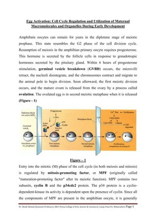
(Figure - 1)
Figure – 1
Entry into the mitotic (M) phase of the cell cycle (in both meiosis and mitosis)
is regulated by mitosis-promoting factor, or MPF (originally called
"maturation-promoting factor" after its meiotic function). MPF contains two
subunits, cyclin B and the p34cdc2 protein. The p34 protein is a cyclin-
dependent-kinase its activity is dependent upon the presence of cyclin. Since all
the components of MPF are present in the amphibian oocyte, it is generally
2. Dr. Shoeb Ahmad (Assistant Professor), AKI’s Poona College of Arts, Science & Commerce, Camp, Pune-01, Maharashtra Page 2
thought that progesterone somehow converts a pre-MPF complex into active
MPF
The mediator of the progesterone signal is the c-mos protein. Progesterone
reinitiates meiosis by causing the egg to polyadenylate the maternal c-mos
mRNA that has been stored in its cytoplasm. This message is translated into a
39-kDa phosphoprotein, known as c-mos. This protein is detectable only during
oocyte maturation and is destroyed quickly upon fertilization. Yet during its
brief lifetime, it plays a major role in releasing the egg from its dormancy. If the
translation of c-mos is inhibited (by injecting c-mos antisense mRNA into the
oocyte), germinal vesicle breakdown and the resumption of oocyte maturation
do not occur. The c-mos protein activates a phosphorylation cascade that
phosphorylates and activates the p34 subunit of MPF. The active MPF allows
the germinal vesicle to break down and the chromosomes to divide.
However, the chromosomes then encounter a second block. MPF can take the
chromosomes through only the first meiotic division and the prophase of the
second meiotic division. The oocyte is arrested again in the metaphase of the
second meiotic division. This metaphase block is caused by the combined
actions of c-mos and another protein, cyclindependent kinase 2 (cdk2). These
two proteins are subunits of cytostatic factor (CSF), which is found in mature
frog eggs, and which can block cell cycles in metaphase. It is thought that CSF
prevents the degradation of cyclin (Figure - 1).
The metaphase block is broken by fertilization. Evidence suggests that the
calcium ion flux attending fertilization enables the calcium-binding protein
calmodulin to become active. Calmodulin, in turn, can activate two enzymes
that inactivate CSF: calmodulin-dependent protein kinase II, which inactivates
3. Dr. Shoeb Ahmad (Assistant Professor), AKI’s Poona College of Arts, Science & Commerce, Camp, Pune-01, Maharashtra Page 3
p34, and calpain II, a calcium-dependent protease that degrades cmos. Without
CSF, cyclin can be degraded, and the meiotic division can be completed