SlideShare a Scribd company logo
1 of 50
ACUTE RENAL FAILURE
By
Soudamini Rath (Tutor),
College of Nursing, Berhampur
Structure of the kidney
•
•
• Specific components are nephrons,
collecting ducts and a unique
microvasculature
Multipapillary kidney of humans – 1 million
nephrons
This number already established
prenatally
– Lost nephrons cannot be replaced
Nephrons
• A renal corpuscle (glomerulus)
– A knot of capillaries
– Connected to a complicated and twisted tubule
– Finally drains into a collecting duct
Urine formation:
Renal physiology
• Prime function – to maintain a stable
milieu interieur
– By selective retention and elimination of
• Water
• Electrolytes
• And other solutes
RENAL FAILURE
• Results when the kidneys cannot remove
the body’s metabolic wastes or perform
their regulatory functions.
•
• The substances normally eliminated in the
urine accumulate in the body fluids as a
result of impaired renal excretion.
• Leading to a disruption in endocrine and
metabolic functions as well as fluid,
electrolyte, and acid-base disturbances.
• I
ACUTE RENAL FAILURE
 A rapid deteriorating condition ofrenal functions,
resulting in accumulation of nitrogenous waste in
the body.
 It is a reversible clinical syndrome where there is a
sudden and almost complete loss of kidney
function (decreased GFR) over a period of hours to
days with failure to excrete nitrogenous waste
products and to maintain fluid and electrolyte
homeostasis.
 ARF manifests as an increase in serum
Creatinine (M=53-132umol/L; F=44-97umol/L) and
BUN (7-20mg/dl). Urine volume may be normal, or
changes may occur. Possible changes include
oliguria (<400ml/day), nonoliguria (>400ml/ day), or
anuria (<50 ml/day)
Acute renal failure can further devided into
1. Prerenal
2. Intrarenal
3. Postrena
according to the etiology
Categories of Acute Renal Failure
1. Prerenal- occurs in 60%-70% of
cases, is the result of impaired blood
flow to that leads to hypoperfusion of
the kidney and a decrease in the
GFR. Common clinical conditions are
volume-depletion states (hemorrhage
or GI losses), impaired cardiac
performance (MI, HF, or cardiogenic
shock), and vasodilation (sepsis or
anaphylaxis)
2. Intrarenal- is the result of actual
parenchymal damage to glomeruli or
kidney tubules. Nephrotoxic agents such as
aminoglycosides and radiocontrast agents
account for 30% of cases of acute tubular
necrosis (ATN), and ischemia due to
decreased renal perfusion accounts for
more than 50% of cases
• Characteristics of ATN are intratubular back
leak (abnormal absorption of filtrate and
decreased urine flow through the tubule),
vasoconstriction, and changes in glomerular
permeability. These processes result in a
decrease of GFR, progressive azotemia, and
impaired fluid and electrolyte balance.

3. Postrenal – is usually the result
of an obstruction somewhere
distal to the kidneys. Pressure
rises in the kidney tubules and
eventually, the GFR decreases
Comparing Categories of Acute Renal Failure
Characteristics Categories
Prerenal Intrarenal Postrenal
Etiology Hypoperfusion Parenchymal
damage
Obstruction
BUN value Increased Increased increased
Creatinine Increased Increased Increased
Urine output Decreased Varies, often
decreased
Varies, may be
decreased, or
sudden anuria
Urine sodium Decreased to < 20
mEq/L
Increased to
>40mEq/L
Varies, often
decreased to
20mEq/L or less
Urinary sediment Normal, few hyaline
casts
Abnormal casts
and debris
Usually normal
Urine osmolality Increased to
500mOsm
About 350 mOsm
similar to serum
Varies, increased
or equal to serum
Urine specific
gravity
Increased Low normal Varies
Caus es of Acute Re nal Failure
A. Prerenal
1. Volume depletion resulting from:
a. hemorrhage
b. renal losses (diuretics)
c. GI losses (vomiting, diarrhea, NG suctioning)
2. Impaired cardiac efficiency resulting from:
a. MI
b. Heart failure
c. Dysrhythmias
d. Cardiogenic shock
3. Vasodilation resulting from:
a. sepsis
b. anaphylaxis
c. antihypertensive medications or
other medications that cause
vasodilation
Caus es of Acute Re nalFailure
B. Intrarenal
1. Prolonged renal ischemia resulting from:
a. pigment nephropathy (associated with the breakdown of
blood cells containing pigments that in turn occlude
kidney structures)
b. Myoglobinuria (trauma, crush injury, burns)
c. Hemoglobinuria (transfusion reaction, hemolytic anemia)
2. Nephrotoxic agents such as:
a. Aminoglycosides antibiotics (gentamicin, tobramycin)
b. Radiopaque contrast media
c. Heavy metals (lead, mercury)
d. Solvents and chemicals (carbon tetrachloride, arsenic)
e. NSAIDs, ACE inhibitors
3. Infectious processes such as:
a. acute pyelonephritis
b. acute GN
Causes of Acute Renal Failure
C. Postrenal
1. Urinary tract obstruction, including:
a. calculi (stones)
b. BPH
c. Strictures
d. Trauma or Blood clots
e. Spinal cord diseases
f. Blader or prostate cancer
Stages in ARF
I. Renal impairment is the first stage of damage:The GFR is 40
to 50% of normal.
II. Renal insufficiency: the 20 to 40% of normal and fuctional
loss also as azotaemia, high BUN and anaemia also occur.
III. Renal failure there is only 10% GFR remaining. Fluid and
electrolyte imbalances occurs and function of kidney
diminished
IV. Uraemia :represent stage of kidney failure I,e .ESRD, High
nitrogenous waste product, multiple clinical manifestation with
blood accumulalated. It is very fatal stage
PATHOPHYSIOLOGY
Related to underlying causes
Damage to the nephron and loss of filtering action, diminished
reabsorption
Loss of ability to produce rennin, erythropoietin and other
substances
As the nephrons destroyed and tubular reabsorption
Increasedsodium concentration and distal tubule stimulates
production of rennin causing vasoconstriction and reducing
renal blood flow,
Cellular edema
Decreased glomerular capillary permeability
Intratubular obstruction
Leakage of glomerular filtrate, which reduce the intra tubular
fluid flow
Preventing ARF
1. Provide adequate hydration to patients at risk of
dehydration:
a. surgical patients before, during and after surgery.
b. Patients undergoing intensive diagnostic studies
requiring fluid restrictions and contrast agents
c. Patients with neoplastic disorders of metabolism
and those receiving chemotherapy
2. Prevent and treat shock promptly with blood and
fluid replacement.
3. Monitor CV and arterial pressures and hourly urine
output of critically ill patients to detect the onset of
renal failure as early as possible.
4. Treat hypotension promptly.
5. Continually assess renal function when appropriate.
Preventing ARF
6. Take precautions to ensure that the
appropriate blood is administered to the
correct patient in order to avoid severe
transfusion reactions, which can precipitate
renal failure.
7. Prevent and treat infections promptly.
Infections can produce progressive renal
damage.
8. Pay special attention to wounds, burns and
other precursors of sepsis
9. To prevent infections from ascending in the
urinary tract, give meticulous care to patients
with indwelling catheters. Remove catheter
ASAP.
10. To prevent toxic drug effects, closely monitor
dosage, duration of use, and blood levels of
all medications metabolized or excreted by
the kidneys.
Medical Management:
1. Pharmacologic therapy
a. hyperkalemia is the most life-threatening of the F/E
changes that occur in RF, the elevated K levels
may be reduced by administering cation-exchange
resins (sodium polystyrene sulfonate [Kayexalate]
orally or by retention enema. It works by
exchanging sodium ions for potassium ions in the
intestinal tract.
b. Sorbitol may be administered in combination with
Kayexalate to induce diarrhea type effect (induce
water loss in the GIT)
c. If hemodynamically unstable, IV dextrose
50%,insulin and calcium replacement may be
administered to shift potassium back into the cells.
d. Diuretics are often administered to control fluid
volume, but they have not been shown to hasten
the recovery form ARF.
Medical Management
2. Nutritional Therapy
a. Dietary proteins are individualized to provide
the maximum benefit. Caloric requirements
are met with high-carbohydrate meals,
because carbohydrates have a protein-
sparing effect.
b. Foods and fluids containing potassium or
phosphorous such as banana, citrus fruits
and juices, coffee are restricted
Nursing Management
•
•
•
•
•
•
Monitoring fluid and electrolyte balance
Maintaining nutrition
Promoting comfort and safety
Preventing infection
Prevent injury
Providing skin care
• facilitate coping
CHRONIC RENAL FAILURE/END S TAGE
RENAL DISEASE (ESRD)
 Is a progressive, irreversible
deterioration in renal function
in which the body’s ability to
maintain metabolic and fluid
and electrolyte balance fails,
resulting in uremia and
azotemia.
ESRD
Causes:
1. DM, HPN, chronic glomerulonephritis,
pyelonephritis, obstruction of the urinary
tract, hereditary lesions as in polycystic
kidney disease, vascular disorders,
infections, medications, or toxic agents.
2. Environmental and occupational agents
that have been implicated in CRF
include lead, mercury and chromium.
Dialysis or kidney transplantation
eventually becomes necessary for
patient’s survival.
Pathophysiology
As renal function declines, the end products of CHON
metabolism (which are normally excreted in urine)
accumulate in the blood. Uremia develops and
adversely affects every system in the body.
Stages of CRF: are based on the GFR. The normal
GFR is 125cc/min/1.73m2
1. Stage 1 = GFR > 90 ml/min/1.73m2. Kidney
damage with normal or increased GFR.
2. Stage 2 = GFR = 60-89 mL/min/1.73m2. Mild
decrease in GFR.
3. Stage 3 = GFR = 30-59 mL/min/1.73m2. Moderate
decrease in GFR.
4. Stage 4 = GFR = 15-29 mL/min/1.73m2. Severe
decrease in GFR.
5. Stage 5 = GFR <15 mL/min/1.73 m2. Kidney failure
Clinical Manifestations
1. CV manifestations:
a. HPN – due to Na and H20 retention or from R-
A-A activation,
b. heart failure and edema - due to fluid overload
c. pericarditis - due to irritation of pericardial lining
by uremic toxins
2. Dermatologic manifestations
a. severe pruritus is common
b. uremic frost, the deposit of urea crystals on the
skin.
3. GI manifestations:
a. anorexia, nausea and vomiting, and hiccups
b. The patient’s breath may have the odor of urine
(uremic fetor); this may be associated with
inadequate dialysis
4. Neurologic manifestations
a. altered LOC, inability to concentrate, muscle
twitching, agitation, confusion and seizures.
b. Peripheral neuropathy, a disorder of the
peripheral NS, is present in some patients
As s es s ment and Diagnos tic
Findings
1. GFR
2. Sodium and water retention
3. acidosis – due to inability of the kidneys to
excrete increased load of acid
4. Anemia
5. calcium and phosphorous imbalance –
hypocalcemia and increase in
phosphorous
Complications
1. Hyperkalemia due to decreased excretion,
metabolic acidosis, catabolism and excessive
intake (diet, meds and fluids)
2. Pericarditis, pericardial effusion and pericardial
tamponade due to retention of uremic waste
products and inadequate dialysis.
and water
the R-A-A
3. Hypertension due to sodium
retention and malfunction of
system
4. Anemia due to decreased erythropoietin
production, decreased RBC life span,
bleeding in the GIT from irritating toxins and
ulcer formation, and blood loss during
hemodialysis
5. Bone disease and metastatic and vascular
calcifications due to retention of phosphorous,
low serum calcium levels, abnormal vitamin D
metabolism and elevated aluminum levels.
Medical Management
1. Pharmacologic Therapy
a. calcium carbonate (Os-cal) or calcium
acetate (Phoslo) are prescribed to treat
hyperphosphatemia and hypocalcemia
b. Antiseizure agents – diazepam (Valium)
or phenytoin (Dilantin)
c. Antihypertensive and CV drugs -
digoxin (Lanoxin) and dobutamine
(Dobutrex)
d. Erythropoietin (Epogen) to treat anemia. It
is initiated to reach a hematocrit of 33%
- 385 and a target hemoglobin of 12g/dl.
2. Nutritional Therapy
a. low sodium, low CHON and low K diet
3. Dialysis
Nursing Management
Nursing Management:
1. Assessing fluid status and
identifying potential sources of
imbalance.
2. implementing a dietary program to
ensure proper nutritional intake
3. promoting positive feelings by
encouraging increased self-care and
greater independence.
4. Provide explanations and information to
the patient and family concerning ESRD,
treatment options and potential
complications.
5. Provide emotional support to the
patient and family.
DIALYSIS
 Is used to substitute some kidney functions
during renal failure.
 It is used to remove fluid and uremic waste
products from the body when the kidneys are unable
to do so.
 It may be indicated to treat patients with edema
that do not respond to treatment.
 Acute dialysis is indicated when there is a high
and increasing level of serum
potassium, fluid overload, or
pulmonary edema, iincreasing
impending
acidosis,
pericarditis and severe confusion. It may also be
used to certain medications or other toxins in the
blood.
PERITONEAL DIALYSIS
TYPES:
2.Intermittent peritoneal dialysis =
acute or chronic renal failure
3.Continuous ambulatory
peritoneal dialysis = chronic
renal failure
4.Continuous cycling peritoneal
dialysis = prolonged dwelling
time
PERITONEAL DIALYSIS
• Indwelling catheter is implanted into the
peritoneum.
• The technique for peritoneal dialysis involves
preparing the patient and setting up the
equipment, inserting the catheter, instilling the
dialysate.
• The catheter insertion may be done in operating
room or at the bedside under local or general
anesthesia.
• The preferred site for insertion is about 3 to 5cm
below the umbilicus, an area which is relatively
avascular and has less fascial resistance.
• The tip of the catheter is usually positioned so that
it lied deeply with the pelvic gutter, the correct
position will often give the patient the urge to
defecate.
• The catheter is generally sutured in place to avoid
accidental dislodgment.
• A connecting tube is attached to the external
end of peritoneal catheter T –tube.
• Plastic bag of dialysate solution is inserted
to the end of T-tube; the other end is recap.
• Dialysate bag is raised to shoulder
level and infused by gravity in the
peritoneal cavity
• Infusion time = 10 minutes/2
liters; dwelling time is 4-6 hours
depending on doctor’s order.
• At the end of dwelling time,
dialysis fluid is drained from the
peritoneal cavity by gravity
• Draining time is 10-20 minutes/2 liters
• Then repeat the procedure when necessary
Peritoneal Dialysis
• Usually for patients with absolutely no
other options of dialysis
• Or as a temporary measure until options
of dialysis sorted out
PERITONEAL DIALYSIS
PERITONEAL DIALYSIS
NURSING RESPONSIBILITIES IN PERITONIAL DIALYSIS:
Before Dialysis
• Prepare the patient emotionally and physically
• See that the consent form is signed
• Weigh the patient
• Check vital signs
• Empty bladder
• Assist with insertion of CVP catheter ECG
monitoring is also employed
• Make the patient comfortable in supine
position and set up the instruments
During Dialysis
• Assist the physician in inserting the peritoneal catheter
• After the procedure, if the fluid is not draining properly, move
the patient from side to side.
• Then the outflow ceases, clamp off the drainage tube and
infuse next exchange.
• Take B.P. and pulse every 15min during the first exchange
and every hour thereafter.
• Maintain a record of patient’s fluid balance.
• Promote patient comfort during dialysis frequent back care
and changing position.
• Observe breathing difficulty, abdominal pain
and leakage arround the catheter.
After Dialysis
• Take the patient’s temperature
• Look for complication
Complications
• Perforation of the bowel
• Puncture of abdominal aorta
• Oedema of the anterior abdominal wall
• Pain
• Plural effusion
• Peritonites
• Hypernatraemia
• Hypokalamia
• Hyperglycaemia
• Disequilbrium
• Pulmonary etc.
HEMODIALYSIS
 it is the process of cleansing the blood of
accumulated waste products.
Patient’s access is prepared and cannulated
surgically
 One needle is inserted to the artery (brachial)
then blood flow is directed to dialyzer (dialysis
machine)
 The machine is equipped with semi-permeable
membrane surrounded with dialysis solution
 Waste products in the blood move to the
dialysis solution passing through the membrane
by means of diffusion
 Excess water is also removed from the blood
by way of ultrafiltration
 The blood is then returned to the vein after it
has been cleansed.
HEMODIALYSIS
HEMODIALYSIS
Patient Access
Vascular catheter
A-V fistula
Synthetic vascular
graft
HEMODIALYSIS
HEMODIALYSIS
NURSING CONSIDERATIONS:
1. The dialyser to be prepared by proper setting, can
be heparinized unless it is contraindicated to
prevent blood clot.
2. Dialysis solution has some electrolytes and
acetate and HCO3 added to achieve proper pH
balance.
3. Methods of circulatory access: AV fistula; AV graft or
U-tube
4. Assess the access site for bruit, signs of infections
and ischemia of the hand.
5. Aseptic technique must be follow.
6. No BP taking on the access site.
7. Cover the access site with adhesive bandage
8. Dietary adjustments of Carbohydrate, Na and fluid intake.
9. Monitor VS regularly
10. Check blood chemistry
11. Constant monitoring of hemodynamic status, electrolytes
and acid-base balance.
13) Observation of patient s pulse and B.P every half an hour and
respiration every hour.
14) Observe for hypothermia, cardiac irregularities, and twitching.
15) Observe the machine a sudden break in the circuit, high
venous pressure due to kinking which leads low
arterial pressure and failure of blood pump.
16) Dietary adjustment of protein, sodium, and
potassium and fluid intake.
17) Psychological and emotional support to the
patient as in stress.
Complication:
• Acute circulatory overload
• Haemorrhage
• Cardiac arrest
• Dialysis disequilibrium syndrome etc
•
THANK YOU

More Related Content

Similar to Arf and crf

GENITO-URINARY SYSTEM DISORDERS PART-1.
GENITO-URINARY SYSTEM DISORDERS PART-1.GENITO-URINARY SYSTEM DISORDERS PART-1.
GENITO-URINARY SYSTEM DISORDERS PART-1.DR .PALLAVI PATHANIA
 
Acute renal failure
Acute renal failureAcute renal failure
Acute renal failureMoheb Faqiri
 
The Medical Assessment and Management of Oliguria
The Medical Assessment and Management of OliguriaThe Medical Assessment and Management of Oliguria
The Medical Assessment and Management of OliguriaLuis Daniel Lugo
 
Chronic kidney disease
Chronic kidney diseaseChronic kidney disease
Chronic kidney diseaseTHUSHARA MOHAN
 
9 hn,rf,transplant 2003
9 hn,rf,transplant 20039 hn,rf,transplant 2003
9 hn,rf,transplant 2003Habrol Afzam
 
GENITO URINARY DISORDERS-2.pptx
GENITO URINARY DISORDERS-2.pptxGENITO URINARY DISORDERS-2.pptx
GENITO URINARY DISORDERS-2.pptxSushil Humane
 
Xenobiotic affect on kidney
Xenobiotic affect on kidneyXenobiotic affect on kidney
Xenobiotic affect on kidneyMysm Al-khattab
 
Acute renal failure.ppt
Acute renal failure.pptAcute renal failure.ppt
Acute renal failure.pptCnetteSLumbo
 
Acute and chronic renal failure
Acute and chronic renal failureAcute and chronic renal failure
Acute and chronic renal failureHizbullah Khan
 
ACUTE RENAL FAILURE OR ACUTE KIDNEY DISEASE
ACUTE RENAL FAILURE OR ACUTE KIDNEY DISEASEACUTE RENAL FAILURE OR ACUTE KIDNEY DISEASE
ACUTE RENAL FAILURE OR ACUTE KIDNEY DISEASEJITENDRAKUMARDAS15
 
acute and chronic r f and types of dialysis.
acute and chronic r f and types of dialysis.acute and chronic r f and types of dialysis.
acute and chronic r f and types of dialysis.REHMAN MEDICAL INSTITUTE
 
Notes on renal failure
Notes on renal failureNotes on renal failure
Notes on renal failureBabitha Devu
 
KIDNEY FAILURE MD5 [Autosaved].pptx
KIDNEY FAILURE MD5 [Autosaved].pptxKIDNEY FAILURE MD5 [Autosaved].pptx
KIDNEY FAILURE MD5 [Autosaved].pptxerickmasele
 
Acute and Chronic Renal Failure.........
Acute and Chronic Renal Failure.........Acute and Chronic Renal Failure.........
Acute and Chronic Renal Failure.........VISHALJADHAV100
 
Ckd and prevalence in India
Ckd and prevalence in IndiaCkd and prevalence in India
Ckd and prevalence in IndiaNilesh Jadhav
 

Similar to Arf and crf (20)

Acute kidney injury
Acute kidney injuryAcute kidney injury
Acute kidney injury
 
GENITO-URINARY SYSTEM DISORDERS PART-1.
GENITO-URINARY SYSTEM DISORDERS PART-1.GENITO-URINARY SYSTEM DISORDERS PART-1.
GENITO-URINARY SYSTEM DISORDERS PART-1.
 
Acute renal failure
Acute renal failureAcute renal failure
Acute renal failure
 
Renal Failure
Renal FailureRenal Failure
Renal Failure
 
The Medical Assessment and Management of Oliguria
The Medical Assessment and Management of OliguriaThe Medical Assessment and Management of Oliguria
The Medical Assessment and Management of Oliguria
 
Chronic kidney disease
Chronic kidney diseaseChronic kidney disease
Chronic kidney disease
 
9 hn,rf,transplant 2003
9 hn,rf,transplant 20039 hn,rf,transplant 2003
9 hn,rf,transplant 2003
 
TOXIC RESPONSES OF KIDNEY
TOXIC RESPONSES OF KIDNEYTOXIC RESPONSES OF KIDNEY
TOXIC RESPONSES OF KIDNEY
 
GENITO URINARY DISORDERS-2.pptx
GENITO URINARY DISORDERS-2.pptxGENITO URINARY DISORDERS-2.pptx
GENITO URINARY DISORDERS-2.pptx
 
Xenobiotic affect on kidney
Xenobiotic affect on kidneyXenobiotic affect on kidney
Xenobiotic affect on kidney
 
AKI AND CKD
AKI AND CKDAKI AND CKD
AKI AND CKD
 
Acute renal failure.ppt
Acute renal failure.pptAcute renal failure.ppt
Acute renal failure.ppt
 
Acute renal failure
Acute renal failureAcute renal failure
Acute renal failure
 
Acute and chronic renal failure
Acute and chronic renal failureAcute and chronic renal failure
Acute and chronic renal failure
 
ACUTE RENAL FAILURE OR ACUTE KIDNEY DISEASE
ACUTE RENAL FAILURE OR ACUTE KIDNEY DISEASEACUTE RENAL FAILURE OR ACUTE KIDNEY DISEASE
ACUTE RENAL FAILURE OR ACUTE KIDNEY DISEASE
 
acute and chronic r f and types of dialysis.
acute and chronic r f and types of dialysis.acute and chronic r f and types of dialysis.
acute and chronic r f and types of dialysis.
 
Notes on renal failure
Notes on renal failureNotes on renal failure
Notes on renal failure
 
KIDNEY FAILURE MD5 [Autosaved].pptx
KIDNEY FAILURE MD5 [Autosaved].pptxKIDNEY FAILURE MD5 [Autosaved].pptx
KIDNEY FAILURE MD5 [Autosaved].pptx
 
Acute and Chronic Renal Failure.........
Acute and Chronic Renal Failure.........Acute and Chronic Renal Failure.........
Acute and Chronic Renal Failure.........
 
Ckd and prevalence in India
Ckd and prevalence in IndiaCkd and prevalence in India
Ckd and prevalence in India
 

Recently uploaded

Introduction to Nonprofit Accounting: The Basics
Introduction to Nonprofit Accounting: The BasicsIntroduction to Nonprofit Accounting: The Basics
Introduction to Nonprofit Accounting: The BasicsTechSoup
 
APM Welcome, APM North West Network Conference, Synergies Across Sectors
APM Welcome, APM North West Network Conference, Synergies Across SectorsAPM Welcome, APM North West Network Conference, Synergies Across Sectors
APM Welcome, APM North West Network Conference, Synergies Across SectorsAssociation for Project Management
 
Accessible design: Minimum effort, maximum impact
Accessible design: Minimum effort, maximum impactAccessible design: Minimum effort, maximum impact
Accessible design: Minimum effort, maximum impactdawncurless
 
Presentation by Andreas Schleicher Tackling the School Absenteeism Crisis 30 ...
Presentation by Andreas Schleicher Tackling the School Absenteeism Crisis 30 ...Presentation by Andreas Schleicher Tackling the School Absenteeism Crisis 30 ...
Presentation by Andreas Schleicher Tackling the School Absenteeism Crisis 30 ...EduSkills OECD
 
9548086042 for call girls in Indira Nagar with room service
9548086042  for call girls in Indira Nagar  with room service9548086042  for call girls in Indira Nagar  with room service
9548086042 for call girls in Indira Nagar with room servicediscovermytutordmt
 
Paris 2024 Olympic Geographies - an activity
Paris 2024 Olympic Geographies - an activityParis 2024 Olympic Geographies - an activity
Paris 2024 Olympic Geographies - an activityGeoBlogs
 
Grant Readiness 101 TechSoup and Remy Consulting
Grant Readiness 101 TechSoup and Remy ConsultingGrant Readiness 101 TechSoup and Remy Consulting
Grant Readiness 101 TechSoup and Remy ConsultingTechSoup
 
Measures of Central Tendency: Mean, Median and Mode
Measures of Central Tendency: Mean, Median and ModeMeasures of Central Tendency: Mean, Median and Mode
Measures of Central Tendency: Mean, Median and ModeThiyagu K
 
1029-Danh muc Sach Giao Khoa khoi 6.pdf
1029-Danh muc Sach Giao Khoa khoi  6.pdf1029-Danh muc Sach Giao Khoa khoi  6.pdf
1029-Danh muc Sach Giao Khoa khoi 6.pdfQucHHunhnh
 
microwave assisted reaction. General introduction
microwave assisted reaction. General introductionmicrowave assisted reaction. General introduction
microwave assisted reaction. General introductionMaksud Ahmed
 
Student login on Anyboli platform.helpin
Student login on Anyboli platform.helpinStudent login on Anyboli platform.helpin
Student login on Anyboli platform.helpinRaunakKeshri1
 
A Critique of the Proposed National Education Policy Reform
A Critique of the Proposed National Education Policy ReformA Critique of the Proposed National Education Policy Reform
A Critique of the Proposed National Education Policy ReformChameera Dedduwage
 
Russian Escort Service in Delhi 11k Hotel Foreigner Russian Call Girls in Delhi
Russian Escort Service in Delhi 11k Hotel Foreigner Russian Call Girls in DelhiRussian Escort Service in Delhi 11k Hotel Foreigner Russian Call Girls in Delhi
Russian Escort Service in Delhi 11k Hotel Foreigner Russian Call Girls in Delhikauryashika82
 
Disha NEET Physics Guide for classes 11 and 12.pdf
Disha NEET Physics Guide for classes 11 and 12.pdfDisha NEET Physics Guide for classes 11 and 12.pdf
Disha NEET Physics Guide for classes 11 and 12.pdfchloefrazer622
 
Q4-W6-Restating Informational Text Grade 3
Q4-W6-Restating Informational Text Grade 3Q4-W6-Restating Informational Text Grade 3
Q4-W6-Restating Informational Text Grade 3JemimahLaneBuaron
 
Software Engineering Methodologies (overview)
Software Engineering Methodologies (overview)Software Engineering Methodologies (overview)
Software Engineering Methodologies (overview)eniolaolutunde
 
Unit-IV- Pharma. Marketing Channels.pptx
Unit-IV- Pharma. Marketing Channels.pptxUnit-IV- Pharma. Marketing Channels.pptx
Unit-IV- Pharma. Marketing Channels.pptxVishalSingh1417
 

Recently uploaded (20)

Mattingly "AI & Prompt Design: Structured Data, Assistants, & RAG"
Mattingly "AI & Prompt Design: Structured Data, Assistants, & RAG"Mattingly "AI & Prompt Design: Structured Data, Assistants, & RAG"
Mattingly "AI & Prompt Design: Structured Data, Assistants, & RAG"
 
Introduction to Nonprofit Accounting: The Basics
Introduction to Nonprofit Accounting: The BasicsIntroduction to Nonprofit Accounting: The Basics
Introduction to Nonprofit Accounting: The Basics
 
APM Welcome, APM North West Network Conference, Synergies Across Sectors
APM Welcome, APM North West Network Conference, Synergies Across SectorsAPM Welcome, APM North West Network Conference, Synergies Across Sectors
APM Welcome, APM North West Network Conference, Synergies Across Sectors
 
Accessible design: Minimum effort, maximum impact
Accessible design: Minimum effort, maximum impactAccessible design: Minimum effort, maximum impact
Accessible design: Minimum effort, maximum impact
 
Código Creativo y Arte de Software | Unidad 1
Código Creativo y Arte de Software | Unidad 1Código Creativo y Arte de Software | Unidad 1
Código Creativo y Arte de Software | Unidad 1
 
INDIA QUIZ 2024 RLAC DELHI UNIVERSITY.pptx
INDIA QUIZ 2024 RLAC DELHI UNIVERSITY.pptxINDIA QUIZ 2024 RLAC DELHI UNIVERSITY.pptx
INDIA QUIZ 2024 RLAC DELHI UNIVERSITY.pptx
 
Presentation by Andreas Schleicher Tackling the School Absenteeism Crisis 30 ...
Presentation by Andreas Schleicher Tackling the School Absenteeism Crisis 30 ...Presentation by Andreas Schleicher Tackling the School Absenteeism Crisis 30 ...
Presentation by Andreas Schleicher Tackling the School Absenteeism Crisis 30 ...
 
9548086042 for call girls in Indira Nagar with room service
9548086042  for call girls in Indira Nagar  with room service9548086042  for call girls in Indira Nagar  with room service
9548086042 for call girls in Indira Nagar with room service
 
Paris 2024 Olympic Geographies - an activity
Paris 2024 Olympic Geographies - an activityParis 2024 Olympic Geographies - an activity
Paris 2024 Olympic Geographies - an activity
 
Grant Readiness 101 TechSoup and Remy Consulting
Grant Readiness 101 TechSoup and Remy ConsultingGrant Readiness 101 TechSoup and Remy Consulting
Grant Readiness 101 TechSoup and Remy Consulting
 
Measures of Central Tendency: Mean, Median and Mode
Measures of Central Tendency: Mean, Median and ModeMeasures of Central Tendency: Mean, Median and Mode
Measures of Central Tendency: Mean, Median and Mode
 
1029-Danh muc Sach Giao Khoa khoi 6.pdf
1029-Danh muc Sach Giao Khoa khoi  6.pdf1029-Danh muc Sach Giao Khoa khoi  6.pdf
1029-Danh muc Sach Giao Khoa khoi 6.pdf
 
microwave assisted reaction. General introduction
microwave assisted reaction. General introductionmicrowave assisted reaction. General introduction
microwave assisted reaction. General introduction
 
Student login on Anyboli platform.helpin
Student login on Anyboli platform.helpinStudent login on Anyboli platform.helpin
Student login on Anyboli platform.helpin
 
A Critique of the Proposed National Education Policy Reform
A Critique of the Proposed National Education Policy ReformA Critique of the Proposed National Education Policy Reform
A Critique of the Proposed National Education Policy Reform
 
Russian Escort Service in Delhi 11k Hotel Foreigner Russian Call Girls in Delhi
Russian Escort Service in Delhi 11k Hotel Foreigner Russian Call Girls in DelhiRussian Escort Service in Delhi 11k Hotel Foreigner Russian Call Girls in Delhi
Russian Escort Service in Delhi 11k Hotel Foreigner Russian Call Girls in Delhi
 
Disha NEET Physics Guide for classes 11 and 12.pdf
Disha NEET Physics Guide for classes 11 and 12.pdfDisha NEET Physics Guide for classes 11 and 12.pdf
Disha NEET Physics Guide for classes 11 and 12.pdf
 
Q4-W6-Restating Informational Text Grade 3
Q4-W6-Restating Informational Text Grade 3Q4-W6-Restating Informational Text Grade 3
Q4-W6-Restating Informational Text Grade 3
 
Software Engineering Methodologies (overview)
Software Engineering Methodologies (overview)Software Engineering Methodologies (overview)
Software Engineering Methodologies (overview)
 
Unit-IV- Pharma. Marketing Channels.pptx
Unit-IV- Pharma. Marketing Channels.pptxUnit-IV- Pharma. Marketing Channels.pptx
Unit-IV- Pharma. Marketing Channels.pptx
 

Arf and crf

  • 1. ACUTE RENAL FAILURE By Soudamini Rath (Tutor), College of Nursing, Berhampur
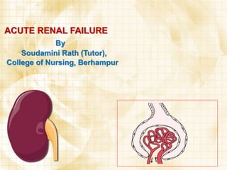
  • 2. Structure of the kidney • • • Specific components are nephrons, collecting ducts and a unique microvasculature Multipapillary kidney of humans – 1 million nephrons This number already established prenatally – Lost nephrons cannot be replaced
  • 3. Nephrons • A renal corpuscle (glomerulus) – A knot of capillaries – Connected to a complicated and twisted tubule – Finally drains into a collecting duct
  • 5. Renal physiology • Prime function – to maintain a stable milieu interieur – By selective retention and elimination of • Water • Electrolytes • And other solutes
  • 6. RENAL FAILURE • Results when the kidneys cannot remove the body’s metabolic wastes or perform their regulatory functions. • • The substances normally eliminated in the urine accumulate in the body fluids as a result of impaired renal excretion. • Leading to a disruption in endocrine and metabolic functions as well as fluid, electrolyte, and acid-base disturbances. • I
  • 7. ACUTE RENAL FAILURE  A rapid deteriorating condition ofrenal functions, resulting in accumulation of nitrogenous waste in the body.  It is a reversible clinical syndrome where there is a sudden and almost complete loss of kidney function (decreased GFR) over a period of hours to days with failure to excrete nitrogenous waste products and to maintain fluid and electrolyte homeostasis.  ARF manifests as an increase in serum Creatinine (M=53-132umol/L; F=44-97umol/L) and BUN (7-20mg/dl). Urine volume may be normal, or changes may occur. Possible changes include oliguria (<400ml/day), nonoliguria (>400ml/ day), or anuria (<50 ml/day)
  • 8. Acute renal failure can further devided into 1. Prerenal 2. Intrarenal 3. Postrena according to the etiology
  • 9. Categories of Acute Renal Failure 1. Prerenal- occurs in 60%-70% of cases, is the result of impaired blood flow to that leads to hypoperfusion of the kidney and a decrease in the GFR. Common clinical conditions are volume-depletion states (hemorrhage or GI losses), impaired cardiac performance (MI, HF, or cardiogenic shock), and vasodilation (sepsis or anaphylaxis)
  • 10. 2. Intrarenal- is the result of actual parenchymal damage to glomeruli or kidney tubules. Nephrotoxic agents such as aminoglycosides and radiocontrast agents account for 30% of cases of acute tubular necrosis (ATN), and ischemia due to decreased renal perfusion accounts for more than 50% of cases • Characteristics of ATN are intratubular back leak (abnormal absorption of filtrate and decreased urine flow through the tubule), vasoconstriction, and changes in glomerular permeability. These processes result in a decrease of GFR, progressive azotemia, and impaired fluid and electrolyte balance. 
  • 11. 3. Postrenal – is usually the result of an obstruction somewhere distal to the kidneys. Pressure rises in the kidney tubules and eventually, the GFR decreases
  • 12. Comparing Categories of Acute Renal Failure Characteristics Categories Prerenal Intrarenal Postrenal Etiology Hypoperfusion Parenchymal damage Obstruction BUN value Increased Increased increased Creatinine Increased Increased Increased Urine output Decreased Varies, often decreased Varies, may be decreased, or sudden anuria Urine sodium Decreased to < 20 mEq/L Increased to >40mEq/L Varies, often decreased to 20mEq/L or less Urinary sediment Normal, few hyaline casts Abnormal casts and debris Usually normal Urine osmolality Increased to 500mOsm About 350 mOsm similar to serum Varies, increased or equal to serum Urine specific gravity Increased Low normal Varies
  • 13. Caus es of Acute Re nal Failure A. Prerenal 1. Volume depletion resulting from: a. hemorrhage b. renal losses (diuretics) c. GI losses (vomiting, diarrhea, NG suctioning) 2. Impaired cardiac efficiency resulting from: a. MI b. Heart failure c. Dysrhythmias d. Cardiogenic shock 3. Vasodilation resulting from: a. sepsis b. anaphylaxis c. antihypertensive medications or other medications that cause vasodilation
  • 14. Caus es of Acute Re nalFailure B. Intrarenal 1. Prolonged renal ischemia resulting from: a. pigment nephropathy (associated with the breakdown of blood cells containing pigments that in turn occlude kidney structures) b. Myoglobinuria (trauma, crush injury, burns) c. Hemoglobinuria (transfusion reaction, hemolytic anemia) 2. Nephrotoxic agents such as: a. Aminoglycosides antibiotics (gentamicin, tobramycin) b. Radiopaque contrast media c. Heavy metals (lead, mercury) d. Solvents and chemicals (carbon tetrachloride, arsenic) e. NSAIDs, ACE inhibitors 3. Infectious processes such as: a. acute pyelonephritis b. acute GN
  • 15. Causes of Acute Renal Failure C. Postrenal 1. Urinary tract obstruction, including: a. calculi (stones) b. BPH c. Strictures d. Trauma or Blood clots e. Spinal cord diseases f. Blader or prostate cancer
  • 16. Stages in ARF I. Renal impairment is the first stage of damage:The GFR is 40 to 50% of normal. II. Renal insufficiency: the 20 to 40% of normal and fuctional loss also as azotaemia, high BUN and anaemia also occur. III. Renal failure there is only 10% GFR remaining. Fluid and electrolyte imbalances occurs and function of kidney diminished IV. Uraemia :represent stage of kidney failure I,e .ESRD, High nitrogenous waste product, multiple clinical manifestation with blood accumulalated. It is very fatal stage
  • 17. PATHOPHYSIOLOGY Related to underlying causes Damage to the nephron and loss of filtering action, diminished reabsorption Loss of ability to produce rennin, erythropoietin and other substances As the nephrons destroyed and tubular reabsorption Increasedsodium concentration and distal tubule stimulates production of rennin causing vasoconstriction and reducing renal blood flow,
  • 18. Cellular edema Decreased glomerular capillary permeability Intratubular obstruction Leakage of glomerular filtrate, which reduce the intra tubular fluid flow
  • 19. Preventing ARF 1. Provide adequate hydration to patients at risk of dehydration: a. surgical patients before, during and after surgery. b. Patients undergoing intensive diagnostic studies requiring fluid restrictions and contrast agents c. Patients with neoplastic disorders of metabolism and those receiving chemotherapy 2. Prevent and treat shock promptly with blood and fluid replacement. 3. Monitor CV and arterial pressures and hourly urine output of critically ill patients to detect the onset of renal failure as early as possible. 4. Treat hypotension promptly. 5. Continually assess renal function when appropriate.
  • 20. Preventing ARF 6. Take precautions to ensure that the appropriate blood is administered to the correct patient in order to avoid severe transfusion reactions, which can precipitate renal failure. 7. Prevent and treat infections promptly. Infections can produce progressive renal damage. 8. Pay special attention to wounds, burns and other precursors of sepsis 9. To prevent infections from ascending in the urinary tract, give meticulous care to patients with indwelling catheters. Remove catheter ASAP. 10. To prevent toxic drug effects, closely monitor dosage, duration of use, and blood levels of all medications metabolized or excreted by the kidneys.
  • 21. Medical Management: 1. Pharmacologic therapy a. hyperkalemia is the most life-threatening of the F/E changes that occur in RF, the elevated K levels may be reduced by administering cation-exchange resins (sodium polystyrene sulfonate [Kayexalate] orally or by retention enema. It works by exchanging sodium ions for potassium ions in the intestinal tract. b. Sorbitol may be administered in combination with Kayexalate to induce diarrhea type effect (induce water loss in the GIT) c. If hemodynamically unstable, IV dextrose 50%,insulin and calcium replacement may be administered to shift potassium back into the cells. d. Diuretics are often administered to control fluid volume, but they have not been shown to hasten the recovery form ARF.
  • 22. Medical Management 2. Nutritional Therapy a. Dietary proteins are individualized to provide the maximum benefit. Caloric requirements are met with high-carbohydrate meals, because carbohydrates have a protein- sparing effect. b. Foods and fluids containing potassium or phosphorous such as banana, citrus fruits and juices, coffee are restricted
  • 23. Nursing Management • • • • • • Monitoring fluid and electrolyte balance Maintaining nutrition Promoting comfort and safety Preventing infection Prevent injury Providing skin care • facilitate coping
  • 24. CHRONIC RENAL FAILURE/END S TAGE RENAL DISEASE (ESRD)  Is a progressive, irreversible deterioration in renal function in which the body’s ability to maintain metabolic and fluid and electrolyte balance fails, resulting in uremia and azotemia.
  • 25. ESRD Causes: 1. DM, HPN, chronic glomerulonephritis, pyelonephritis, obstruction of the urinary tract, hereditary lesions as in polycystic kidney disease, vascular disorders, infections, medications, or toxic agents. 2. Environmental and occupational agents that have been implicated in CRF include lead, mercury and chromium. Dialysis or kidney transplantation eventually becomes necessary for patient’s survival.
  • 26. Pathophysiology As renal function declines, the end products of CHON metabolism (which are normally excreted in urine) accumulate in the blood. Uremia develops and adversely affects every system in the body. Stages of CRF: are based on the GFR. The normal GFR is 125cc/min/1.73m2 1. Stage 1 = GFR > 90 ml/min/1.73m2. Kidney damage with normal or increased GFR. 2. Stage 2 = GFR = 60-89 mL/min/1.73m2. Mild decrease in GFR. 3. Stage 3 = GFR = 30-59 mL/min/1.73m2. Moderate decrease in GFR. 4. Stage 4 = GFR = 15-29 mL/min/1.73m2. Severe decrease in GFR. 5. Stage 5 = GFR <15 mL/min/1.73 m2. Kidney failure
  • 27. Clinical Manifestations 1. CV manifestations: a. HPN – due to Na and H20 retention or from R- A-A activation, b. heart failure and edema - due to fluid overload c. pericarditis - due to irritation of pericardial lining by uremic toxins 2. Dermatologic manifestations a. severe pruritus is common b. uremic frost, the deposit of urea crystals on the skin. 3. GI manifestations: a. anorexia, nausea and vomiting, and hiccups b. The patient’s breath may have the odor of urine (uremic fetor); this may be associated with inadequate dialysis 4. Neurologic manifestations a. altered LOC, inability to concentrate, muscle twitching, agitation, confusion and seizures. b. Peripheral neuropathy, a disorder of the peripheral NS, is present in some patients
  • 28. As s es s ment and Diagnos tic Findings 1. GFR 2. Sodium and water retention 3. acidosis – due to inability of the kidneys to excrete increased load of acid 4. Anemia 5. calcium and phosphorous imbalance – hypocalcemia and increase in phosphorous
  • 29. Complications 1. Hyperkalemia due to decreased excretion, metabolic acidosis, catabolism and excessive intake (diet, meds and fluids) 2. Pericarditis, pericardial effusion and pericardial tamponade due to retention of uremic waste products and inadequate dialysis. and water the R-A-A 3. Hypertension due to sodium retention and malfunction of system 4. Anemia due to decreased erythropoietin production, decreased RBC life span, bleeding in the GIT from irritating toxins and ulcer formation, and blood loss during hemodialysis 5. Bone disease and metastatic and vascular calcifications due to retention of phosphorous, low serum calcium levels, abnormal vitamin D metabolism and elevated aluminum levels.
  • 30. Medical Management 1. Pharmacologic Therapy a. calcium carbonate (Os-cal) or calcium acetate (Phoslo) are prescribed to treat hyperphosphatemia and hypocalcemia b. Antiseizure agents – diazepam (Valium) or phenytoin (Dilantin) c. Antihypertensive and CV drugs - digoxin (Lanoxin) and dobutamine (Dobutrex) d. Erythropoietin (Epogen) to treat anemia. It is initiated to reach a hematocrit of 33% - 385 and a target hemoglobin of 12g/dl. 2. Nutritional Therapy a. low sodium, low CHON and low K diet 3. Dialysis
  • 31. Nursing Management Nursing Management: 1. Assessing fluid status and identifying potential sources of imbalance. 2. implementing a dietary program to ensure proper nutritional intake 3. promoting positive feelings by encouraging increased self-care and greater independence. 4. Provide explanations and information to the patient and family concerning ESRD, treatment options and potential complications. 5. Provide emotional support to the patient and family.
  • 32. DIALYSIS  Is used to substitute some kidney functions during renal failure.  It is used to remove fluid and uremic waste products from the body when the kidneys are unable to do so.  It may be indicated to treat patients with edema that do not respond to treatment.  Acute dialysis is indicated when there is a high and increasing level of serum potassium, fluid overload, or pulmonary edema, iincreasing impending acidosis, pericarditis and severe confusion. It may also be used to certain medications or other toxins in the blood.
  • 33. PERITONEAL DIALYSIS TYPES: 2.Intermittent peritoneal dialysis = acute or chronic renal failure 3.Continuous ambulatory peritoneal dialysis = chronic renal failure 4.Continuous cycling peritoneal dialysis = prolonged dwelling time
  • 34. PERITONEAL DIALYSIS • Indwelling catheter is implanted into the peritoneum. • The technique for peritoneal dialysis involves preparing the patient and setting up the equipment, inserting the catheter, instilling the dialysate. • The catheter insertion may be done in operating room or at the bedside under local or general anesthesia. • The preferred site for insertion is about 3 to 5cm below the umbilicus, an area which is relatively avascular and has less fascial resistance. • The tip of the catheter is usually positioned so that it lied deeply with the pelvic gutter, the correct position will often give the patient the urge to defecate. • The catheter is generally sutured in place to avoid accidental dislodgment.
  • 35. • A connecting tube is attached to the external end of peritoneal catheter T –tube. • Plastic bag of dialysate solution is inserted to the end of T-tube; the other end is recap. • Dialysate bag is raised to shoulder level and infused by gravity in the peritoneal cavity • Infusion time = 10 minutes/2 liters; dwelling time is 4-6 hours depending on doctor’s order. • At the end of dwelling time, dialysis fluid is drained from the peritoneal cavity by gravity • Draining time is 10-20 minutes/2 liters • Then repeat the procedure when necessary
  • 36. Peritoneal Dialysis • Usually for patients with absolutely no other options of dialysis • Or as a temporary measure until options of dialysis sorted out
  • 39. NURSING RESPONSIBILITIES IN PERITONIAL DIALYSIS: Before Dialysis • Prepare the patient emotionally and physically • See that the consent form is signed • Weigh the patient • Check vital signs • Empty bladder • Assist with insertion of CVP catheter ECG monitoring is also employed • Make the patient comfortable in supine position and set up the instruments
  • 40. During Dialysis • Assist the physician in inserting the peritoneal catheter • After the procedure, if the fluid is not draining properly, move the patient from side to side. • Then the outflow ceases, clamp off the drainage tube and infuse next exchange. • Take B.P. and pulse every 15min during the first exchange and every hour thereafter. • Maintain a record of patient’s fluid balance. • Promote patient comfort during dialysis frequent back care and changing position. • Observe breathing difficulty, abdominal pain and leakage arround the catheter. After Dialysis • Take the patient’s temperature • Look for complication
  • 41. Complications • Perforation of the bowel • Puncture of abdominal aorta • Oedema of the anterior abdominal wall • Pain • Plural effusion • Peritonites • Hypernatraemia • Hypokalamia • Hyperglycaemia • Disequilbrium • Pulmonary etc.
  • 42. HEMODIALYSIS  it is the process of cleansing the blood of accumulated waste products. Patient’s access is prepared and cannulated surgically  One needle is inserted to the artery (brachial) then blood flow is directed to dialyzer (dialysis machine)  The machine is equipped with semi-permeable membrane surrounded with dialysis solution  Waste products in the blood move to the dialysis solution passing through the membrane by means of diffusion  Excess water is also removed from the blood by way of ultrafiltration  The blood is then returned to the vein after it has been cleansed.
  • 44. HEMODIALYSIS Patient Access Vascular catheter A-V fistula Synthetic vascular graft
  • 46. HEMODIALYSIS NURSING CONSIDERATIONS: 1. The dialyser to be prepared by proper setting, can be heparinized unless it is contraindicated to prevent blood clot. 2. Dialysis solution has some electrolytes and acetate and HCO3 added to achieve proper pH balance. 3. Methods of circulatory access: AV fistula; AV graft or U-tube 4. Assess the access site for bruit, signs of infections and ischemia of the hand. 5. Aseptic technique must be follow. 6. No BP taking on the access site.
  • 47. 7. Cover the access site with adhesive bandage 8. Dietary adjustments of Carbohydrate, Na and fluid intake. 9. Monitor VS regularly 10. Check blood chemistry 11. Constant monitoring of hemodynamic status, electrolytes and acid-base balance.
  • 48. 13) Observation of patient s pulse and B.P every half an hour and respiration every hour. 14) Observe for hypothermia, cardiac irregularities, and twitching. 15) Observe the machine a sudden break in the circuit, high venous pressure due to kinking which leads low arterial pressure and failure of blood pump. 16) Dietary adjustment of protein, sodium, and potassium and fluid intake. 17) Psychological and emotional support to the patient as in stress.
  • 49. Complication: • Acute circulatory overload • Haemorrhage • Cardiac arrest • Dialysis disequilibrium syndrome etc