SlideShare a Scribd company logo
1 of 41
Human 
Anatomy 
& Physiology 
SEVENTH EDITION 
Elaine N. Marieb 
Katja Hoehn 
Copyright © 2006 Pearson Education, Inc., publishing as Benjamin Cummings 
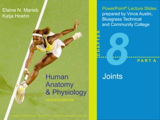
PowerPoint® Lecture Slides 
prepared by Vince Austin, 
Bluegrass Technical 
and Community College 
C H A P T E R 
8 
Joints 
P A R T A
Joints (Articulations) 
 Weakest parts of the skeleton 
 Articulation – site where two or more bones meet 
 Functions of joints 
 Give the skeleton mobility 
 Hold the skeleton together 
Copyright © 2006 Pearson Education, Inc., publishing as Benjamin Cummings
Classification of Joints: Structural 
 Structural classification focuses on the material 
binding bones together and whether or not a joint 
cavity is present 
 The three structural classifications are: 
 Fibrous 
 Cartilaginous 
 Synovial 
Copyright © 2006 Pearson Education, Inc., publishing as Benjamin Cummings
Classification of Joints: Functional 
 Functional classification is based on the amount of 
movement allowed by the joint 
 The three functional classes of joints are: 
 Synarthroses – immovable 
 Amphiarthroses – slightly movable 
 Diarthroses – freely movable 
Copyright © 2006 Pearson Education, Inc., publishing as Benjamin Cummings
Fibrous Structural Joints 
 The bones are joined by fibrous tissues 
 There is no joint cavity 
 Most are immovable 
 There are three types – sutures, syndesmoses, and 
gomphoses 
Copyright © 2006 Pearson Education, Inc., publishing as Benjamin Cummings
Fibrous Structural Joints: Sutures 
 Occur between the bones of the skull 
 Comprised of interlocking junctions completely 
filled with connective tissue fibers 
 Bind bones tightly together, but allow for growth 
during youth 
 In middle age, skull bones fuse and are called 
synostoses 
Copyright © 2006 Pearson Education, Inc., publishing as Benjamin Cummings
Fibrous Structural Joints: Sutures 
Copyright © 2006 Pearson Education, Inc., publishing as Benjamin Cummings 
Figure 8.1a
Fibrous Structural Joints: Syndesmoses 
 Bones are connected by a fibrous tissue ligament 
 Movement varies from immovable to slightly 
variable 
 Examples include the connection between the tibia 
and fibula, and the radius and ulna 
Copyright © 2006 Pearson Education, Inc., publishing as Benjamin Cummings
Fibrous Structural Joints: Syndesmoses 
Copyright © 2006 Pearson Education, Inc., publishing as Benjamin Cummings 
Figure 8.1b
Fibrous Structural Joints: Gomphoses 
 The peg-in-socket fibrous joint between a tooth 
and its alveolar socket 
 The fibrous connection is the periodontal ligament 
Copyright © 2006 Pearson Education, Inc., publishing as Benjamin Cummings
Cartilaginous Joints 
 Articulating bones are united by cartilage 
 Lack a joint cavity 
 Two types – synchondroses and symphyses 
Copyright © 2006 Pearson Education, Inc., publishing as Benjamin Cummings
Cartilaginous Joints: Synchondroses 
 A bar or plate of hyaline cartilage unites the bones 
 All synchondroses are synarthrotic 
 Examples include: 
 Epiphyseal plates of children 
 Joint between the costal cartilage of the first rib 
and the sternum 
Copyright © 2006 Pearson Education, Inc., publishing as Benjamin Cummings
Cartilaginous Joints: Synchondroses 
Copyright © 2006 Pearson Education, Inc., publishing as Benjamin Cummings 
Figure 8.2a, b
Cartilaginous Joints: Symphyses 
 Hyaline cartilage covers the articulating surface of 
the bone and is fused to an intervening pad of 
fibrocartilage 
 Amphiarthrotic joints designed for strength and 
flexibility 
 Examples include intervertebral joints and the 
pubic symphysis of the pelvis 
Copyright © 2006 Pearson Education, Inc., publishing as Benjamin Cummings
Cartilaginous Joints: Symphyses 
Copyright © 2006 Pearson Education, Inc., publishing as Benjamin Cummings 
Figure 8.2c
Synovial Joints 
 Those joints in which the articulating bones are 
separated by a fluid-containing joint cavity 
 All are freely movable diarthroses 
 Examples – all limb joints, and most joints of the 
body 
Copyright © 2006 Pearson Education, Inc., publishing as Benjamin Cummings
Synovial Joints: General Structure 
 Synovial joints all have the following 
 Articular cartilage 
 Joint (synovial) cavity 
 Articular capsule 
 Synovial fluid 
 Reinforcing ligaments 
Copyright © 2006 Pearson Education, Inc., publishing as Benjamin Cummings
Synovial Joints: General Structure 
Copyright © 2006 Pearson Education, Inc., publishing as Benjamin Cummings 
Figure 8.3a, b
Copyright © 2006 Pearson Education, Inc., publishing as Benjamin Cummings Table 8.2.1
Copyright © 2006 Pearson Education, Inc., publishing as Benjamin Cummings Table 8.2.2
Copyright © 2006 Pearson Education, Inc., publishing as Benjamin Cummings Table 8.2.3
Synovial Joints: Friction-Reducing Structures 
 Bursae – flattened, fibrous sacs lined with synovial 
membranes and containing synovial fluid 
 Common where ligaments, muscles, skin, tendons, 
or bones rub together 
 Tendon sheath – elongated bursa that wraps 
completely around a tendon 
Copyright © 2006 Pearson Education, Inc., publishing as Benjamin Cummings
Synovial Joints: Friction-Reducing Structures 
Copyright © 2006 Pearson Education, Inc., publishing as Benjamin Cummings 
Figure 8.4
Synovial Joints: Stability 
 Stability is determined by: 
 Articular surfaces – shape determines what 
movements are possible 
 Ligaments – unite bones and prevent excessive or 
undesirable motion 
Copyright © 2006 Pearson Education, Inc., publishing as Benjamin Cummings
Synovial Joints: Stability 
 Muscle tone is accomplished by: 
 Muscle tendons across joints acting as stabilizing 
factors 
 Tendons that are kept tight at all times by muscle 
tone 
Copyright © 2006 Pearson Education, Inc., publishing as Benjamin Cummings
Synovial Joints: Movement 
 The two muscle attachments across a joint are: 
 Origin – attachment to the immovable bone 
 Insertion – attachment to the movable bone 
 Described as movement along transverse, frontal, 
or sagittal planes 
Copyright © 2006 Pearson Education, Inc., publishing as Benjamin Cummings
Synovial Joints: Range of Motion 
 Nonaxial – slipping movements only 
 Uniaxial – movement in one plane 
 Biaxial – movement in two planes 
 Multiaxial – movement in or around all three 
planes 
Copyright © 2006 Pearson Education, Inc., publishing as Benjamin Cummings
Gliding Movements 
 One flat bone surface glides or slips over another 
similar surface 
 Examples – intercarpal and intertarsal joints, and 
between the flat articular processes of the vertebrae 
Copyright © 2006 Pearson Education, Inc., publishing as Benjamin Cummings
Angular Movement 
 Flexion — bending movement that decreases the 
angle of the joint 
 Extension — reverse of flexion; joint angle is 
increased 
 Dorsiflexion and plantar flexion — up and down 
movement of the foot 
Copyright © 2006 Pearson Education, Inc., publishing as Benjamin Cummings
Angular Movement 
 Abduction — movement away from the midline 
 Adduction — movement toward the midline 
 Circumduction — movement describes a cone in 
space 
Copyright © 2006 Pearson Education, Inc., publishing as Benjamin Cummings
Gliding Movement 
Copyright © 2006 Pearson Education, Inc., publishing as Benjamin Cummings 
Figure 8.5a
Angular Movement 
Copyright © 2006 Pearson Education, Inc., publishing as Benjamin Cummings 
Figure 8.5b
Angular Movement 
Copyright © 2006 Pearson Education, Inc., publishing as Benjamin Cummings 
Figure 8.5c, d
Angular Movement 
Copyright © 2006 Pearson Education, Inc., publishing as Benjamin Cummings 
Figure 8.5e, f
Rotation 
 The turning of a bone 
around its own long axis 
 Examples 
 Between first two 
vertebrae 
 Hip and shoulder joints 
Copyright © 2006 Pearson Education, Inc., publishing as Benjamin Cummings 
Figure 8.5g
Special Movements 
 Supination and pronation 
 Inversion and eversion 
 Protraction and retraction 
 Elevation and depression 
 Opposition 
Copyright © 2006 Pearson Education, Inc., publishing as Benjamin Cummings
Special Movements 
Copyright © 2006 Pearson Education, Inc., publishing as Benjamin Cummings 
Figure 8.6a
Special Movements 
Copyright © 2006 Pearson Education, Inc., publishing as Benjamin Cummings 
Figure 8.6b
Special Movements 
Copyright © 2006 Pearson Education, Inc., publishing as Benjamin Cummings 
Figure 8.6c
Special Movements 
Copyright © 2006 Pearson Education, Inc., publishing as Benjamin Cummings 
Figure 8.6d
Special Movements 
Copyright © 2006 Pearson Education, Inc., publishing as Benjamin Cummings 
Figure 8.6e

More Related Content

What's hot

Ch06 a.bone
Ch06 a.boneCh06 a.bone
Ch06 a.bone
TheSlaps
 
Ch06 b.bones
Ch06 b.bonesCh06 b.bones
Ch06 b.bones
TheSlaps
 
Dr. B Ch 11_lecture_presentation
Dr. B Ch 11_lecture_presentationDr. B Ch 11_lecture_presentation
Dr. B Ch 11_lecture_presentation
TheSlaps
 
Dr. B Ch 10_lecture_presentation
Dr. B Ch 10_lecture_presentationDr. B Ch 10_lecture_presentation
Dr. B Ch 10_lecture_presentation
TheSlaps
 
Ch07 d.skeletal
Ch07 d.skeletalCh07 d.skeletal
Ch07 d.skeletal
TheSlaps
 
Ch14 Digestive System Overview
Ch14 Digestive System OverviewCh14 Digestive System Overview
Ch14 Digestive System Overview
aescobed
 

What's hot (20)

Biology in Focus - Chapter 6
Biology in Focus - Chapter 6Biology in Focus - Chapter 6
Biology in Focus - Chapter 6
 
07 [chapter 7 the skeletal system the axial skeleton]
07 [chapter 7 the skeletal system   the axial skeleton]07 [chapter 7 the skeletal system   the axial skeleton]
07 [chapter 7 the skeletal system the axial skeleton]
 
06 [chapter 6 the skeletal system bone tissue]
06 [chapter 6 the skeletal system   bone tissue]06 [chapter 6 the skeletal system   bone tissue]
06 [chapter 6 the skeletal system bone tissue]
 
Ch06 a.bone
Ch06 a.boneCh06 a.bone
Ch06 a.bone
 
Chapter 9 Muscular System
Chapter 9   Muscular SystemChapter 9   Muscular System
Chapter 9 Muscular System
 
Chapter 7 Skeletal System
Chapter 7   Skeletal SystemChapter 7   Skeletal System
Chapter 7 Skeletal System
 
Skeletol system advanced
Skeletol system advancedSkeletol system advanced
Skeletol system advanced
 
Chapter 3 Cells
Chapter 3  CellsChapter 3  Cells
Chapter 3 Cells
 
Biology in Focus - Chapter 9
Biology in Focus - Chapter 9Biology in Focus - Chapter 9
Biology in Focus - Chapter 9
 
Anatomy of skeletal system
Anatomy of skeletal systemAnatomy of skeletal system
Anatomy of skeletal system
 
Biology in Focus Chapter 5
Biology in Focus Chapter 5Biology in Focus Chapter 5
Biology in Focus Chapter 5
 
Ch06 b.bones
Ch06 b.bonesCh06 b.bones
Ch06 b.bones
 
Dr. B Ch 09_lecture_presentation
Dr. B Ch 09_lecture_presentationDr. B Ch 09_lecture_presentation
Dr. B Ch 09_lecture_presentation
 
08 [chapter 8 the skeletal system appendicular skeleton]
08 [chapter 8 the skeletal system   appendicular skeleton]08 [chapter 8 the skeletal system   appendicular skeleton]
08 [chapter 8 the skeletal system appendicular skeleton]
 
Dr. B Ch 11_lecture_presentation
Dr. B Ch 11_lecture_presentationDr. B Ch 11_lecture_presentation
Dr. B Ch 11_lecture_presentation
 
10 [chapter 10 muscular tissue]
10 [chapter 10 muscular tissue]10 [chapter 10 muscular tissue]
10 [chapter 10 muscular tissue]
 
Biology in Focus Chapter 2
Biology in Focus Chapter 2Biology in Focus Chapter 2
Biology in Focus Chapter 2
 
Dr. B Ch 10_lecture_presentation
Dr. B Ch 10_lecture_presentationDr. B Ch 10_lecture_presentation
Dr. B Ch 10_lecture_presentation
 
Ch07 d.skeletal
Ch07 d.skeletalCh07 d.skeletal
Ch07 d.skeletal
 
Ch14 Digestive System Overview
Ch14 Digestive System OverviewCh14 Digestive System Overview
Ch14 Digestive System Overview
 

Viewers also liked

Emg for sports medicine providers2010
Emg for sports medicine providers2010Emg for sports medicine providers2010
Emg for sports medicine providers2010
EsserHealth
 
Ch 08 active_lecture_questions
Ch 08 active_lecture_questionsCh 08 active_lecture_questions
Ch 08 active_lecture_questions
ebellebell
 
Ch 08 lecture_outline_a
Ch 08 lecture_outline_aCh 08 lecture_outline_a
Ch 08 lecture_outline_a
ebellebell
 
Physiology CH 8 lecture notes
Physiology CH 8 lecture notesPhysiology CH 8 lecture notes
Physiology CH 8 lecture notes
bfurlong
 
Ch 08 lecture_outline_b
Ch 08 lecture_outline_bCh 08 lecture_outline_b
Ch 08 lecture_outline_b
ebellebell
 
Ch 17_lecture_presentation
 Ch 17_lecture_presentation Ch 17_lecture_presentation
Ch 17_lecture_presentation
TheSlaps
 
Ch 26_lecture_presentation
 Ch 26_lecture_presentation Ch 26_lecture_presentation
Ch 26_lecture_presentation
TheSlaps
 
Ch 15_lecture_presentation
 Ch 15_lecture_presentation Ch 15_lecture_presentation
Ch 15_lecture_presentation
TheSlaps
 
Ch 27_lecture_presentation
 Ch 27_lecture_presentation Ch 27_lecture_presentation
Ch 27_lecture_presentation
TheSlaps
 
163 ch 12_lecture_presentation
163 ch 12_lecture_presentation163 ch 12_lecture_presentation
163 ch 12_lecture_presentation
gwrandall
 
Ch 25_lecture_presentation
 Ch 25_lecture_presentation Ch 25_lecture_presentation
Ch 25_lecture_presentation
TheSlaps
 
Ch 23_lecture_presentation
 Ch 23_lecture_presentation Ch 23_lecture_presentation
Ch 23_lecture_presentation
TheSlaps
 
Ch 21_lecture_presentation
 Ch 21_lecture_presentation Ch 21_lecture_presentation
Ch 21_lecture_presentation
TheSlaps
 
Ch 18_lecture_presentation
 Ch 18_lecture_presentation Ch 18_lecture_presentation
Ch 18_lecture_presentation
TheSlaps
 
Ch 13_lecture_presentation
 Ch 13_lecture_presentation Ch 13_lecture_presentation
Ch 13_lecture_presentation
TheSlaps
 
Ch 22_lecture_presentation
 Ch 22_lecture_presentation Ch 22_lecture_presentation
Ch 22_lecture_presentation
TheSlaps
 

Viewers also liked (20)

Jointsrev
JointsrevJointsrev
Jointsrev
 
Lower ext pain
Lower ext painLower ext pain
Lower ext pain
 
Emg for sports medicine providers2010
Emg for sports medicine providers2010Emg for sports medicine providers2010
Emg for sports medicine providers2010
 
Ch 08 active_lecture_questions
Ch 08 active_lecture_questionsCh 08 active_lecture_questions
Ch 08 active_lecture_questions
 
8.4
8.48.4
8.4
 
Ch 08 lecture_outline_a
Ch 08 lecture_outline_aCh 08 lecture_outline_a
Ch 08 lecture_outline_a
 
Physiology CH 8 lecture notes
Physiology CH 8 lecture notesPhysiology CH 8 lecture notes
Physiology CH 8 lecture notes
 
Ch 08 lecture_outline_b
Ch 08 lecture_outline_bCh 08 lecture_outline_b
Ch 08 lecture_outline_b
 
Ch 17_lecture_presentation
 Ch 17_lecture_presentation Ch 17_lecture_presentation
Ch 17_lecture_presentation
 
Dr. B Ch 19_lecture_presentation
Dr. B Ch 19_lecture_presentationDr. B Ch 19_lecture_presentation
Dr. B Ch 19_lecture_presentation
 
Ch 26_lecture_presentation
 Ch 26_lecture_presentation Ch 26_lecture_presentation
Ch 26_lecture_presentation
 
Ch 15_lecture_presentation
 Ch 15_lecture_presentation Ch 15_lecture_presentation
Ch 15_lecture_presentation
 
Ch 27_lecture_presentation
 Ch 27_lecture_presentation Ch 27_lecture_presentation
Ch 27_lecture_presentation
 
163 ch 12_lecture_presentation
163 ch 12_lecture_presentation163 ch 12_lecture_presentation
163 ch 12_lecture_presentation
 
Ch 25_lecture_presentation
 Ch 25_lecture_presentation Ch 25_lecture_presentation
Ch 25_lecture_presentation
 
Ch 23_lecture_presentation
 Ch 23_lecture_presentation Ch 23_lecture_presentation
Ch 23_lecture_presentation
 
Ch 21_lecture_presentation
 Ch 21_lecture_presentation Ch 21_lecture_presentation
Ch 21_lecture_presentation
 
Ch 18_lecture_presentation
 Ch 18_lecture_presentation Ch 18_lecture_presentation
Ch 18_lecture_presentation
 
Ch 13_lecture_presentation
 Ch 13_lecture_presentation Ch 13_lecture_presentation
Ch 13_lecture_presentation
 
Ch 22_lecture_presentation
 Ch 22_lecture_presentation Ch 22_lecture_presentation
Ch 22_lecture_presentation
 

Similar to Ch08 a.joint

Ch08 b.joints
Ch08 b.jointsCh08 b.joints
Ch08 b.joints
TheSlaps
 
10 a.muscular.s
10 a.muscular.s10 a.muscular.s
10 a.muscular.s
TheSlaps
 
10 d. muscles system
10 d. muscles system10 d. muscles system
10 d. muscles system
TheSlaps
 
10 d. muscles system
10 d. muscles system10 d. muscles system
10 d. muscles system
TheSlaps
 
Ch09 a. muscles tissue
Ch09 a. muscles tissueCh09 a. muscles tissue
Ch09 a. muscles tissue
TheSlaps
 
Chapter 7b appendicular skeleton
Chapter 7b   appendicular skeletonChapter 7b   appendicular skeleton
Chapter 7b appendicular skeleton
TheSlaps
 
Bone formation and development 14
Bone formation and development 14Bone formation and development 14
Bone formation and development 14
Mathew Ngugi
 
Ch07 b.skeletal
Ch07 b.skeletalCh07 b.skeletal
Ch07 b.skeletal
TheSlaps
 

Similar to Ch08 a.joint (20)

Ch08 b.joints
Ch08 b.jointsCh08 b.joints
Ch08 b.joints
 
CVA A&P - Chapter 5a: Standard Bone Tissue
CVA A&P - Chapter 5a: Standard Bone TissueCVA A&P - Chapter 5a: Standard Bone Tissue
CVA A&P - Chapter 5a: Standard Bone Tissue
 
Lesson-4.-Skeletal-System.ppt
Lesson-4.-Skeletal-System.pptLesson-4.-Skeletal-System.ppt
Lesson-4.-Skeletal-System.ppt
 
CVA A&P - Chapter 5a: Honors Bone Tissue
CVA A&P - Chapter 5a: Honors Bone TissueCVA A&P - Chapter 5a: Honors Bone Tissue
CVA A&P - Chapter 5a: Honors Bone Tissue
 
CVA A&P - Chapter 6c: Muscle Movements and Major Muscles
CVA A&P - Chapter 6c: Muscle Movements and Major MusclesCVA A&P - Chapter 6c: Muscle Movements and Major Muscles
CVA A&P - Chapter 6c: Muscle Movements and Major Muscles
 
10 a.muscular.s
10 a.muscular.s10 a.muscular.s
10 a.muscular.s
 
10 a.muscular.s
10 a.muscular.s10 a.muscular.s
10 a.muscular.s
 
CVA A&P - Chapter 5d: Joints
CVA A&P - Chapter 5d: JointsCVA A&P - Chapter 5d: Joints
CVA A&P - Chapter 5d: Joints
 
anatomy_apendicular_skeleton.ppt
anatomy_apendicular_skeleton.pptanatomy_apendicular_skeleton.ppt
anatomy_apendicular_skeleton.ppt
 
CVA A&P - Chapter 6a: Muscle Types and Tissue
CVA A&P - Chapter 6a: Muscle Types and TissueCVA A&P - Chapter 6a: Muscle Types and Tissue
CVA A&P - Chapter 6a: Muscle Types and Tissue
 
3468448.pptddddddddddddddddceedddddddddddd
3468448.pptddddddddddddddddceedddddddddddd3468448.pptddddddddddddddddceedddddddddddd
3468448.pptddddddddddddddddceedddddddddddd
 
10 d. muscles system
10 d. muscles system10 d. muscles system
10 d. muscles system
 
10 d. muscles system
10 d. muscles system10 d. muscles system
10 d. muscles system
 
8.4
8.48.4
8.4
 
Ch09 a. muscles tissue
Ch09 a. muscles tissueCh09 a. muscles tissue
Ch09 a. muscles tissue
 
Chapter 7b appendicular skeleton
Chapter 7b   appendicular skeletonChapter 7b   appendicular skeleton
Chapter 7b appendicular skeleton
 
anatomy_axial_skeleton_pearson.ppt
anatomy_axial_skeleton_pearson.pptanatomy_axial_skeleton_pearson.ppt
anatomy_axial_skeleton_pearson.ppt
 
Bone formation and development 14
Bone formation and development 14Bone formation and development 14
Bone formation and development 14
 
CVA A&P - Chapter 5c: Standard Appendicular Skeleton
CVA A&P - Chapter 5c: Standard Appendicular SkeletonCVA A&P - Chapter 5c: Standard Appendicular Skeleton
CVA A&P - Chapter 5c: Standard Appendicular Skeleton
 
Ch07 b.skeletal
Ch07 b.skeletalCh07 b.skeletal
Ch07 b.skeletal
 

More from TheSlaps (20)

Med Surg Chapter 020
Med Surg Chapter 020Med Surg Chapter 020
Med Surg Chapter 020
 
Med Surg Chapter 019
Med Surg Chapter 019Med Surg Chapter 019
Med Surg Chapter 019
 
Med Surg Chapter 018
Med Surg Chapter 018Med Surg Chapter 018
Med Surg Chapter 018
 
Adams ch09 lecture
Adams ch09 lectureAdams ch09 lecture
Adams ch09 lecture
 
Adams ch08 lecture
Adams ch08 lectureAdams ch08 lecture
Adams ch08 lecture
 
Adams ch10 lecture
Adams ch10 lectureAdams ch10 lecture
Adams ch10 lecture
 
Adams ch05 lecture
Adams ch05 lectureAdams ch05 lecture
Adams ch05 lecture
 
Adams ch07 lecture
Adams ch07 lectureAdams ch07 lecture
Adams ch07 lecture
 
Adams ch06 lecture
Adams ch06 lectureAdams ch06 lecture
Adams ch06 lecture
 
Adams ch04 lecture
Adams ch04 lectureAdams ch04 lecture
Adams ch04 lecture
 
Adams ch02 lecture
Adams ch02 lectureAdams ch02 lecture
Adams ch02 lecture
 
Adams ch01 lecture
Adams ch01 lectureAdams ch01 lecture
Adams ch01 lecture
 
Adams ch03 lecture
Adams ch03 lectureAdams ch03 lecture
Adams ch03 lecture
 
Pathophysiology Chapter 52
Pathophysiology Chapter 52Pathophysiology Chapter 52
Pathophysiology Chapter 52
 
Pathophysiology Chapter 51
Pathophysiology Chapter 51Pathophysiology Chapter 51
Pathophysiology Chapter 51
 
Pathophysiology Chapter 47
Pathophysiology Chapter 47Pathophysiology Chapter 47
Pathophysiology Chapter 47
 
Pathophysiology Chapter 45
Pathophysiology Chapter 45Pathophysiology Chapter 45
Pathophysiology Chapter 45
 
Pathophysiology Chapter 44
Pathophysiology Chapter 44Pathophysiology Chapter 44
Pathophysiology Chapter 44
 
Pathophysiology Chapter 36
Pathophysiology Chapter 36Pathophysiology Chapter 36
Pathophysiology Chapter 36
 
Pathophysiology Chapter 37
Pathophysiology Chapter 37Pathophysiology Chapter 37
Pathophysiology Chapter 37
 

Ch08 a.joint

  • 1. Human Anatomy & Physiology SEVENTH EDITION Elaine N. Marieb Katja Hoehn Copyright © 2006 Pearson Education, Inc., publishing as Benjamin Cummings PowerPoint® Lecture Slides prepared by Vince Austin, Bluegrass Technical and Community College C H A P T E R 8 Joints P A R T A
  • 2. Joints (Articulations)  Weakest parts of the skeleton  Articulation – site where two or more bones meet  Functions of joints  Give the skeleton mobility  Hold the skeleton together Copyright © 2006 Pearson Education, Inc., publishing as Benjamin Cummings
  • 3. Classification of Joints: Structural  Structural classification focuses on the material binding bones together and whether or not a joint cavity is present  The three structural classifications are:  Fibrous  Cartilaginous  Synovial Copyright © 2006 Pearson Education, Inc., publishing as Benjamin Cummings
  • 4. Classification of Joints: Functional  Functional classification is based on the amount of movement allowed by the joint  The three functional classes of joints are:  Synarthroses – immovable  Amphiarthroses – slightly movable  Diarthroses – freely movable Copyright © 2006 Pearson Education, Inc., publishing as Benjamin Cummings
  • 5. Fibrous Structural Joints  The bones are joined by fibrous tissues  There is no joint cavity  Most are immovable  There are three types – sutures, syndesmoses, and gomphoses Copyright © 2006 Pearson Education, Inc., publishing as Benjamin Cummings
  • 6. Fibrous Structural Joints: Sutures  Occur between the bones of the skull  Comprised of interlocking junctions completely filled with connective tissue fibers  Bind bones tightly together, but allow for growth during youth  In middle age, skull bones fuse and are called synostoses Copyright © 2006 Pearson Education, Inc., publishing as Benjamin Cummings
  • 7. Fibrous Structural Joints: Sutures Copyright © 2006 Pearson Education, Inc., publishing as Benjamin Cummings Figure 8.1a
  • 8. Fibrous Structural Joints: Syndesmoses  Bones are connected by a fibrous tissue ligament  Movement varies from immovable to slightly variable  Examples include the connection between the tibia and fibula, and the radius and ulna Copyright © 2006 Pearson Education, Inc., publishing as Benjamin Cummings
  • 9. Fibrous Structural Joints: Syndesmoses Copyright © 2006 Pearson Education, Inc., publishing as Benjamin Cummings Figure 8.1b
  • 10. Fibrous Structural Joints: Gomphoses  The peg-in-socket fibrous joint between a tooth and its alveolar socket  The fibrous connection is the periodontal ligament Copyright © 2006 Pearson Education, Inc., publishing as Benjamin Cummings
  • 11. Cartilaginous Joints  Articulating bones are united by cartilage  Lack a joint cavity  Two types – synchondroses and symphyses Copyright © 2006 Pearson Education, Inc., publishing as Benjamin Cummings
  • 12. Cartilaginous Joints: Synchondroses  A bar or plate of hyaline cartilage unites the bones  All synchondroses are synarthrotic  Examples include:  Epiphyseal plates of children  Joint between the costal cartilage of the first rib and the sternum Copyright © 2006 Pearson Education, Inc., publishing as Benjamin Cummings
  • 13. Cartilaginous Joints: Synchondroses Copyright © 2006 Pearson Education, Inc., publishing as Benjamin Cummings Figure 8.2a, b
  • 14. Cartilaginous Joints: Symphyses  Hyaline cartilage covers the articulating surface of the bone and is fused to an intervening pad of fibrocartilage  Amphiarthrotic joints designed for strength and flexibility  Examples include intervertebral joints and the pubic symphysis of the pelvis Copyright © 2006 Pearson Education, Inc., publishing as Benjamin Cummings
  • 15. Cartilaginous Joints: Symphyses Copyright © 2006 Pearson Education, Inc., publishing as Benjamin Cummings Figure 8.2c
  • 16. Synovial Joints  Those joints in which the articulating bones are separated by a fluid-containing joint cavity  All are freely movable diarthroses  Examples – all limb joints, and most joints of the body Copyright © 2006 Pearson Education, Inc., publishing as Benjamin Cummings
  • 17. Synovial Joints: General Structure  Synovial joints all have the following  Articular cartilage  Joint (synovial) cavity  Articular capsule  Synovial fluid  Reinforcing ligaments Copyright © 2006 Pearson Education, Inc., publishing as Benjamin Cummings
  • 18. Synovial Joints: General Structure Copyright © 2006 Pearson Education, Inc., publishing as Benjamin Cummings Figure 8.3a, b
  • 19. Copyright © 2006 Pearson Education, Inc., publishing as Benjamin Cummings Table 8.2.1
  • 20. Copyright © 2006 Pearson Education, Inc., publishing as Benjamin Cummings Table 8.2.2
  • 21. Copyright © 2006 Pearson Education, Inc., publishing as Benjamin Cummings Table 8.2.3
  • 22. Synovial Joints: Friction-Reducing Structures  Bursae – flattened, fibrous sacs lined with synovial membranes and containing synovial fluid  Common where ligaments, muscles, skin, tendons, or bones rub together  Tendon sheath – elongated bursa that wraps completely around a tendon Copyright © 2006 Pearson Education, Inc., publishing as Benjamin Cummings
  • 23. Synovial Joints: Friction-Reducing Structures Copyright © 2006 Pearson Education, Inc., publishing as Benjamin Cummings Figure 8.4
  • 24. Synovial Joints: Stability  Stability is determined by:  Articular surfaces – shape determines what movements are possible  Ligaments – unite bones and prevent excessive or undesirable motion Copyright © 2006 Pearson Education, Inc., publishing as Benjamin Cummings
  • 25. Synovial Joints: Stability  Muscle tone is accomplished by:  Muscle tendons across joints acting as stabilizing factors  Tendons that are kept tight at all times by muscle tone Copyright © 2006 Pearson Education, Inc., publishing as Benjamin Cummings
  • 26. Synovial Joints: Movement  The two muscle attachments across a joint are:  Origin – attachment to the immovable bone  Insertion – attachment to the movable bone  Described as movement along transverse, frontal, or sagittal planes Copyright © 2006 Pearson Education, Inc., publishing as Benjamin Cummings
  • 27. Synovial Joints: Range of Motion  Nonaxial – slipping movements only  Uniaxial – movement in one plane  Biaxial – movement in two planes  Multiaxial – movement in or around all three planes Copyright © 2006 Pearson Education, Inc., publishing as Benjamin Cummings
  • 28. Gliding Movements  One flat bone surface glides or slips over another similar surface  Examples – intercarpal and intertarsal joints, and between the flat articular processes of the vertebrae Copyright © 2006 Pearson Education, Inc., publishing as Benjamin Cummings
  • 29. Angular Movement  Flexion — bending movement that decreases the angle of the joint  Extension — reverse of flexion; joint angle is increased  Dorsiflexion and plantar flexion — up and down movement of the foot Copyright © 2006 Pearson Education, Inc., publishing as Benjamin Cummings
  • 30. Angular Movement  Abduction — movement away from the midline  Adduction — movement toward the midline  Circumduction — movement describes a cone in space Copyright © 2006 Pearson Education, Inc., publishing as Benjamin Cummings
  • 31. Gliding Movement Copyright © 2006 Pearson Education, Inc., publishing as Benjamin Cummings Figure 8.5a
  • 32. Angular Movement Copyright © 2006 Pearson Education, Inc., publishing as Benjamin Cummings Figure 8.5b
  • 33. Angular Movement Copyright © 2006 Pearson Education, Inc., publishing as Benjamin Cummings Figure 8.5c, d
  • 34. Angular Movement Copyright © 2006 Pearson Education, Inc., publishing as Benjamin Cummings Figure 8.5e, f
  • 35. Rotation  The turning of a bone around its own long axis  Examples  Between first two vertebrae  Hip and shoulder joints Copyright © 2006 Pearson Education, Inc., publishing as Benjamin Cummings Figure 8.5g
  • 36. Special Movements  Supination and pronation  Inversion and eversion  Protraction and retraction  Elevation and depression  Opposition Copyright © 2006 Pearson Education, Inc., publishing as Benjamin Cummings
  • 37. Special Movements Copyright © 2006 Pearson Education, Inc., publishing as Benjamin Cummings Figure 8.6a
  • 38. Special Movements Copyright © 2006 Pearson Education, Inc., publishing as Benjamin Cummings Figure 8.6b
  • 39. Special Movements Copyright © 2006 Pearson Education, Inc., publishing as Benjamin Cummings Figure 8.6c
  • 40. Special Movements Copyright © 2006 Pearson Education, Inc., publishing as Benjamin Cummings Figure 8.6d
  • 41. Special Movements Copyright © 2006 Pearson Education, Inc., publishing as Benjamin Cummings Figure 8.6e