SlideShare une entreprise Scribd logo
1  sur  8
Télécharger pour lire hors ligne
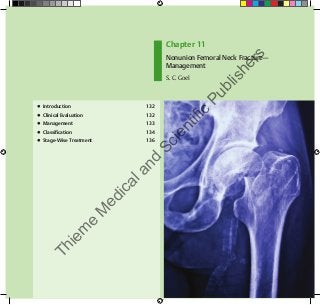
Chapter 11
Nonunion Femoral Neck Fracture—
Management
S. C. Goel
●  Introduction	 132
●  Clinical Evaluation	 132
●  Management	 133
●  Classification	 134
●  Stage-Wise Treatment	 136
Thiem
e
M
edicaland
Scientific
Publishers
132
Chapter 11
11  Nonunion Femoral Neck Fracture—Management
fractures more than 3 weeks old.6
Meyers et al7
arbitrarily
classified fracture of the neck of the femur treated more than
30 days after injury as having delayed treatment. Fractures
being operated more than 3 weeks after injury have remained
displaced for a considerable time, resulting in damage to vas-
cular supply of the head and hence are more prone to avas-
cular necrosis (AVN). As the fracture fragments are not in
contact, reparative process does not start and the fracture
ends tend to become smooth with absorption and sclerosis.
Some of the patients have been walking on this fracture, lead-
ing to formation of a pseudarthrosis and cavitation at frac-
tures ends. These fractures cannot be expected to unite by a
closed reduction and internal fixation due to lack of osteo-
genic potential and avascularity. When the fracture neck of
the femur is of more than 3 weeks’ duration, internal fixation
alone has a very high failure rate. Internal fixation has to be
combined with some type of bone graft or osteotomy, par-
ticularly in young patients below the age of 50 years (or 55
years) in whom it is desirable to preserve the joint.8
In developing countries, many patients are treated under
compromised situations, suboptimal operation theater con-
ditions, and improper implants and they report with nonun-
ion. These patients are treated in the same way as neglected
fractures.
██ Clinical Evaluation
Patients with nonunion of femoral neck fracture usually pre-
sent with painful limp. This pain generally appears within
few months after fracture, whereas pain from AVN generally
presents 1 to 2 years after fracture. Neglected femoral neck
fracture presents with shortening, severe external rotation of
the lower extremity, upward displacement of the trochanter,
with or without soft tissue contracture. Head and neck would
have undergone variable degree of absorption.
██ Introduction
Nonunion is the most common complication of fracture neck
of femur. The incidence of nonunion of femoral neck fracture
is up to18.5% for elderly patients and 8% for young patients.1,2
If patient’s age is more or the displacement is significant,
chances of nonunion may increase.3
The best end result after
a femoral neck fracture is the patient’s own healed femoral
neck and head. Much attention has been paid to the problem
of fracture of the neck of the femur in elderly patients, but
few of the published articles make any comment with regard
to the same lesion in younger patients.
Fracture of the neck of the femur has multiple factors,
leading to difficulties in union. Both biological and mechani-
cal parameters contribute to the development of union com-
plications. On the biological side, the lack of cambium layer of
the periosteum of femoral neck and the presence of synovial
fluid at the fracture site inhibit fracture union. Mechanical
parameters include the amount of vertical inclination of the
fracture line, quality of reduction, stability of fixation, and
integrity of the posterior cortex. These mechanical factors
lead to an unstable fracture.
Time interval between injury and operation is an impor-
tant factor in results of treatment of femoral neck fractures.
It is recommended that early anatomical reduction and rigid
internal fixation should be performed preferably on the day
of injury.4,5
Delayed treatment of the femoral neck fractures is
not as simple as those of fresh fracture. In developing coun-
tries, patients tend to visit first the local bone setter. There
are additional problems of transport, communication, lack of
early diagnosis, and so forth, leading to delay in seeking treat-
ment, which makes it more difficult to achieve osteosynthesis.
There is lot of confusion regarding the definition of unu-
nited fracture. Many authors have suggested against closed
reduction and internal fixation of displaced untreated
Thiem
e
M
edicaland
Scientific
Publishers
133
Nonunion Femoral Neck Fracture—Management
Plain X-rays are adequate to make a clinical diagnosis and
stage the neglected femoral neck fracture. Radiographic signs
of nonunion9
present at 3 months that indicate femoral neck
fracture nonunion are as follows:
1.	 Change in fracture position by 10 mm,
2.	 Change in screw position by 5%,
3.	 Backing out of the screws by 20 mm, and
4.	 Perforation of the screws into the hip joint.
Computed tomography (CT) scanning provides the best
assessment of fracture union. CT can be done even with inter-
nal fixation implant inside. CT scan may be useful to see the
bony appearance of stippled area and bony sclerosis, trabecu-
lar resorption, microfracture and subchondral collapse, and
presence of AVN. Radiographic changes often are not present
with AVN until the disease has reached an advanced stage.
Therefore, bone scintigraphy or magnetic resonance imaging
(MRI) may be considered to assess femoral head viability.
Infection must be ruled out as a cause or differential diagno-
sis for nonunion. History about any constitutional symptoms
should be taken and the skin incision should be inspected for
any signs of infection.
Preoperative evaluation must include white blood cell
count, erythrocyte sedimentation rate, and C-reactive pro-
tein. Intraoperative tissue samples from the nonunion site
should be obtained for frozen section at the time of any surgi-
cal intervention.
██ Management
Many different procedures have been proposed for the treat-
ment of nonunion of fracture of the neck of the femur. Albee10
reported using tibial bone peg graft in ununited femoral neck
fractures. Henderson11
used tibial or fibula grafts at Mayo clinic
in 77 young patients of ununited femoral neck fractures after
making a channel with a drill with good results. King6
and Nagi
et al12
have used fibular graft with internal fixation with good
results. Lindequist13
reported introduction of cancellous bone
graft through a drilled channel in femoral neck without expos-
ing the fracture after fixation by multiple pins.
Various workers have advised use of displacement and
angulation osteotomies in management of nonunion of fem-
oral neck fractures. McMurray14
proposed a displacement
osteotomy in which the femur is placed medially beneath the
femoral head following an oblique osteotomy from the base
of the greater trochanter to the upper end of the lesser tro-
chanter. He used it with good results in nonunion and fresh
fractures of femoral neck. The union occurred between three
fragments and the head remained viable. In a later series,
although the functional results had been good but union at
the fracture site had not been obtained always, shortening
has been a constant feature and incidence of AVN has been
between 18.2 and 22%.15
Pauwels16
demonstrated that the problem is essentially
biomechanical. He described an abduction osteotomy at the
intertrochanteric level, which shifts line of weight bearing
medially and converts the shearing force at the nonunion site
into a compression force as the fracture line becomes horizon-
tal. However, extreme valgus position shortens the lever arm,
thus placing increased pressure on the head of femur during
walking. He fixed the nonunion with graft and removed a
wedge from lateral side to realign the femur in valgus posi-
tion. Muller et al17
modified it and fixed the osteotomy with a
blade-plate. This concept has been popular in Europe.
Dickson18
devised a geometric angulation osteotomy com-
bined with an osteosynthesis. This osteotomy changes the
angle of weight bearing by 45 degrees. Marti et al19
got 76%
good or excellent results in 50 ununited femoral neck frac-
tures after valgus osteotomy. Many other reports have shown
good results with this procedure.20–22
Judet23
in 1962 described quadratus femoris–based muscle
pedicle bone graft for femoral neck fractures. Later, Meyers et
al24
and Baksi25
popularized this method.
Elderly patients with nonunion of femoral neck fracture are
not expected to show union with any osteosynthetic procedure
and they are treated by appropriate arthroplasty procedure.
Thiem
e
M
edicaland
Scientific
Publishers
134
Chapter 11
The factors on which the management depends are as
follows:
1.	 Age of the patient
2.	 Vascularity
3.	 Remaining bone quality
4.	 Status of the articular surface and sphericity of the
femoral head
5.	 Alignment of the neck and shaft
6.	 Potential limb length discrepancy
██ Classification
The classifications described for fresh fractures cannot be
used reliably to predict the outcome in cases of nonunion
of femoral neck fracture as there is absorption of femoral
neck; fracture surfaces get smoothened; the fracture gap is
increased; and the head of the femur can develop AVN. These
changes occur in variable severity depending on the length
of neglect. Classifications for fresh fracture like the Pauwels
and Garden classification require proper fracture surfaces to
see the displacement, which is difficult if there is absorption.
To see these changes good-quality X-ray of pelvis including
both hip joints in as identical position as possible should be
taken. The length of the proximal fragment is measured from
upper margin of fovea centralis to the midpoint of the fracture
margin. CT scan or MRI of pelvis can be extremely useful in
accurately measuring the gap between the fragments and the
size of the proximal fragment. Sometimes, the absorption of
the proximal fragment is more marked in the center than on
the periphery, giving it the shape of a cup or a moon. This may
not be clearly seen on routine AP view X-ray of the hip and
can be better appreciated on CT scan or MRI. AVN of the head
of the femur may be seen earlier on MRI/CT scan than on plain
X-ray of the hip.
Based on these changes, Sandhu26
has classified the unu-
nited femoral neck in following three stages.
Stage I (Fig. 11.1)
A.	 Fracture surfaces are still irregular (Fresh)
B.	 The size of proximal fragment is 2.5 cm or more
C.	 Gap between the fragments is 1 cm or less
D.	 Head of the femur is viable; there is no sign of AVN on
X-ray picture or MRI or CT scan
Stage II (Fig. 11.2)
A.	 Fracture surfaces are smoothened out
B.	 The size of the proximal fragment is 2.5 cm or more
C.	 The gap between the fragments is more than 1 cm but
less than 2.5 cm
D.	 The head of the femur is viable
If either of the feature a or c is present, it is allocated to
Stage II.
Fig. 11.1  Stage I.
Thiem
e
M
edicaland
Scientific
Publishers
135
Nonunion Femoral Neck Fracture—Management
Fig. 11.2  Stage II.
Fig. 11.3  (A) Stage III (B) Head of femur in same
patient after removal, looking only a shell.
Stage III (Fig. 11.3)
A.	 Fracture surfaces are smoothened out
B.	 The size of the proximal fragment is less than 2.5 cm
C.	 The gap between the fragments is more than 2.5 cm
D.	 The head of the femur shows signs of AVN
If any of the feature b, c, or d is present, the fracture is allo-
cated to Stage III.
A
B
Thiem
e
M
edicaland
Scientific
Publishers
136
Chapter 11
██ Stage-Wise Treatment
The goal of treatment in neglected fracture neck of femur
is to achieve a painless, mobile, and stable hip.27
The treat-
ment depends on the age and physical status of the patient,
duration of nonunion, viability and spherocity of the femoral
head, amount of resorption of the femoral neck, and potential
limb length inequality. Early anatomical reduction and stable
internal fixation restore the vascularity and reduce the inci-
dence of AVN.3
Nonunion and AVN of the femoral head are
the main complications following femoral neck fractures.
Unfortunately, in developing countries, patients traditionally
are, often, treated by local bone setters initially and report to
hospital late due to difficulties of finance, communication, and
transport. Even if they seek medical advice, many times the
diagnosis is missed or a poor technique or improper implant is
used, leading to nonunion. The fractures presenting late have
their own problems of management. Even in developed coun-
tries, the diagnosis is missed sometimes especially in elderly
people.28
If the fracture is more than 6 weeks old, accurate
close reduction and firm fixation to achieve union is difficult.
Management options being used currently are grouped as
follows:
1.	 Osteosynthesis with or without vascularized or non-
vascularized bone grafting
2.	 Osteotomy, displacement, or angulation type
3.	 Osteosynthesis with muscle pedicle bone grafting
4.	 Replacement (hemiarthroplasty, HA or total hip
replacement) (Figs. 11.4 and 11.5)
These are to be used in different stages as follows:29
Stage I
1.	 Closed reduction and internal fixation
2.	 Closed reduction and internal fixation with one screw
and double fibular graft or two screws and one fibular
graft
Fig. 11.4  (A) Radiograph of a 50-year-old female showing failure of fixation and nonunion; (B) treated with bipolar arthroplasty.
A B
Thiem
e
M
edicaland
Scientific
Publishers
137
Nonunion Femoral Neck Fracture—Management
A B C
Fig. 11.5  (A) Fracture neck of femur in a 45-year-old male; (B) failed internal fixation with nonunion; (C) managed by cemented total hip
arthroplasty.
3.	 Closed reduction or open reduction and bone muscle
pedicle graft based on quadratus femoris or sartorius
or tensor fascia femoris
4.	 Abduction osteotomy and osteosynthesis with DHS or
135 degree angled blade plate or 120 degree double-
angle blade plate
Stage II
1.	 Open reduction, freshening of fracture surfaces, and
internal fixation with two screws and one free fibular
graft
2.	 Open reduction and internal fixation with multiple
screws and muscle pedicle bone graft
3.	 Valgus osteotomy
Stage III
1.	 Total hip arthroplasty
2.	 Bipolar or HA
3.	 Valgus osteotomy
These procedures are discussed in detail in later chapters.
References
1. Bhandari M, Devereaux PJ, Swiontkowski
MF, et al. Internal fixation compared with arthroplasty for
displaced fractures of the femoral neck. A meta-analysis.
J Bone Joint Surg Am 2003;85-A(9):1673–1681
2. Haidukewych GJ, Rothwell WS, Jacofsky DJ, Torchia ME, Berry
DJ. Operative treatment of femoral neck fractures in patients
between the ages of fifteen and fifty years. J Bone Joint Surg
Am 2004;86-A(8):1711–1716
3. Parker MJ. Prediction of fracture union after internal fixation
of intracapsular femoral neck fractures. Injury 1994;25(Suppl
2):B3–B6
4. Deyerle WM. Multiple pin peripheral fixation in the fracture
of the neck of the femur: Immediate weight bearing. Clin
Orthop Relat Res 1965;39(39):135–156
5. Tooke SM, Favero KJ. Femoral neck fractures in skeletally
mature patients, fifty years old or less. J Bone Joint Surg Am
1985;67(8):1255–1260
Thiem
e
M
edicaland
Scientific
Publishers
138
Chapter 11
6. King T. The closed operation for intracapsular fracture of the
neck of the femur. Br J Surg 1939;26:721–748
7. Meyers MH, Harvey JP Jr, Moore TM. Treatment of displaced
subcapital and transcervical fractures of the femoral neck by
muscle-pedicle-bone graft and internal fixation. A preliminary
report on one hundred and fifty cases. J Bone Joint Surg Am
1973;55(2):257–274
8. Sandhu HS. Management of fracture neck of femur. Ind J
Othop 2005;39:130–136
9. Alho A, Benterud JG, Solovieva S. Internally fixed femoral neck
fractures. Early prediction of failure in 203 elderly patients
with displaced fractures. Acta Orthop Scand 1999;70(2):141–
144
10. Albee FH. The bone graft peg in treatment of fracture of the
neck of the femur. Ann Surg 1915;62(1):85–91
11. Henderson MS. Ununited fracture of the neck of the femur
treated by the aid of bone graft. J Bone Joint Surg 1940;22:97–
106
12. Nagi ON, Dhillon MS, Goni VG. Open reduction, internal
fixation and fibular autografting for neglected fracture of the
femoral neck. J Bone Joint Surg Br 1998;80(5):798–804
13. Lindequist S. Bone grafting in femoral neck fractures: results
in 28 cases operated on with multiple pinning and cancellous
bone grafting. Arch Orthop Trauma Surg 1989;108(2):116–
118
14. McMurry TP. Treatment of fracture of the neck of the femur by
oblique osteotomy. BMJ 1936;1:330
15. Goel SC, Srivastava AN, Goel MK, Kacker JN, Singh OP. Role of
McMurray’s osteotomy in treatment of intracapsular fracture
of the femoral neck. Indian J Orthop 1980;14:32–37
16. Pauwels F. Der schenkenholsbruck em mechanisches
problem. Grundlagen des heilungsvorganges. prognose und
kausale therapie. Stuttgard, Beilageheft zur zeitschrift fur
orthopaedische chirurgie, Ferninand enke. 1935.
17. Muller ME, Allgower M, Willinegger H. Manual of Internal
Fixation. Berlin: Springer-Verlag. 1979.
18. Dickson JA. The high geometric osteotomy, with rotation and
bone graft, for ununited fractures of the neck of the femur; a
preliminary report. J Bone Joint Surg Am 1947;29(4):1005–
1018
19. Marti RK, Schüller HM, Raaymakers ELFB. Intertrochanteric
osteotomy for non-union of the femoral neck. J Bone Joint
Surg Br 1989;71(5):782–787
20. Huang CH. Treatment of neglected femoral neck fractures in
young adults. Clin Orthop Relat Res 1986; (206):117–126
21. Magu NK, Rohilla R, Singh R, Tater R. Modified Pauwels’
intertrochanteric osteotomy in neglected femoral neck
fracture. Clin Orthop Relat Res 2009;467(4):1064–1073
22. Kalra M, Anand S. Valgus intertrochanteric osteotomy for
neglected femoral neck fractures in young adults. Int Orthop
2001;25(6):363–366
23. Judet R. Traitement des fractures du col du femur par greffe
pediculle. Acta Scand Orthop. 1962;32:421–427
24. Meyers MH, Harvey JP Jr, Moore TM. Delayed treatment of
subcapital and transcervical fractures of the neck of the femur
with internal fixation and a muscle pedicle bone graft. Orthop
Clin North Am 1974;5(4):743–756
25. Baksi DP. Internal fixation of ununited femoral neck fractures
combined with muscle-pedicle bone grafting. J Bone Joint
Surg Br 1986;68(2):239–245
26. Sandhu HS, Sandhu PS, Kapoor A. Neglected fractured neck
of the femur: a predictive classification and treatment by
osteosynthesis. Clin Orthop Relat Res 2005; (431):14–20
27. Kainth GS, Yuvarajan P, Maini L, Kumar V. Neglected
femoral neck fractures in adults. J Orthop Surg (Hong Kong)
2011;19(1):13–17
28. Eastwood HD. Delayed diagnosis of femoral-neck fractures in
the elderly. Age Ageing 1987;16(6):378–382
29. Jain AK, Mukunth R, Srivastava A. Treatment of neglected
femoral neck fracture. Indian J Orthop 2015;49(1):17–27
Thiem
e
M
edicaland
Scientific
Publishers

Contenu connexe

Plus de Thieme Medical and Scientific Publishers, India (6)

Anatomy for Dental Medicine, 2/e by Baker
Anatomy for Dental Medicine, 2/e by BakerAnatomy for Dental Medicine, 2/e by Baker
Anatomy for Dental Medicine, 2/e by Baker
 
Guidelines in Fracture Management - Proximal Tibia by Babhulkar
Guidelines in Fracture Management - Proximal Tibia by BabhulkarGuidelines in Fracture Management - Proximal Tibia by Babhulkar
Guidelines in Fracture Management - Proximal Tibia by Babhulkar
 
Progress in Clinical Neurosciences Vol 28, Chapter 1
Progress in Clinical Neurosciences Vol 28, Chapter 1Progress in Clinical Neurosciences Vol 28, Chapter 1
Progress in Clinical Neurosciences Vol 28, Chapter 1
 
Recent Human Anatomy: Volume 1 by Prasad
Recent Human Anatomy: Volume 1 by PrasadRecent Human Anatomy: Volume 1 by Prasad
Recent Human Anatomy: Volume 1 by Prasad
 
Principles of Medical Physiology by Sircar
Principles of Medical Physiology by SircarPrinciples of Medical Physiology by Sircar
Principles of Medical Physiology by Sircar
 
A Manual of Essential Pediatrics-Meharban Singh
A Manual of Essential Pediatrics-Meharban SinghA Manual of Essential Pediatrics-Meharban Singh
A Manual of Essential Pediatrics-Meharban Singh
 

Dernier

Advance Directives and Advance Care Planning: Ensuring Patient Voices Are Heard
Advance Directives and Advance Care Planning: Ensuring Patient Voices Are HeardAdvance Directives and Advance Care Planning: Ensuring Patient Voices Are Heard
Advance Directives and Advance Care Planning: Ensuring Patient Voices Are HeardVITASAuthor
 
Artificial Intelligence Robotics & Computational Fluid Dynamics
Artificial Intelligence Robotics & Computational Fluid DynamicsArtificial Intelligence Robotics & Computational Fluid Dynamics
Artificial Intelligence Robotics & Computational Fluid DynamicsParag Kothawade
 
办理西安大略大学毕业证成绩单|购买加拿大UWO文凭证书
办理西安大略大学毕业证成绩单|购买加拿大UWO文凭证书办理西安大略大学毕业证成绩单|购买加拿大UWO文凭证书
办理西安大略大学毕业证成绩单|购买加拿大UWO文凭证书zdzoqco
 
Biology class 12 assignment neet level practise chapter wise
Biology class 12 assignment neet level practise chapter wiseBiology class 12 assignment neet level practise chapter wise
Biology class 12 assignment neet level practise chapter wiseNAGKINGRAPELLY
 
Low Vision Case (Nisreen mokhanawala).pptx
Low Vision Case (Nisreen mokhanawala).pptxLow Vision Case (Nisreen mokhanawala).pptx
Low Vision Case (Nisreen mokhanawala).pptxShubham
 
Globalny raport: „Prawdziwe piękno 2024" od Dove
Globalny raport: „Prawdziwe piękno 2024" od DoveGlobalny raport: „Prawdziwe piękno 2024" od Dove
Globalny raport: „Prawdziwe piękno 2024" od Doveagatadrynko
 
Critical Advancements in Healthcare Software Development | smartData Enterpri...
Critical Advancements in Healthcare Software Development | smartData Enterpri...Critical Advancements in Healthcare Software Development | smartData Enterpri...
Critical Advancements in Healthcare Software Development | smartData Enterpri...amynickle2106
 
Incentive spirometry powerpoint presentation
Incentive spirometry powerpoint presentationIncentive spirometry powerpoint presentation
Incentive spirometry powerpoint presentationpratiksha ghimire
 
Text Neck Syndrome and its probable way out.pptx
Text Neck Syndrome and its probable way out.pptxText Neck Syndrome and its probable way out.pptx
Text Neck Syndrome and its probable way out.pptxProf. Satyen Bhattacharyya
 
Mobile Health And Apps (mhealth) How to design Application for medical App.pptx
Mobile Health And Apps (mhealth) How to design Application for medical App.pptxMobile Health And Apps (mhealth) How to design Application for medical App.pptx
Mobile Health And Apps (mhealth) How to design Application for medical App.pptxMahesh Chopra
 
Preventing Common Nutritional Deficiencies In Poultry Flocks (PPT).pdf
Preventing Common Nutritional Deficiencies In Poultry Flocks (PPT).pdfPreventing Common Nutritional Deficiencies In Poultry Flocks (PPT).pdf
Preventing Common Nutritional Deficiencies In Poultry Flocks (PPT).pdfAditiAlishetty
 
Lipid Profile test & Cardiac Markers for MBBS, Lab. Med. and Nursing.pptx
Lipid Profile test & Cardiac Markers for MBBS, Lab. Med. and Nursing.pptxLipid Profile test & Cardiac Markers for MBBS, Lab. Med. and Nursing.pptx
Lipid Profile test & Cardiac Markers for MBBS, Lab. Med. and Nursing.pptxRajendra Dev Bhatt
 
Clinical Education Presentation at Accelacare
Clinical Education Presentation at AccelacareClinical Education Presentation at Accelacare
Clinical Education Presentation at Accelacarepablor40
 
Single Assessment Framework - What We Know So Far
Single Assessment Framework - What We Know So FarSingle Assessment Framework - What We Know So Far
Single Assessment Framework - What We Know So FarCareLineLive
 
2024 HCAT Healthcare Technology Insights
2024 HCAT Healthcare Technology Insights2024 HCAT Healthcare Technology Insights
2024 HCAT Healthcare Technology InsightsHealth Catalyst
 

Dernier (20)

Advance Directives and Advance Care Planning: Ensuring Patient Voices Are Heard
Advance Directives and Advance Care Planning: Ensuring Patient Voices Are HeardAdvance Directives and Advance Care Planning: Ensuring Patient Voices Are Heard
Advance Directives and Advance Care Planning: Ensuring Patient Voices Are Heard
 
Artificial Intelligence Robotics & Computational Fluid Dynamics
Artificial Intelligence Robotics & Computational Fluid DynamicsArtificial Intelligence Robotics & Computational Fluid Dynamics
Artificial Intelligence Robotics & Computational Fluid Dynamics
 
办理西安大略大学毕业证成绩单|购买加拿大UWO文凭证书
办理西安大略大学毕业证成绩单|购买加拿大UWO文凭证书办理西安大略大学毕业证成绩单|购买加拿大UWO文凭证书
办理西安大略大学毕业证成绩单|购买加拿大UWO文凭证书
 
Coping with Childhood Cancer - How Does it Hurt Today
Coping with Childhood Cancer - How Does it Hurt TodayCoping with Childhood Cancer - How Does it Hurt Today
Coping with Childhood Cancer - How Does it Hurt Today
 
Biology class 12 assignment neet level practise chapter wise
Biology class 12 assignment neet level practise chapter wiseBiology class 12 assignment neet level practise chapter wise
Biology class 12 assignment neet level practise chapter wise
 
Low Vision Case (Nisreen mokhanawala).pptx
Low Vision Case (Nisreen mokhanawala).pptxLow Vision Case (Nisreen mokhanawala).pptx
Low Vision Case (Nisreen mokhanawala).pptx
 
Globalny raport: „Prawdziwe piękno 2024" od Dove
Globalny raport: „Prawdziwe piękno 2024" od DoveGlobalny raport: „Prawdziwe piękno 2024" od Dove
Globalny raport: „Prawdziwe piękno 2024" od Dove
 
Critical Advancements in Healthcare Software Development | smartData Enterpri...
Critical Advancements in Healthcare Software Development | smartData Enterpri...Critical Advancements in Healthcare Software Development | smartData Enterpri...
Critical Advancements in Healthcare Software Development | smartData Enterpri...
 
Incentive spirometry powerpoint presentation
Incentive spirometry powerpoint presentationIncentive spirometry powerpoint presentation
Incentive spirometry powerpoint presentation
 
Check Your own POSTURE & treat yourself.pptx
Check Your own POSTURE & treat yourself.pptxCheck Your own POSTURE & treat yourself.pptx
Check Your own POSTURE & treat yourself.pptx
 
Text Neck Syndrome and its probable way out.pptx
Text Neck Syndrome and its probable way out.pptxText Neck Syndrome and its probable way out.pptx
Text Neck Syndrome and its probable way out.pptx
 
Mobile Health And Apps (mhealth) How to design Application for medical App.pptx
Mobile Health And Apps (mhealth) How to design Application for medical App.pptxMobile Health And Apps (mhealth) How to design Application for medical App.pptx
Mobile Health And Apps (mhealth) How to design Application for medical App.pptx
 
Dr Sujit Chatterjee Hiranandani Hospital Kidney.pdf
Dr Sujit Chatterjee Hiranandani Hospital Kidney.pdfDr Sujit Chatterjee Hiranandani Hospital Kidney.pdf
Dr Sujit Chatterjee Hiranandani Hospital Kidney.pdf
 
Preventing Common Nutritional Deficiencies In Poultry Flocks (PPT).pdf
Preventing Common Nutritional Deficiencies In Poultry Flocks (PPT).pdfPreventing Common Nutritional Deficiencies In Poultry Flocks (PPT).pdf
Preventing Common Nutritional Deficiencies In Poultry Flocks (PPT).pdf
 
Lipid Profile test & Cardiac Markers for MBBS, Lab. Med. and Nursing.pptx
Lipid Profile test & Cardiac Markers for MBBS, Lab. Med. and Nursing.pptxLipid Profile test & Cardiac Markers for MBBS, Lab. Med. and Nursing.pptx
Lipid Profile test & Cardiac Markers for MBBS, Lab. Med. and Nursing.pptx
 
Clinical Education Presentation at Accelacare
Clinical Education Presentation at AccelacareClinical Education Presentation at Accelacare
Clinical Education Presentation at Accelacare
 
Single Assessment Framework - What We Know So Far
Single Assessment Framework - What We Know So FarSingle Assessment Framework - What We Know So Far
Single Assessment Framework - What We Know So Far
 
DELIRIUM psychiatric delirium is a organic mental disorder
DELIRIUM  psychiatric  delirium is a organic mental disorderDELIRIUM  psychiatric  delirium is a organic mental disorder
DELIRIUM psychiatric delirium is a organic mental disorder
 
Kidney Transplant At Hiranandani Hospital
Kidney Transplant At Hiranandani HospitalKidney Transplant At Hiranandani Hospital
Kidney Transplant At Hiranandani Hospital
 
2024 HCAT Healthcare Technology Insights
2024 HCAT Healthcare Technology Insights2024 HCAT Healthcare Technology Insights
2024 HCAT Healthcare Technology Insights
 

Guidelines in Fracture Management - Fracture of the Neck of the Femur

  • 1. Chapter 11 Nonunion Femoral Neck Fracture— Management S. C. Goel ●  Introduction 132 ●  Clinical Evaluation 132 ●  Management 133 ●  Classification 134 ●  Stage-Wise Treatment 136 Thiem e M edicaland Scientific Publishers
  • 2. 132 Chapter 11 11  Nonunion Femoral Neck Fracture—Management fractures more than 3 weeks old.6 Meyers et al7 arbitrarily classified fracture of the neck of the femur treated more than 30 days after injury as having delayed treatment. Fractures being operated more than 3 weeks after injury have remained displaced for a considerable time, resulting in damage to vas- cular supply of the head and hence are more prone to avas- cular necrosis (AVN). As the fracture fragments are not in contact, reparative process does not start and the fracture ends tend to become smooth with absorption and sclerosis. Some of the patients have been walking on this fracture, lead- ing to formation of a pseudarthrosis and cavitation at frac- tures ends. These fractures cannot be expected to unite by a closed reduction and internal fixation due to lack of osteo- genic potential and avascularity. When the fracture neck of the femur is of more than 3 weeks’ duration, internal fixation alone has a very high failure rate. Internal fixation has to be combined with some type of bone graft or osteotomy, par- ticularly in young patients below the age of 50 years (or 55 years) in whom it is desirable to preserve the joint.8 In developing countries, many patients are treated under compromised situations, suboptimal operation theater con- ditions, and improper implants and they report with nonun- ion. These patients are treated in the same way as neglected fractures. ██ Clinical Evaluation Patients with nonunion of femoral neck fracture usually pre- sent with painful limp. This pain generally appears within few months after fracture, whereas pain from AVN generally presents 1 to 2 years after fracture. Neglected femoral neck fracture presents with shortening, severe external rotation of the lower extremity, upward displacement of the trochanter, with or without soft tissue contracture. Head and neck would have undergone variable degree of absorption. ██ Introduction Nonunion is the most common complication of fracture neck of femur. The incidence of nonunion of femoral neck fracture is up to18.5% for elderly patients and 8% for young patients.1,2 If patient’s age is more or the displacement is significant, chances of nonunion may increase.3 The best end result after a femoral neck fracture is the patient’s own healed femoral neck and head. Much attention has been paid to the problem of fracture of the neck of the femur in elderly patients, but few of the published articles make any comment with regard to the same lesion in younger patients. Fracture of the neck of the femur has multiple factors, leading to difficulties in union. Both biological and mechani- cal parameters contribute to the development of union com- plications. On the biological side, the lack of cambium layer of the periosteum of femoral neck and the presence of synovial fluid at the fracture site inhibit fracture union. Mechanical parameters include the amount of vertical inclination of the fracture line, quality of reduction, stability of fixation, and integrity of the posterior cortex. These mechanical factors lead to an unstable fracture. Time interval between injury and operation is an impor- tant factor in results of treatment of femoral neck fractures. It is recommended that early anatomical reduction and rigid internal fixation should be performed preferably on the day of injury.4,5 Delayed treatment of the femoral neck fractures is not as simple as those of fresh fracture. In developing coun- tries, patients tend to visit first the local bone setter. There are additional problems of transport, communication, lack of early diagnosis, and so forth, leading to delay in seeking treat- ment, which makes it more difficult to achieve osteosynthesis. There is lot of confusion regarding the definition of unu- nited fracture. Many authors have suggested against closed reduction and internal fixation of displaced untreated Thiem e M edicaland Scientific Publishers
  • 3. 133 Nonunion Femoral Neck Fracture—Management Plain X-rays are adequate to make a clinical diagnosis and stage the neglected femoral neck fracture. Radiographic signs of nonunion9 present at 3 months that indicate femoral neck fracture nonunion are as follows: 1. Change in fracture position by 10 mm, 2. Change in screw position by 5%, 3. Backing out of the screws by 20 mm, and 4. Perforation of the screws into the hip joint. Computed tomography (CT) scanning provides the best assessment of fracture union. CT can be done even with inter- nal fixation implant inside. CT scan may be useful to see the bony appearance of stippled area and bony sclerosis, trabecu- lar resorption, microfracture and subchondral collapse, and presence of AVN. Radiographic changes often are not present with AVN until the disease has reached an advanced stage. Therefore, bone scintigraphy or magnetic resonance imaging (MRI) may be considered to assess femoral head viability. Infection must be ruled out as a cause or differential diagno- sis for nonunion. History about any constitutional symptoms should be taken and the skin incision should be inspected for any signs of infection. Preoperative evaluation must include white blood cell count, erythrocyte sedimentation rate, and C-reactive pro- tein. Intraoperative tissue samples from the nonunion site should be obtained for frozen section at the time of any surgi- cal intervention. ██ Management Many different procedures have been proposed for the treat- ment of nonunion of fracture of the neck of the femur. Albee10 reported using tibial bone peg graft in ununited femoral neck fractures. Henderson11 used tibial or fibula grafts at Mayo clinic in 77 young patients of ununited femoral neck fractures after making a channel with a drill with good results. King6 and Nagi et al12 have used fibular graft with internal fixation with good results. Lindequist13 reported introduction of cancellous bone graft through a drilled channel in femoral neck without expos- ing the fracture after fixation by multiple pins. Various workers have advised use of displacement and angulation osteotomies in management of nonunion of fem- oral neck fractures. McMurray14 proposed a displacement osteotomy in which the femur is placed medially beneath the femoral head following an oblique osteotomy from the base of the greater trochanter to the upper end of the lesser tro- chanter. He used it with good results in nonunion and fresh fractures of femoral neck. The union occurred between three fragments and the head remained viable. In a later series, although the functional results had been good but union at the fracture site had not been obtained always, shortening has been a constant feature and incidence of AVN has been between 18.2 and 22%.15 Pauwels16 demonstrated that the problem is essentially biomechanical. He described an abduction osteotomy at the intertrochanteric level, which shifts line of weight bearing medially and converts the shearing force at the nonunion site into a compression force as the fracture line becomes horizon- tal. However, extreme valgus position shortens the lever arm, thus placing increased pressure on the head of femur during walking. He fixed the nonunion with graft and removed a wedge from lateral side to realign the femur in valgus posi- tion. Muller et al17 modified it and fixed the osteotomy with a blade-plate. This concept has been popular in Europe. Dickson18 devised a geometric angulation osteotomy com- bined with an osteosynthesis. This osteotomy changes the angle of weight bearing by 45 degrees. Marti et al19 got 76% good or excellent results in 50 ununited femoral neck frac- tures after valgus osteotomy. Many other reports have shown good results with this procedure.20–22 Judet23 in 1962 described quadratus femoris–based muscle pedicle bone graft for femoral neck fractures. Later, Meyers et al24 and Baksi25 popularized this method. Elderly patients with nonunion of femoral neck fracture are not expected to show union with any osteosynthetic procedure and they are treated by appropriate arthroplasty procedure. Thiem e M edicaland Scientific Publishers
  • 4. 134 Chapter 11 The factors on which the management depends are as follows: 1. Age of the patient 2. Vascularity 3. Remaining bone quality 4. Status of the articular surface and sphericity of the femoral head 5. Alignment of the neck and shaft 6. Potential limb length discrepancy ██ Classification The classifications described for fresh fractures cannot be used reliably to predict the outcome in cases of nonunion of femoral neck fracture as there is absorption of femoral neck; fracture surfaces get smoothened; the fracture gap is increased; and the head of the femur can develop AVN. These changes occur in variable severity depending on the length of neglect. Classifications for fresh fracture like the Pauwels and Garden classification require proper fracture surfaces to see the displacement, which is difficult if there is absorption. To see these changes good-quality X-ray of pelvis including both hip joints in as identical position as possible should be taken. The length of the proximal fragment is measured from upper margin of fovea centralis to the midpoint of the fracture margin. CT scan or MRI of pelvis can be extremely useful in accurately measuring the gap between the fragments and the size of the proximal fragment. Sometimes, the absorption of the proximal fragment is more marked in the center than on the periphery, giving it the shape of a cup or a moon. This may not be clearly seen on routine AP view X-ray of the hip and can be better appreciated on CT scan or MRI. AVN of the head of the femur may be seen earlier on MRI/CT scan than on plain X-ray of the hip. Based on these changes, Sandhu26 has classified the unu- nited femoral neck in following three stages. Stage I (Fig. 11.1) A. Fracture surfaces are still irregular (Fresh) B. The size of proximal fragment is 2.5 cm or more C. Gap between the fragments is 1 cm or less D. Head of the femur is viable; there is no sign of AVN on X-ray picture or MRI or CT scan Stage II (Fig. 11.2) A. Fracture surfaces are smoothened out B. The size of the proximal fragment is 2.5 cm or more C. The gap between the fragments is more than 1 cm but less than 2.5 cm D. The head of the femur is viable If either of the feature a or c is present, it is allocated to Stage II. Fig. 11.1  Stage I. Thiem e M edicaland Scientific Publishers
  • 5. 135 Nonunion Femoral Neck Fracture—Management Fig. 11.2  Stage II. Fig. 11.3  (A) Stage III (B) Head of femur in same patient after removal, looking only a shell. Stage III (Fig. 11.3) A. Fracture surfaces are smoothened out B. The size of the proximal fragment is less than 2.5 cm C. The gap between the fragments is more than 2.5 cm D. The head of the femur shows signs of AVN If any of the feature b, c, or d is present, the fracture is allo- cated to Stage III. A B Thiem e M edicaland Scientific Publishers
  • 6. 136 Chapter 11 ██ Stage-Wise Treatment The goal of treatment in neglected fracture neck of femur is to achieve a painless, mobile, and stable hip.27 The treat- ment depends on the age and physical status of the patient, duration of nonunion, viability and spherocity of the femoral head, amount of resorption of the femoral neck, and potential limb length inequality. Early anatomical reduction and stable internal fixation restore the vascularity and reduce the inci- dence of AVN.3 Nonunion and AVN of the femoral head are the main complications following femoral neck fractures. Unfortunately, in developing countries, patients traditionally are, often, treated by local bone setters initially and report to hospital late due to difficulties of finance, communication, and transport. Even if they seek medical advice, many times the diagnosis is missed or a poor technique or improper implant is used, leading to nonunion. The fractures presenting late have their own problems of management. Even in developed coun- tries, the diagnosis is missed sometimes especially in elderly people.28 If the fracture is more than 6 weeks old, accurate close reduction and firm fixation to achieve union is difficult. Management options being used currently are grouped as follows: 1. Osteosynthesis with or without vascularized or non- vascularized bone grafting 2. Osteotomy, displacement, or angulation type 3. Osteosynthesis with muscle pedicle bone grafting 4. Replacement (hemiarthroplasty, HA or total hip replacement) (Figs. 11.4 and 11.5) These are to be used in different stages as follows:29 Stage I 1. Closed reduction and internal fixation 2. Closed reduction and internal fixation with one screw and double fibular graft or two screws and one fibular graft Fig. 11.4  (A) Radiograph of a 50-year-old female showing failure of fixation and nonunion; (B) treated with bipolar arthroplasty. A B Thiem e M edicaland Scientific Publishers
  • 7. 137 Nonunion Femoral Neck Fracture—Management A B C Fig. 11.5  (A) Fracture neck of femur in a 45-year-old male; (B) failed internal fixation with nonunion; (C) managed by cemented total hip arthroplasty. 3. Closed reduction or open reduction and bone muscle pedicle graft based on quadratus femoris or sartorius or tensor fascia femoris 4. Abduction osteotomy and osteosynthesis with DHS or 135 degree angled blade plate or 120 degree double- angle blade plate Stage II 1. Open reduction, freshening of fracture surfaces, and internal fixation with two screws and one free fibular graft 2. Open reduction and internal fixation with multiple screws and muscle pedicle bone graft 3. Valgus osteotomy Stage III 1. Total hip arthroplasty 2. Bipolar or HA 3. Valgus osteotomy These procedures are discussed in detail in later chapters. References 1. Bhandari M, Devereaux PJ, Swiontkowski MF, et al. Internal fixation compared with arthroplasty for displaced fractures of the femoral neck. A meta-analysis. J Bone Joint Surg Am 2003;85-A(9):1673–1681 2. Haidukewych GJ, Rothwell WS, Jacofsky DJ, Torchia ME, Berry DJ. Operative treatment of femoral neck fractures in patients between the ages of fifteen and fifty years. J Bone Joint Surg Am 2004;86-A(8):1711–1716 3. Parker MJ. Prediction of fracture union after internal fixation of intracapsular femoral neck fractures. Injury 1994;25(Suppl 2):B3–B6 4. Deyerle WM. Multiple pin peripheral fixation in the fracture of the neck of the femur: Immediate weight bearing. Clin Orthop Relat Res 1965;39(39):135–156 5. Tooke SM, Favero KJ. Femoral neck fractures in skeletally mature patients, fifty years old or less. J Bone Joint Surg Am 1985;67(8):1255–1260 Thiem e M edicaland Scientific Publishers
  • 8. 138 Chapter 11 6. King T. The closed operation for intracapsular fracture of the neck of the femur. Br J Surg 1939;26:721–748 7. Meyers MH, Harvey JP Jr, Moore TM. Treatment of displaced subcapital and transcervical fractures of the femoral neck by muscle-pedicle-bone graft and internal fixation. A preliminary report on one hundred and fifty cases. J Bone Joint Surg Am 1973;55(2):257–274 8. Sandhu HS. Management of fracture neck of femur. Ind J Othop 2005;39:130–136 9. Alho A, Benterud JG, Solovieva S. Internally fixed femoral neck fractures. Early prediction of failure in 203 elderly patients with displaced fractures. Acta Orthop Scand 1999;70(2):141– 144 10. Albee FH. The bone graft peg in treatment of fracture of the neck of the femur. Ann Surg 1915;62(1):85–91 11. Henderson MS. Ununited fracture of the neck of the femur treated by the aid of bone graft. J Bone Joint Surg 1940;22:97– 106 12. Nagi ON, Dhillon MS, Goni VG. Open reduction, internal fixation and fibular autografting for neglected fracture of the femoral neck. J Bone Joint Surg Br 1998;80(5):798–804 13. Lindequist S. Bone grafting in femoral neck fractures: results in 28 cases operated on with multiple pinning and cancellous bone grafting. Arch Orthop Trauma Surg 1989;108(2):116– 118 14. McMurry TP. Treatment of fracture of the neck of the femur by oblique osteotomy. BMJ 1936;1:330 15. Goel SC, Srivastava AN, Goel MK, Kacker JN, Singh OP. Role of McMurray’s osteotomy in treatment of intracapsular fracture of the femoral neck. Indian J Orthop 1980;14:32–37 16. Pauwels F. Der schenkenholsbruck em mechanisches problem. Grundlagen des heilungsvorganges. prognose und kausale therapie. Stuttgard, Beilageheft zur zeitschrift fur orthopaedische chirurgie, Ferninand enke. 1935. 17. Muller ME, Allgower M, Willinegger H. Manual of Internal Fixation. Berlin: Springer-Verlag. 1979. 18. Dickson JA. The high geometric osteotomy, with rotation and bone graft, for ununited fractures of the neck of the femur; a preliminary report. J Bone Joint Surg Am 1947;29(4):1005– 1018 19. Marti RK, Schüller HM, Raaymakers ELFB. Intertrochanteric osteotomy for non-union of the femoral neck. J Bone Joint Surg Br 1989;71(5):782–787 20. Huang CH. Treatment of neglected femoral neck fractures in young adults. Clin Orthop Relat Res 1986; (206):117–126 21. Magu NK, Rohilla R, Singh R, Tater R. Modified Pauwels’ intertrochanteric osteotomy in neglected femoral neck fracture. Clin Orthop Relat Res 2009;467(4):1064–1073 22. Kalra M, Anand S. Valgus intertrochanteric osteotomy for neglected femoral neck fractures in young adults. Int Orthop 2001;25(6):363–366 23. Judet R. Traitement des fractures du col du femur par greffe pediculle. Acta Scand Orthop. 1962;32:421–427 24. Meyers MH, Harvey JP Jr, Moore TM. Delayed treatment of subcapital and transcervical fractures of the neck of the femur with internal fixation and a muscle pedicle bone graft. Orthop Clin North Am 1974;5(4):743–756 25. Baksi DP. Internal fixation of ununited femoral neck fractures combined with muscle-pedicle bone grafting. J Bone Joint Surg Br 1986;68(2):239–245 26. Sandhu HS, Sandhu PS, Kapoor A. Neglected fractured neck of the femur: a predictive classification and treatment by osteosynthesis. Clin Orthop Relat Res 2005; (431):14–20 27. Kainth GS, Yuvarajan P, Maini L, Kumar V. Neglected femoral neck fractures in adults. J Orthop Surg (Hong Kong) 2011;19(1):13–17 28. Eastwood HD. Delayed diagnosis of femoral-neck fractures in the elderly. Age Ageing 1987;16(6):378–382 29. Jain AK, Mukunth R, Srivastava A. Treatment of neglected femoral neck fracture. Indian J Orthop 2015;49(1):17–27 Thiem e M edicaland Scientific Publishers