SlideShare une entreprise Scribd logo
1  sur  33
Télécharger pour lire hors ligne
ANOLYTE AND CATHOLYTE AS DISINFECTANTS IN A POULTRY
PROCESSING PLANT
Prof T E Cloete, Dept of Microbiology and Plant Pathology, University of
Pretoria
1. INTRODUCTION
Poultry is a important part of the animal food market and production is increasing
to satisfy public demand world-wide (Bryan 1980; Anand et al., 1989). Poultry
and its products are also a major dietary item for the South African population
(Bok et al., 1986). According to the Directorate of Veterinary Public Health, 4.4
billion rands worth of poultry meat products were purchased by South Africans in
1994 (AFMA, 1996; SAPA, 1996). Therefore, it becomes necessary to maintain
absolute hygiene and strict control at different stages of processing to produce a
safe and wholesome chicken product.
Healthy chickens ready for processing harbour a tremendous amount and variety
of bacteria. These bacteria are present on the surfaces of feet, feathers, skin
and also in the intestines. During processing, a high proportion of these
organisms will be removed, but further contamination can occur at any stage of
the processing operation. The procedure for converting a live, healthy bird into a
safe and wholesome poultry product provides many opportunities for micro-
organisms to colonise on the surface of the carcase. During the various
processing operations, opportunities exist for the contamination of the carcases
from the environment, the process in the plant itself, contamination via knives,
equipment, the hands of workers and also by cross-contamination from carcase
to carcase. Some processing operations encourage an increase of
contamination or even multiplication of contaminating organisms. As a result,
the microbial population changes from mainly Gram-positive rods and micrococci
on the outside of the live chicken to Gram-negative micro-organisms on the
finished product (Bryan, 1980; Thomas et al., 1980; Eustace, 1981; Roberts,
1982; Grau, 1986; Bailey et al., 1987; Connor et al., 1987; Banwart, 1989; Mead,
1989). Poultry processing has a number of unique features which make control
of microbial contamination more difficult than the processing of any other
conventional meat animal. Among them is the rapid rate of processing in some
processing plants, a condition which favours the spread of micro-organisms.
The carcase must be kept whole throughout the process and the viscera have to
be removed rapidly through a small opening in the abdomen without breakage,
to minimise contamination of the carcase with intestinal organisms. After
defeathering, the skin provides a complex surface with many holes which are
capable of trapping bacteria (Mead, 1982; Grau, 1986; Mead, 1989).
The micro-organisms are widely distributed over the carcases under normal
circumstances and are spread over the skin during scalding and defeathering
and on the inner and outer surfaces during evisceration and further processing
(Bailey et al., 1987). Efforts should be made to prevent the build-up of
contamination peaks during processing. Rinsing of the carcases, especially
during defeathering and evisceration is therefore of great importance (McMeekin
et al., 1979a; Brown et al., 1982; Mead, 1982; Anand et al., 1989; Mead, 1989).
Spoilage bacteria grow mainly on the skin surfaces, in the feather follicles and on
cut muscle surfaces under the skin. The nature and rate of attachment of the
micro-organisms depends upon several factors including the bacteria involved
and their concentration and also the conditions under which attachment occurs,
namely, pH, temperature and contact-time. It was also found that Pseudomonas
strains attach to meat surfaces more rapidly than any other bacteria
(Firstenberg-Eden, 1981). The structure of the skin also has a crucial influence
on attachment of bacteria. The organisms adhere by way of flagella and fimbrae
and cannot easily be removed by rinsing, especially after a delay. There is still
some disagreement on the role and importance of flagella in the attachment
process of bacteria to meat. Research also shows that mesophilic bacteria are
more heat-resistant when attached to skin than are the same bacteria not
attached. (Barnes et al., 1973; Green, 1974; Notermans et al., 1974; Notermans
et al., 1975; Harrigan, 1976; Firstenberg-Eden, 1981; Thomas et al., 1981; Faber
et al., 1984; Lillard, 1985). The skin serves as a barrier to micro-organisms that
might otherwise contaminate the underlying muscle and therefore the deep
muscles are normally free of bacteria (Bryan, 1980; Mead, 1982). The few
bacteria found in the deep muscle are of types that can only multiply slowly or
not at all at low temperatures. The important microbiological changes take place
on the surfaces of the carcases. It appears that some parts of the carcase are
more favourable than others for bacterial growth, depending on the type of
muscle and pH. Studies conducted over the last few years show that the sites
most heavily contaminated are the neck skin and less frequently on the back and
the area around the vent. Fewer organisms are found around the breast, legs
and under the wings. Acinetobacter and Alteromonas grow better in leg muscle
where pH is 6.4 to 6.7 than in breast muscle where pH is 5.7 to 5.9.
Pseudomonas spp. can grow well at both pH ranges (Patterson, 1972; Barnes et
al., 1973; Green, 1974; McMeekin et al., 1979a; Bryan, 1980; Thomas et al.,
1981; Mead, 1982; Gill, 1983; Grau, 1986; Anand et al., 1989). The presumable
reason for the neck skin being the most heavily contaminated is that the
washings from the rest of the carcase run down the neck while the carcase
hangs on the conveyor (Patterson, 1972; Connor et al., 1987).
2. LITERATURE REVIEW
2.1 Microbiology of poultry
Contaminants may be micro-organisms that cause spoilage of the product or
organisms of public health significance. Pathogens associated with poultry are
Salmonella, Staphylococcus aureus, Clostridium perfringens and Escherichia
coli. Listeria monocytogenes and Campylobacter jejuni have also been isolated
from poultry. Spoilage bacteria most frequently associated with poultry are
Pseudomonas spp., Acinetobacter, Moraxella, Alteromonas putrefaciens,
Aeromonas spp., Corynebacterium, Flavobacterium, Micrococcaceae and
Enterobacteriaceae. Poultry is a common vehicle of foodborne illness (See
Table 1.1) (Bryan, 1980; Todd, 1980; Smeltzer, 1981; Brown et al., 1982; Mead,
1982; Roberts, 1982; Ralph et al., 1984; Evans, 1986; Gill, 1986; Grau, 1986;
Silliker et al., 1986; Cunningham 1987; Banwart, 1989; Mead, 1989; Zottola
et al., 1990; Jones et al., 1991).
2.2 Pathogens
Salmonella
Salmonella are the main cause of food poisoning from poultry meat (Dougherty,
1976; Todd, 1980). Little is known about the incidence of Salmonella in South
Africa although figures have been reported by Bok et al., 1986 and Geornaras et
al., 1994. There are many sources from which poultry may obtain Salmonella,
the main sources being from cross-contamination during breeding, hatching and
intensive rearing operations. Salmonellas are not part of the normal intestinal
microflora of poultry, but are acquired from the farm environment via insects,
rodents and birds. Feed is also an important source of salmonellas through
contamination of various components of the feed mix. The organisms occur
more often in the caecum than in any other region of the gut from where they
may be excreted for varying periods, without the host showing any sign of
disease ( Morris et al., 1970; Mead, 1982; Grau, 1986; Silliker et al., 1986; Mead,
1989; Zottola et al., 1990; Jones et al., 1991). Salmonellas from one flock can
contaminate another, usually during conditions of intensive rearing and also
when there is inadequate cleaning and disinfecting of the multi-cage
transportation lorries used to convey the birds to the abattoir. Studies have also
shown that live poultry transported from the farm often introduce Salmonella into
the processing plant. Such contamination may result in considerable scattering
of salmonellae during processing especially in the plucking machines and the
scalding tank and may lead to contamination of the final product (McBride et al.,
1980; Mead, 1982; Mead, 1989; James et al., 1992).
Clostridium perfringens
Clostridium perfringens is considered to be more widespread in the environment
than any other pathogenic bacteria. This organism is commonly present in the
intestinal tract of many warm-blooded animals and has been isolated from faecal
matter, soil and dust. Raw poultry meat is normally stored at temperatures too
low (< 15°C) to permit Clostridium perfringens to grow. Therefore, there seems
little risk of multiplication in the processing plant. Clostridium perfringens is
mainly present on processed poultry as spores ( Bryan, 1980; Todd, 1980;
Mead, 1982; Bailey et al., 1987; Mead, 1989). Only type A strains are normally
involved in human food poisoning and these may be haemolytic, with heat-
sensitive spores or non-haemolytic, with spores that are highly heat resistant.
These heat-resistant strains can survive normal cooking procedures and if the
cooked meat is held under favourable conditions, the organism can multiply to
hazardous levels (Todd, 1980; Mead, 1989; Zottola et al., 1990).
Staphylococcus aureus
Food poisoning from poultry meat caused by Staphylococcus aureus is much
less common than that due to salmonellas or Clostridium perfringens (Todd,
1980; Mead, 1982). Staphylococcus is important in relation to poultry meat,
because it can produce enterotoxins which may cause food poisoning in humans
(Notermans et al., 1982). Live poultry carry Staphylococcus aureus on skin
surfaces and in nasal cavities, but low numbers are also present in the intestinal
tract (Todd, 1980; Evans, 1986; Grau, 1986; Mead, 1989). Isolates of
Staphylococcus aureus from poultry can be subdivided into human, non-human
and intermediate types (Gibbs et al., 1978; Mead 1989). It appears that
Staphylococcus aureus may also be obtained from human sources after
hatching and during processing of the carcases (Gibbs et al., 1978; Mead,
1982). Notermans et al., 1982 indicated that after processing, contamination of
carcases with this organism increased to >103
g-1
of skin. Defeathering
machinery in particular may support the build-up of Staphylococcus aureus.
Evisceration and chilling are also processing stages which have been
incriminated in contaminating carcases with Staphylococcus aureus (Gibbs et
al., 1978; Todd, 1980; Mead, 1982; Notermans et al., 1982; Mead, 1989).
Campylobacter jejuni
Campylobacter is widely spread in nature and is isolated from wild and domestic
animals as well as from the environment. Poultry is a major reservoir of
Campylobacter jejuni. Many commercial poultry flocks appear to be
symptomless carriers of C. jejuni, with up to 107
.g-1
of gut content being
demonstrated in the ileum and caeca of infected poultry and similar levels in the
faeces (Genigeorgis et al., 1986; Mead, 1989; Zottola et al., 1990). Some
poultry flocks that are negative before slaughter will therefore become
contaminated during processing. Campylobacter is microaerophilic with a
relative high minimum growth temperature (30 C) and there seems little
likelihood of them multiplying in the processing plant or on the raw, processed
product. The main problem in processing is that of cross-contamination (Zottola
et al., 1990; Smeltzer, 1981). Campylobacter spp. are more sensitive than many
other organisms to the adverse effects of environmental conditions (drying,
freezing and cold storage). For this reason, attention has been given to factors
influencing the survival of campylobacters in processing. Although freezing is
harmfull to Campylobacter, it does not eliminate this organism from poultry.
Nevertheless, the contamination rate tends to be higher in fresh than in frozen
carcases. Campylobacter spp. are also more sensitive to chlorine than E. coli,
but are not eliminated from poultry carcases by immersion chilling in chlorinated
water. On the contrary, cooling-water seems to be an important reservoir of this
organism: 100-3000 CFU.ml
-1
were demonstrated and survival over long periods
at low temperatures is possible. Campylobacter was also isolated from air
samples as well as equipment (Cunningham, 1987; Mead, 1989; Zottola et al.,
1990).
Listeria monocytogenes
Listeria monocytogenes is widely distributed in nature and the environment.
These organisms are isolated from soil, vegetation and faeces of humans and
animals, with poultry often being contaminated. Studies also indicated that 57%
(20 of 35 samples) and 33% (17 of 51 samples) of market poultry, respectively,
contained L. monocytogenes. L. monocytogenes can multiply at refrigeration
temperatures. Data also suggests that L. monocytogenes is more heat resistant
in meat than Salmonella. The necessity of proper hygiene procedures in
handling, processing and packaging of poultry is therefore emphasised (Zottola
et al., 1990).
2.3 Spoilage organisms
The spoilage of raw poultry meat is invariably due to the growth and metabolic
activities of specific types of bacteria, the psychrotrophs (Ralph et al., 1984 Kraft,
1986; Mead 1989). Psychrotrophs most frequently associated with poultry are
Acinetobacter, Moraxella, Alteromonas putrefaciens, Aeromonas spp.,
Flavobacterium spp., Corynebacterium, Micrococcaceae, Enterobacteriaceae,
Serratia liquefaciens, the pigmented and non-pigmented Pseudomonas spp. and
also yeast and moulds (Bryan, 1980; Kraft, 1986; Mead, 1989). The bacteria
which usually predominate on spoiled carcases held below 10°C are the
Pseudomonas (P.) spp., especially P. fluorescens, P. putida and P. fragi and
also Acinetobacter and Moraxella (Bryan, 1980; Lahellec et al., 1981; Ralph,
1984; Mead, 1989). Some spoilage bacteria originate from the rearing
environment and these organisms are carried in large numbers on the feet and
feathers of poultry. These bacteria are not found in the intestines of poultry
(Mead, 1982; Grau, 1986; Bailey et al., 1987). Prior to slaughter, the incoming
chickens are contaminated with a large number of spoilage bacteria, but most
are destroyed when passing through the scald tank, such as Acinetobacter,
Moraxella, Pseudomonas, Corynebacterium and Flavobacterium (Lahellec et al.,
1979; Mead, 1989). Pseudomonas, however, form a small proportion of
psychrotrophic flora on the outside of the chicken (Mead, 1982).
Recontamination occurs during various processing stages, because the
organisms multiply on all wet surfaces, including the carcases (Bryan, 1980;
Mead, 1989). Another possible source of spoilage bacteria is also the
processing plant water-supply. The Pseudomonas are more resistant to chlorine
than Escherichia coli and therefore may survive normal water treatment in the
processing plant. Pseudomonas can be eliminated by super-chlorination of
water at the processing plant The quality of water in the processing plant is
therefore of great importance. Essential steps to prevent excessive levels of
contamination include prompt washing and chilling of eviscerated poultry and
effective cleaning and disinfection procedures for equipment and working
surfaces at the end of the processing day, prior to the next days production
(Lahellec et al., 1979; Mead, 1989).
The growth of spoilage bacteria and thus the shelf-life of raw poultry meat,
stored under chill conditions, will depend on the numbers and types of spoilage
organisms present immediately after processing, the storage time and
temperature, the type of tissue (skin or muscle), the pH, the redox potential, the
type of packaging and the presence or absence of carbon dioxide (Bryan, 1980;
Ralph et al., 1984; Mead, 1989).
2.4 Time and Temperature
Psychrotrophs can grow at temperatures of -3°C, but most do not multiply above
34 C (Mead, 1989). Psychrotrophic Pseudomonas become the predominant
flora on the aerobic surfaces of poultry stored at low temperatures and they can
multiply the entire time carcases are held at commonly used refrigerator
temperatures (Bryan, 1980). There is a simple relationship between storage
temperature and shelf-life under aerobic conditions and for any given chill
temperature this is related to the doubling time of the spoilage organisms
(Barnes, 1976). The differential effect of storage temperature on the microbial
growth rates influences the composition of the ultimate spoilage organisms.
Pseudomonas predominated at spoilage when poultry carcases were held at
1°C. Above 10 C, however the predominant organisms comprised mainly
Acinetobacter spp. and Enterobacter (McMeekin, 1975; Mead, 1982). Fewer
organisms are capable of growth at 4°C and those which do so often undergo a
lengthy lag phase (Mead, 1982).
2.5 Meat pH and type of muscle
Although most carcase contaminants are found on the skin and over the inner
surface of the visceral cavity, the growth of spoilage bacteria during chill storage
conditions occurs primarily on cut muscle tissue and in the feather holes (Mead,
1982; Grau, 1986; Mead, 1989). Some parts of the chicken carcase appear to
be more favourable than others for bacterial growth, depending on the muscle
type and pH (Barnes, 1976). Pigmented and non-pigmented strains of
Pseudomonas spp. grew equally well in the breast (pH 5,7 - 5,9) and in leg
muscle (pH 6,4 - 6,7)(Mead, 1982). Acinetobacter - Moraxella spp. grew better
in leg muscle, but not in breast muscle, whilst Shewanella grew faster in leg than
in breast (Mead, 1982; Grau, 1986). Therefore one might expect to find different
bacteria growing on the various cut muscle surfaces of spoiling carcases. The
possibility also exists that spoilage may be more rapid in the high pH areas
(Barnes, 1976).
2.6 Packaging and carbon dioxide
Apart from the tendency to retain moisture, the most important property of
packaging film in relation to shelf-life is the permeability to oxygen and carbon
dioxide (Mead, 1982). It was also shown that chicken carcases stored at 1°C in
impermeable vacuum packs (vinylidene chloride-vinyl chloride copolymer) kept
for ca. 5 days longer than those packed in gas-permeable polyethylene (Barnes,
1976; Mead, 1982). Pseudomonas spp. are the principal causes of spoilage on
carcases packed in oxygen-permeable films, while Shewanella putrefaciens
were the principal cause of spoilage of poultry carcases packed in oxygen
impermeable films (Barnes, 1976; Bryan, 1980; Gill, 1983).
2.7 Influence of processing on poultry
The main operations in processing poultry are as follows: birds are removed
from crates, hung by the feet on shackles on a conveyor, stunned by a low
voltage electric shock in a water bath and killed by exsanguination following
slitting of the neck and severing the carotid arteries. They are then scalded,
defeathered and washed. Heads, feet and the viscera are removed. The
carcases are then washed and chilled in cold water or in humidified air. After
chilling, the carcases are further processed or packaged and stored chilled or
frozen (Fig. 1.1) (McMeekin et al., 1979; Bailey et al., 1987; Bryan, 1980, Mead,
1982; Grau, 1986). During each stage of the process, opportunity exists for the
contamination of the carcases with micro-organisms from the environment of the
poultry processing plant or by cross-contamination from other birds (McMeekin et
al., 1979). Numbers of bacteria on carcase surfaces vary considerably at
different stages of processing and increases and decreases in numbers have
been demonstrated (Thomas et al., 1980). Defeathering and evisceration are
the two stages where bacterial contamination mostly takes place (Mead, 1982;
Grau, 1986).
Pre-slaughter handling and transportation
For transportation to the processing plant, birds are usually caged in batches.
However, stress caused by transport, crowding and exposure to weather
conditions may lead to an increased frequency of defecation and discharge of
ceacal contents (Grau, 1986; Mead, 1982; Parry, 1989). In the little space
available, birds tend to stand in an accumulation of their own droppings. Cages
with solid floors used during transportation enable birds to sit in accumulated
droppings. On the other hand, cages with perforated floors allow birds at higher
levels to contaminate birds at lower levels (Mead, 1982; Grau, 1986; Mead,
1989). There is evidence that stress occurring during transportation can
increase the proportion of birds which are intestinal carriers of Salmonella
(Mead, 1982). It is therefore usual to starve birds before slaughter in order to
minimise faecal contamination of carcases during transportation and processing
(Anand et al., 1989; Mead, 1989). During unloading, it is inevitable that some
birds will struggle and flap their wings as they are hung on the shackles, and this
results in a considerable scattering of dust and micro-organisms. The only
effective control in preventing the spread of airborne contaminants is the
complete separation of this area from the rest of the processing plant (Mead,
1982; Mead, 1989).
Scalding
Carcases are scalded to loosen the feathers by immersion in a hot water tank, at
either 50 - 52 C (soft scalding) or at 56°C to 60°C (hard scalding) (Bailey et al.,
1987; Mead, 1989). During scalding micro-organisms on the skin and feathers
and in the faeces of the birds are washed from the birds and continually released
into the water of the scald tank. Aerobic plate counts of scald water however,
are usually less than 5x10
4
cfu ml
-1
of scald water (Mulder et al., 1974; Bryan,
1980). The survival of Enterobacteriaceae and mesophiles is higher at low scald
temperatures of 50°C to 54°C than at higher temperatures (Grau, 1986; Anand
et al., 1989). At a scald temperature of 61°C, reductions of more than 1000-fold
can be obtained, whereas at scald temperatures of 53°C to 55,5°C the counts
are reduced by 10 to 100-fold (McBride et al., 1980; Notermans et al., 1980;
Grau, 1986). The accumulation and survival of micro-organisms in the scald
tank during processing is influenced by the temperature of scalding and the rate
at which fresh water is added (Mead, 1982; Bryan, 1986; Bailey et al., 1987).
The great reduction in counts during scalding and the absence of Pseudomonas
indicate that scald water contamination plays a relative minor role in spoilage of
chicken carcases (Bailey et al., 1987). Scald temperatures have little effect on
the spores of Clostridium perfringens in the water (Mead, 1982; Bailey et al.,
1987). Evidence also indicates that the shelf-life of carcases is reduced by
scalding at temperatures above 58°C. This can be attributed to the fact that
scalding at about 58°C - 60°C (hard scalding) and above, followed by
mechanical plucking results in removal of the outer epidermal layer (cuticle),
whereas scalding at 52°C - 53°C (soft scalding) does not. The cuticle free skin
of the carcases serves as a more suitable substrate for spoilage organisms and
in particular Pseudomonas (Bryan, 1980; Bailey et al., 1987).
Defeathering
During defeathering there is a considerable scattering of micro-organisms from
carcase to carcase and also from the defeathering equipment itself. The warm,
moist conditions under which these operations take place also favour microbial
growth. There are two aspects to the contaminating effect of defeathering. One
arises from the extensive aerial scattering of micro-organisms in the vicinity of
the machines, and is due to their mechanical action (Mead, 1989). It is therefore
necessary to ensure complete separation of the plucking and scalding area from
the clean areas of processing (Zottola et al., 1990; Mead, 1989). The other
aspect of defeathering hygiene is the nature of the machines themselves, and
their siting next to the scald tank, which helps to maintain a warm moist
environment suitable for microbial growth. The rubber "fingers" used to remove
the feathers harbour micro-organisms and are not easily cleaned and disinfected
(Mead, 1982; Grau, 1986). Micro-organisms can persist in cracks and other
imperfections even after vigorous cleaning (Gibbs et al., 1978; Grau, 1986). Up
to 10
6
Staphylococcus aureus cm
-2
can be found on the rubber "fingers" of
defeathering machines and treatment with 100ppm chlorine for 30min may
reduce the counts by only ca. tenfold (Gibbs et al., 1978). The counts of both
aerobic mesophiles and psychrotrophs on poultry skin can increase during
defeathering and also the numbers of Enterobacteriaceae (Lahellec et al., 1979;
Thomas et al., 1980). Salmonella are also more frequently isolated from
carcases after defeathering, than following any other processing operation
(McBride et al., 1980). Following a hot or hard scalding, defeathering damages
and removes the epidermal layer and exposes a new surface layer. This cuticle-
free skin serves as a very suitable substrate for spoilage organisms and the
organisms become trapped in the skin follicles and folds (Thomas et al., 1980;
Grau, 1986; Connor et al., 1987; Mead, 1989).
Evisceration
During evisceration the opportunity exists for contamination with
Enterobacteriaceae from the intestinal contents. Careless manual opening of
the body cavities and manual evisceration leads to contamination of carcases,
especially when the intestines are cut or the vent is inadequately loosened.
Cross-contamination can also occur due to workers' hands, evisceration
implements and other slaughter equipment (Mead, 1982; Grau, 1986; Mead,
1989). No difference was found between plants using manual evisceration and
those with automatic equipment, although automatic evisceration can cause
considerable damage to carcases due to rupturing of the intestines when
carcases in a particular batch varies in size (Mead, 1989). Aerobic mesophiles
on the carcases usually do not increase significantly during evisceration, but the
numbers of Enterobacteriaceae and the frequency of contamination with
Salmonella often increase (Notermans et al., 1980; Grau, 1986). Significant
contamination with Staphylococcus aureus can occur even though
Staphylococcus aureus is not detected in the intestinal tract. This contamination
comes from sources other than the bird and the contaminating strains also
appear to be endemic to the processing plant (Notermans et al., 1982).
Washing of carcases after evisceration and before chilling removes organic
matter and some of the micro-organisms acquired during evisceration. The
visceral cavities also become contaminated during evisceration, especially when
the intestines are cut and it is less easily reached by washing with conventional
washing equipment (Notermans et al., 1980; Mead, 1982; Connor et al., 1987;
Jones et al., 1991). However, strategically sited spray-washers with high-
pressure and the use of water containing at least 40ppm available chlorine are
effective in reducing the number of bacteria and 70ppm chlorine almost totally
eliminated build-up of bacteria (Notermans et al., 1980; Bailey et al., 1987;
Mead, 1989).
Chilling
In many processing plants, the rate of processing is such that there is little loss
of heat from the carcases before if reaches the chilling stage. The deep muscle
temperature of the freshly eviscerated carcases is 30°C and to prevent and
limit the growth of spoilage bacteria and pathogens it is necessary that the
carcases must be chilled rapidly and efficiently after evisceration to a keep
temperature of below 10°C (McMeekin et al.a, 1979; Eustace, 1981; Mead,
1989). Two methods of chilling are in common use, one involving dry chilling in
cold air and the other immersion of carcases in ice-chilled water (Mead, 1982;
Mead, 1989). Continuous immersion chilling is the most widely used method
and comprises one or more units, each consisting of a large tank capable of
holding many hundreds of carcases, through which water flows continuously.
The water can flow with or against the direction taken by the carcases (Bryan,
1980; Mead, 1982). In through-flow systems carcases move in the same
direction as the water flow, whereas in counter-flow chillers the birds are moved
mechanically in the opposite direction to the flow of in-coming water (Mead,
1982). Hygienic operation of immersion chillers requires measures to prevent a
build-up of microbial contaminants in the cooling medium and this depends on
the water usage and temperature control. Adequate use of fresh water aids the
cooling process and prevents the chiller temperature from reaching a point when
bacterial growth becomes a problem (Mead, 1989). The water temperature at
the carcase entry and exit points must not exceed 16°C and 4 C respectively
(Mead, 1982). Counter-flow immersion chilling (in which carcases at the end of
the chilling process come into contact with the cleanest water) effectively
decreases counts on carcases and minimises cross-contamination (Bryan,
1980).
Air-chilling, whether as a batch process in a chill room or by continuous air-blast,
requires the use of low scald temperatures of ca. 50°C. This is to avoid skin
damage and colour change of the carcases (Bryan, 1980; Mead, 1989). Air-
chilled carcases are always likely to have higher bacterial counts than those
chilled in properly controlled immersion systems. Several studies have
confirmed this supposition, although the differences are relatively small and
usually less than 10-fold (Mead, 1989). Air-chilling is less likely to cause cross-
contamination than water immersion, but micro-organisms may circulate in the
currents of cold air and usually there is some degree of contact between
individual birds in the chiller (Bryan, 1980; Mead, 1989).
Post-chilling handling
Bacterial counts can increase after chilling, because of the transfer of micro-
organisms during weighing and packaging. Even at this stage contamination
with salmonellas can occur and therefore, the final product should be frozen or
transferred to a chill store without delay (Bryan, 1980; Mead, 1989).
Table 1.1 Micro-organisms associated with poultry.
Pathogens Spoilage Bacteria
Salmonella Acinetobacter
Clostridium perfringens Shewanella
Staphylococcus aureus Pseudomonas spp.
Yersinia Flavobacterium
Campylobacter Moraxella
Escherichia coli Aeromonas
Listeria monocytogenes Enterobacter spp.
Corynebactera
Micrococcus
Receiving Stunning Slaughter
BleedingScaldingDefeathering
Head and
Neck Rem oval
Spray W ashing
Hock and Vent
Cutting
EviscerationSpray W ashingChilling
Packaging
Fig. 1.1. Flowsheet of stages in a typical poultry processing plant.
3. MATERIALS AND METHODS
3.1 Disinfectant solutions
3.1.1 Laboratory evaluation (Anolyte)
Anolyte for minimum inhibitory concentration (MIC) determinations under
laboratory conditions was supplied by Radical Waters. The MIC was
determined on the same day the Anolyte solution was obtained.
3.1.2 Poultry Plant evaluation (Anolyte)
Fresh Anolyte solution was prepared on the day of evaluation for a particular
application.
3.1.3 Catholyte evaluation during scalding
Catholyte was supplied by Radical Waters, for evaluation during scalding.
NOTE: 0,01 % Sodium thioglycolate was added to all test solutions to
neutralize the anolyte, before conducting microbiological analysis.
3.2 Applications, sample collection and preparation
3.2.1 Applications and sample collection
- Pre-evisceration spray application: During this application, chicken
carcases were sprayed with 100 ml of a 1:10 Anolyte solution.
- Post-evisceration spray application: During this application, chicken
carcases were sprayed with 1000 ml of a 1:10 Anolyte solution.
- Spinchiller application: Chicken carcases were submerged in a 1:10
Anolyte solution at 10°C for 30 min.
- Catholyte scalding application: Feathered chickens were submerged
in a 100 % Catholyte solution for 7 min at a temperature of 54°C +
2°C.
For each of the above applications, control samples of chickens were randomly
taken off the processing line.
3.2.2 Sample preparation
A composite meat and skin sample was prepared, by taking samples from the
neck, back between the wings and under the wing 5g of sample was added to
45 ml of sterile Ringers solution to give a 1:10 dilution. From this a dilution
series (10
-1
- 10-6
) was prepared for plating.
3.3 Microbiological analysis
This composite sample was placed into quarter strength Ringers solution in a 1+9
mass/volume ratio based on exact mass. Each composite sample was
homogenised for 20 min (Seward Medical 400 Stomacher Lab Blender). Tenfold
serial dilution’s in Ringers solution were plated out in duplicate on Nutrient Agar for
the total bacteria count and on McConkey agar for the coliform bacteria count,
using the spread-plate technique (ICMSF, 1978; Busta et al., 1984; von Holy et al.,
1992) and incubated aerobically at 37 C for 48 h. Plates showing between 30
and 300 colony forming units (cfu) (or the highest number if below 30) were
counted. Bacterial counts obtained from the plates of each duplicate set were
meaned.
4. RESULTS
Table 1 Microbiological quality of chicken carcass sprayed with 100 ml
of anolyte immediately after defeathering, compared to the
standard practise
Anolyte Total bacteria cfu/g
1. 200 000 - 200 000
2. 60 000 80 000 70 000
3. 16 200 10 600 13 400
4. 10 800 7 600 9 200
5. 240 000 360 000 300 000
6. 220 000 130 000 17 5000
7. 20 000 15 000 17 500
8. 1 400 1 400
9. 820 000 820 000
10. 220 000 190 000 205 000
x
Rang
e
1,81 x 10⁵
1,4 x 10
3
- 8,2 x 10
5
Control Total bacteria cfu/g
1. 590 000 800 000 695 000
2. 580 000 600 000 590 000
3. 90 000 150 000 120 000
4. 160 000 180 000 170 000
5. 230 000 260 000 245 000
6. 30 000 46 000 38 000
7. 38 600 54 600 46 600
8. 990 000 1150 000 1070 000
9.
10. 700 000 50 000 375 000
x
Rang
e
3,7 x 10⁵
3,8 x 10
4
- 1,07 x 10
6
Spraying the chickens with anolyte immediately after defeathering, resulted in
a 1 log difference in the bacterial number on average compared to the
standard practise. This was considered a significant difference.
Anolyte Coliform bacteria cfu/g
1. 11 100 9 300 10 200
2. 7 800 8 300 8 050
3. 30 500 30 000 32 500
4. 130 000 17 000 73 500
5. 12 500 12 200 12 350
6. 100 000 130 000 115 000
7. 7 100 6 300 6 700
8. 18 000 14 500 16 250
9. 840 000 770 000 805 000
10. 20 000 ND 20 000
x 1.09 x 10
5
Rang
e
8,05 x 10
3
- 8,05 x
10
5
Control Coliform bacteria cfu/g
1. 590 000 800 000 695 000
2. 11 900 8 900 10 400
3. 110 000 90 000 100 000
4. 40 600 38 900 39 750
5. 16 200 17 800 17 000
6. - - -
7. 90 000 280 000 185 000
8. - - -
9. 10 200 11 000 10 600
10. - 180 000 180 000
x
s
1,54 x 10
5
1,0 x 10⁴
- 6,95 x
10
5
Figure 1 Microbiological quality of chicken carcass sprayed with 100 ml
of anolyte immediately after defeathering, compared to the
standard practise
No significant difference in the number coliform bacteria was observed
between the anolyte spray and the control (Figure 1).
4
5
6
Logcfu/g
Total bacteria Coliforms
Anolyte
Control
Table 2 Microbiological quality of chicken carcases using anolyte in a
simulated spinchiller application compared to the standard
practise
Anolyte Total bacteria cfu/g
1. 250 000 340 000 295 000
2. 440 000 460 000 450 000
3. 140 000 - 140 000
4. 70 000 - 70 000
5. 16 200 - 16 200
6. 60 000 - 60 000
7. 20 800 18 200 19 500
8. 10 400 14 500 12 450
x
Rang
e
1,32 x 10⁵
1,24 x 10
4
- 4,5 x 10
5
Control Total bacteria cfu/g
1. 15 500 - 15 500
2. 490 000 - 490 000
3. 23 800 - 23 800
4. 14 500 - 10 750
5. 8 000 7 000 7 500
6. 13 300 8 300 10 800
7. 9 900 - 9 600
8. 27 500 9 300 18 400
9. 5 100 19 300 12 200
x
Rang
e
6,66 x 10
4
9,6 x 10³
- 4,9 x 10⁵
During the simulated spinchiller application, the average total number of
bacteria in the control was lower than the anolyte treatment. The anolyte
treatment was nevertheless within the same range and hence not considered
significantly different.
Anolyte Coliform bacteria cfu/g
1. 2 000 2 000 2 000
2. 6 500 ND 6 500
3. 5 100 4 000 4 550
4. ND 8 000 8 000
5. ND 6 000 6 000
6. 9 600 8 800 9 200
X
Rang
e
6,04 x 10
3
2,5 x 10
3
- 8,0 x 10
3
Control Coliform bacteria cfu/g
1. ND 5 800 5 800
2. - 4 200 4 200
3. 19 100 14 900 17 000
4. 11 600 - 11 600
5. 8 000 6 900 7 450
6. 4 500 7 500 6 000
7. 4 700 2 700 3 700
x
Rang
e
7,96 x 10
3
3,7 x 10
3
- 1,1 x 10
4
Figure 2 Microbiological quality of chicken carcases using anolyte in a
simulated spinchiller application compared to the standard
practise
As with the total bacteria count, no significant difference was noted between
the anolyte treatment and control (Figure 2).
3
4
5
6
Logcfu/g
Total bacteria Coliforms
Anolyte
Control
Table 3 Microbiologial quality of chicken carcases sprayed with 1 000ml
of anolyte, compared to the standard practise
Anolyte Total bacteria cfu/g
1. 28 400 ND 28 400
2. 300 000 130 000 215 000
3. 670 000 980 000 825 000
4. 920 000 1000 000 960 000
5. 39 000 49 800 44 400
6. 19 900 12 100 16 000
7. 840 000 910 000 875 000
8. 37 500 42 800 40 150
9. 24 600 36 200 30 400
10. 77 000 42 000 59 500
x
Rang
e
3,09 x 10
5
2,84 x 10
4
- 9,6 x 10
5
Control Total bacteria cfu/ml
1. 43 600 ND 43 600
2. 17 000 19 100 18 050
3. 39 000 25 600 32 300
4. 110 000 20 000 65 000
5. 270 000 280 000 275 000
6. 29 600 300 000 164 800
7. 2600 000 270 000 1435 000
8. 980 000 ND 980 000
9. 110 000 270 000 190 000
10. 24 000 27 300 25 650
x
Rang
e
3,22 x 10
5
1,80 x 10⁴
- 1.4 x 10⁶
Anolyte Coliform bacteria cfu/ml
1. 2 500 2 200 2 350
2. 72 000 72 000
3. 22 000 26 000 24 000
4. 4 000 4 200 4 100
5. 7 100 8 400 7 750
6. 1 600 3 300 2 450
7. 41 000 39 400 40 200
8. 22 100 17 900 20 000
9. 11 600 16 200 13 900
10. 14 300 12 700 13 500
x
Rang
e
2,00 x 10
4
4,1 x 10
3
- 7,0 x 10
4
Control Coliform bacteria cfu/ml
1. 16 600 19 600 18 100
2. 7 000 1 900 4 450
3. 11 200 8 800 10 000
4. 6 200 6 100 6 150
5. 18 600 ND 18 600
6. 8 600 18 800 13 700
7. 25 500 20 300 22 900
8. 240 000 15 000 127 500
9. 11 700 12 000 11 850
10. 12 300 ND 12 300
x
Rang
e
2,45 x 10
4
4,45 x 10³
- 1,95 x
10
5
Figure 3 Microbiologial quality of chicken carcases sprayed with 1 000ml
of anolyte, compared to the standard practise
Neither the total bacteria number, or the coliform bacteria number differed
significantly between the anolyte treatment and the control (Figure 3).
4
5
6
Logcfu/g
Total bacteria Coliforms
Anolyte
Contol
Table 4 Microbiological quality of chicken skin after scalding at 54°C in
catholyte solution, compared to the standard practice
Catholyte Total bacteria cfu/g
1. 200 000 150 000 175 000
2. 690 000 590 000 640 000
3. 460 000 580 000 520 000
x
Rang
e
4,45 x 10
5
1,75 x 10
5
- 6,4 x 10
5
Control Total bacteria cfu/g
1. 960 000 1280 000 1120 000
2. 350 000 220 000 285 000
3. 590 000 - 590 000
4. 240 000 - 240 000
5. 800 000 630 000 715 000
x
Rang
e
5,9 x 10
5
2,4 x 10⁵
- 1,2 x 10⁶
Catholyte Coliform bacteria cfu/g
1. 700 000 920 000 810 000
2. 9 700 910 500 460 100
3. 150 000 180 000 165 000
4. 15 000 90 000 52 500
x
Rang
e
3,7 x 10
5
5,3 x 10
4
- 8,1 x 10
5
Control Coliform bacteria cfu/g
1. 260 000 230 000 245 000
2. 510 000 760 000 635 000
3. 6 000 5 900 5 950
4. 20 000 40 000 30 000
5. 3 600 7 000 5 300
x
Rang
e
1,84 x 10
5
5,3 x 10
3
- 6,35 x 10
5
Figure 4 Microbiological quality of chicken skin after scalding at 54°C in
catholyte solution, compared to the standard practice
Both the total bacteria and coliform bacteria numbers on the skin of chicken
were concluded, that no significant difference existed between the two
treatments (Figure 4).
Table 5 Microbiological quality of feathers after scalding at 52°C in
catholyte compared to the standard practice
Catholyte Total bacteria cfu/g
1. 60 000 80 000 70 000
2. 1140 000 320 000 730 000
3. 1500 000 1500 000 1500 000
4. 15 000 20 000 17 500
x
Rang
e
5,79 x 10
5
1,75 x 10⁴
- 1,5 x 10⁶
Control Total bacteria cfu/g
1. 500 000 340 000 420 000
2. 330 000 780 000 550 000
3. 270 000 500 000 385 000
x
Rang
e
4,5 x 10
5
4,2 x 10
5
- 3,85 x 10
5
4
5
6
Logcfu/g
Total bacteria Coliforms
Catholyte
Control
The average number of bacteria for the catholyte treatment compared to the
control was exactly the same. The range was also similar for the two
treatments.
Catholyte Coliform bacteria cfu/g
1. <10 <10 <10
2. <10 <10 <10
3. <10 <10 <10
4. <10 <10 <10
5. <10 <10 <10
x
Rang
e
<10
<10
Control Coliform bacteria cfu/g
1. 15 000 15 000 15 000
2. 7 200 10 200 8 700
3. 8 900 13 400 11 150
4. 6 100 15 200 10 650
5. 15 000 60 000 37 500
x
Rang
e
1,66 x 10⁴
8,7 x 10³
- 3,75 x 10⁴
Figure 5 Microbiological quality of feathers after scalding at 52°C in
catholyte compared to the standard practice
0
1
2
3
4
5
6
Logcfu/ml
Total bacteria Coliforms
Catholyte
Control
No coliform bacteria were detected on the feathers, treated with the catholyte,
in contrast to the control. This suggested, that the catholyte, either
despersed all the coliforms, or killed all the coliforms on the feathers (Figure
5).
5. DISCUSSION
During the laboratory evaluation of the anolyte solution, a 1:10 dilution gave
the best result, using E.coli as test organism. The 1:20 and lower dilutions,
did not result in a significant decrease of E.coli, using ca. 10
6
organisms as a
challenge. Hence, it was decided to use the 1:10 dilution in our evaluation.
The anolyte spray compared very favourably with the current practise in all
applications. The most promising result was obtained immediately after
defeathering, where the anolyte resulted in a significant decrease in the
bacterial numbers compared to the control. It is highly recommended that this
practise be followed, since it will reduce the bacterial numbers right at the
outset and very early on during processing.
Both the post-eviration spray application and the spinchiller application
compared favorably with the current practice.
Catholyte used during the scalding application, did not result in an improved
result, with regards to the bacterial numbers on the skin, nor on the feathers.
It should also be noted, that should the catholyte disperse the bacteria during
scalding, without also killing the bacteria, it might result in a higher level of
contamination, because bacteria attached to the feather will be dispersed,
which may not be case with the current practise.
6. CONCLUSIONS
It was concluded, that the anolyte solution (1:10 dilution) could replace the
current practise and especially if it is introduced directly after defeathering it
could have major benefits.
The use of catholyte during the scalding application is not recommended.
7. RECOMMENDATIONS
1. The cost-effectivity of the anolyte solution (1:10 dilution) should be
determined. If it is not comparable to the current practise, a
comparable dilution of the anolyte should be evaluated.
2. A study should be conducted to determine the consistency of the
anolyte product, with respect to its disinfection capability between
different batches.
2. The antimicrobial properties of the catholyte solution should be
determined.
8. REFERENCES
1. AFMA. 1996 Braaikuikenbedryf in Suid-Afrika. AFMA MATRIX 5(3): 27
2. Anand S.K., Mahapatra C.M., Pandey N.K. and Verma S.S. 1989 Microbial
changes on chicken carcasses during processing. Indian J. Poult. Sci. 24(3): 203 -
209
3. Bailey J.S., Thomson J.E. and Cox N.A. 1987 Contamination of poultry during
processing. [Edited by Cunningham F.E. and N.A. Cox] In The microbiology of
poultry meat products. Academic Press, Orlando. 193 - 211.
4. Banwart George J. 1989 Basic Food Microbiology. 2nd ed. Published by Van
Nostrand Reinhold.
5. Barnes Ella M., Impey C.S. and Parry R.T. 1973 The Sampling of Chickens,
Turkeys, Ducks and Game Birds. [Edited by Board R.G. and D.W. Lovelock] In
Sampling - Microbiological Monitoring of Environments, Society of Applied
Bacteriology Technical Series No.7, Academic Press, London. 63 -73
6. Bok H.E., Holzapfel W.H., Odendaal E.S. and van der Linde H.J. 1986 Incidence
of foodborne pathogens on retail broilers. International Journal of Food Microbiology
3: 273 - 285
7. Brenner, D.J. 1984 Family Enterobacteriaceae. [Edited by Murray R.G.E., Brenner
D.J., Bryant M.P., Holt J.G., Krieg N.R., Moulder J.W., Pfennig N., Sneath
P.H.A. and Staley J.T.] In Bergey's Manual of Systematic Bacteriology, Vol.
I. Williams and Wilkins, Baltimore, USA. 409 - 498.
8. Brown M.H. and Baird-Parker A.C. 1982 The microbiological examination of meat.
[Edited by Brown M.H.] In Meat Microbiology. Applied Science Publishers,
London. 423 -520
9. Bryan F.L. 1980 Poultry and meat products. [Edited by Silliker J.H., Elliot R.P.,
Baird-Parker A.C., Bryan F.L., Christian J.H.B., Clark D.S., Olson J.C. and T.A.
Roberts] In Microbial Ecology of Foods, Vol.2: Food Commodities. (ICMSF) 410 -
458
10. Busta F.F., Peterson E.H., Adams D.M. and Johnson M.G. 1984 Colony counts
methods. [Edited by Speck M.L.] In Compedium of Methods for the Microbiological
Examination of Foods, 2nd ed. American Public Health Association (APHA)
Washington, DC. 62 - 83
11. Codex Alimentarius Commission (CAC) 1976 Recommended International Code of
Hygiene Practice for Poultry Processing. CAC/RCP 14-1976, 2
nd
edn.
12. Connor J.T., McMeekin T.A. and Patterson J.T. 1987 Prevention of microbial
contamination in the poultry processing plant. [Edited by Smulders F.J.M.] In
Elimination of Pathogenic Organisms from Meat and Poultry. Proceedings of the
International Symposium: Prevention of Contamination, and Decontamination in
the Meat Industry. Zeist, The Netherlands, 2 - 4 June 1986. Elsevier Science
Publishers. 163 - 175
13. Cunningham F.E. 1987 Types of microorganisms associated with poultry
carcasses. [Edited by Cunningham F.E. and N.A. Cox] In The microbiology of
poultry meat products. Academic Press, Orlando. 29 - 42.
14. Dougherty T.J. 1976 A study of Salmonalla contamination in broiler flocks. Poultry
Science 55: 1811 - 1815
15. Eley, 1992 Microbial Food Poisoning. Chapman and Hall, London
16. Eustace I.J. 1981 Control of bacterial contamination of meat during processing.
Food Technology in Australia 33(1): 28 - 32
17. Evans J.B. 1986 Staphylococci. [Edited by Pearson A.M. and T.R. Dutson] In
Advances in Meat Research, Vol.6: Meat and Health. Elsevier Science Publishers
Ltd. 157 - 183
18. Farber J.M. and Idziak E.S. 1984 Attachment of psychrotrophic meat spoilage
bacteria to muscle surfaces. Journal of Food Protection 47(2): 92 - 95
19. Firstenberg-Eden R. 1981 Attachment of bacteria to meat surfaces: A review.
Journal of Food Protection 44(8): 602 - 607
20. Genigeorgis C., Hassuneh M. and Collins P. 1986 Campylobacter jejuni infection
on poultry farms and its effect on poultry meat contamination during slaughter.
Journal of Food Protection 49: 895 - 903
21. Geornaras I., Dykes G.A. and von Holy A. 1994 Microbial populations associated
with refrigerated poultry. South African Journal of Science 90: 579 - 582
22. Goernaras I., de Jesus A.E., van Zyl E. and von Holy A. 1996 Bacterial populations
associated with poultry processing in a South African abattoir. Food Microbiology
13: 457 - 465
23. Goernaras I., de Jesus A.E., van Zyl E. and von Holy A. 1997 Bacterial populations
of different sample types from carcasses in the dirty area of a South African poultry
abattoir. Journal of Food Protection 60: 551 - 554
24. Genstat 5 Committee of the Statistics Department. 1990 Rothamsted
Experimental Station Genstat 5 Reference Manual, Release 2.2. Clarendon Press,
Oxford.
25. Gibbs P.A., Patterson J.T. and Thompson J.K. 1978 The distribution of
Staphylococcus aureus in a poultry processing plant. Journal of Applied
Bacteriology 44: 401 - 410
26. Gill C.O. 1983 Meat spoilage and evaluation of the potential storage life of fresh
meat. Journal of Food Protection 46(5): 444 - 452
27. Gill C.O. 1986 The control of microbial spoilage in fresh meats. [Edited by Pearson
A.M. and T.R. Dutson] In Advances in Meat Research, Vol.2: Meat and
Poultry Microbiology. Avi Publishing Company, Inc. 49 - 88
28. Government Gazette, No. 11881, May 1989, No. R. 1028. Animal Slaughter, Meat
and Animal Products Hygiene Act, 1967 (Act. No. 87 of 1967) - Grades and
Requirements for Abattoirs pp. 5 - 9. Republic of South Africa Government Printers.
29. Government Gazette, No. 2540, October 1969, No. R. 3505. Standing Regulations
under the Animal Slaughter, Meat and Animal Products Hygiene Act, 1967 (Act
No. 87 of 1967) Part XVIII pp. 34 - 44. Republic of South Africa Government
Printers.
30. Grau F.H. 1986 Microbial ecology of meat and poultry. [Edited by Pearson A.M.
and T.R. Dutson] In Advances in Meat Research, Vol.2: Meat and Poultry
Microbiology. Avi Publishing Company, Inc. 1 - 36
31. Green S. 1974 Microbiology in the poultry processing industry. Process
Biochemistry 9(1): 27 - 28
32. Hardie J.M. 1986 Genus Streptococcus. [Edited by Murray R.G.E., Brenner D.J.,
Bryant M.P., Holt J.G., Krieg N.R., Moulder J.W., Pfennig N., Sneath P.H.A. and
Staley J.T.] In Bergey's Manual of Systematic Bacteriology, Vol. II. Williams and
Wilkins, Baltimore, USA. 1043 - 1071.
33. Harrigan W.F. and McCane M.E. 1976 Laboratory methods in food and diary
microbiology. Academic Press Inc. London
34. Holmes B., Owen R.J. and McMeekin T.J. 1984 Genus Flavobacterium. [Edited by
Murray R.G.E., Brenner D.J., Bryant M.P., Holt J.G., Krieg N.R., Moulder J.W.,
Pfennig N., Sneath P.H.A. and Staley J.T.] In Bergey's Manual of Systematic
Bacteriology, Vol. I. Williams and Wilkins, Baltimore, USA. 353 - 360.
35. International Commision on Microbiology Specifications for foods (ICMSF) 1978
Micro-organisms in Foods. 1 Their Significance and Methods of Enumeration, 2nd
ed. University of Toronto Press, Canada
36. James W.O., Williams O.W., Prucha J.C., Johnston R. and Chritensen W. 1992
Profile of selected bacterial counts and Salmonella prevalence on raw poultry in a
poultry slaughter establishment. JAVMA 200(1): 57 - 59
37. Jones F.T., Axtell R.C., Rives D.V., Scheideler S.E., Tarver F.R., Walker R.L. and
Wineland M.J. 1991 A survey of Salmonella contamination in modern broiler
production. Journal of Food Protection. 54(7): 502 - 513
38. Juni E. 1984 Genus Acinetobacter. [Edited by Murray R.G.E., Brenner D.J., Bryant
M.P., Holt J.G., Krieg N.R., Moulder J.W., Pfennig N., Sneath P.H.A. and Staley
J.T.] In Bergey's Manual of Systematic Bacteriology, Vol. I. Williams and Wilkins,
Baltimore, USA. 303 - 307.
39. Kandler O. and Weiss N. 1986 Genus Lactobacillus. [Edited by Murray R.G.E.,
Brenner D.J., Bryant M.P., Holt J.G., Krieg N.R., Moulder J.W., Pfennig N., Sneath
P.H.A. and Staley J.T.] In Bergey's Manual of Systematic Bacteriology, Vol. II.
Williams and Wilkins, Baltimore, USA. 1209 - 1234
40. Keddie R.M., Collins M.D. and Jones D. 1986 Genus Arthrobacter. [Edited by
Murray R.G.E., Brenner D.J., Bryant M.P., Holt J.G., Krieg N.R., Moulder J.W.,
Pfennig N., Sneath P.H.A. and Staley J.T.] In Bergey's Manual of Systematic
Bacteriology, Vol. II. Williams and Wilkins, Baltimore, USA. 1288 - 1301.
41. Kersters K. and de Ley J. 1984 Genus Agrobacterium. [Edited by Murray R.G.E.,
Brenner D.J., Bryant M.P., Holt J.G., Krieg N.R., Moulder J.W., Pfennig N., Sneath
P.H.A. and Staley J.T.] In Bergey's Manual of Systematic Bacteriology, Vol. Vol. I.
Williams and Wilkins, Baltimore, USA. 244 - 254.
42. Kilsby D.C. 1982 Sampling schemes and limits. [Edited by Brown M.H.] In Meat
Microbiology. Applied Science Publishers, London. 387 - 421
43. Kloos E.W. and Schleifer K.H. 1986 Genus Staphylococcus. [Edited by Murray
R.G.E., Brenner D.J., Bryant M.P., Holt J.G., Krieg N.R., Moulder J.W., Pfennig N.,
Sneath P.H.A. and Staley J.T.] In Bergey's Manual of Systematic Bacteriology,
Vol. II. Williams and Wilkins, Baltimore, USA. 1013 - 1035.
44. Kocur M. 1986 Genus Micrococcus. [Edited by Murray R.G.E., Brenner D.J.,
Bryant M.P., Holt J.G., Krieg N.R., Moulder J.W., Pfennig N., Sneath P.H.A. and
Staley J.T.] In Bergey's Manual of Systematic Bacteriology, Vol. II. Williams and
Wilkins, Baltimore, USA. 1004 - 1008.
45. Komagata K. and Suzuki K. 1986 Genus Aureobacterium. [Edited by Murray
R.G.E., Brenner D.J., Bryant M.P., Holt J.G., Krieg N.R., Moulder J.W., Pfennig N.,
Sneath P.H.A. and Staley J.T.] In Bergey's Manual of Systematic Bacteriology,
Vol. II. Williams and Wilkins, Baltimore, USA. 1323 - 1325.
46. Kraft A.A. 1986 Psycrotrophic organisms. [Edited by Pearson A.M. and T.R.
Dutson] In Advances in Meat Research, Vol.2: Meat and Poultry
Microbiology. Avi Publishing Company, Inc. 191 - 208
47. Lahellec C. and Colin P. 1981 Pseudomonas as spoilage agents in poultry:
Observations concerning strains isolated at different stages of processing and
during storage. [Edited by Roberts T.A., Hobbs G., Christian J.H.B. and N.
Skovgaard] In Psychrotrophic Micro-organisms in Spoilage and Pathogenicity.
Academic Press, London. 269 - 274
48. Lahellec C. and Colin P. 1979 Bacterial flora of poultry: Changes due to variations
in ecological conditions during processing and storage. Archiv Für
Lebensmittelhygiene 30 : 95 - 98
49. Lillard H.S. 1985 Bacterial cell characteristics and conditions influencing their
adhesion to poultry skin. Journal of Food Protection 48(9): 803 - 807
50. McBride G.B., Skura B.J., Yada R.Y. and Bowmer E.J. 1980 Relationship between
incidence of Salmonella contamination among pre-scalded, eviscerated and post-
chilled chickens in a poultry processing plant. Journal of Food Protection 43(7):
538 - 542
51. McMeekin T.A. 1975 Spoilage association of chicken breast muscle. Applied
Microbiology 29(1): 44 - 47
52a. McMeekin T.A. and Thomas C.J. 1979 Aspects of the microbial ecology of poultry
processing and storage: A Review. Food Technology in Australia 31: 35 - 43
52b. McMeekin T.A., Thomas C.J. and McCall D. 1979 Scanning electron microscopy
of micro-organisms on chicken skin. Journal of Applied Bacteriology 46:195 - 200
53. Microbiology and Food Safety Committee of the National Food Processors
Association, 1993 Implementation of HACCP in a food processing plant. Journal of Food
Protection 56 (6): 548 - 554
54. Mead G.C. 1989 Hygiene problems and control of process contamination. [Edited
by Mead G.C.] In Processing of Poultry. Elsevier Science Publishers Ltd. 183 -
220
55. Mead G.C. 1982 Microbiology of poultry and game birds. [Edited by Brown M.H.]
In Meat Microbiology. Applied Science Publishers Ltd. 67 - 101
56. Morris G.K. and Wells J.G. 1970 Salmonella contamination in a poultry-processing
plant. Applied Microbiology 19 (15): 795 - 799
57. Mulder R.W.A.W. and Veerkamp C.H. 1974 Improvements in poultry
slaughterhouse as a result of cleaning before cooling. Poultry Science 53:
1690 - 1974
58. Mulder R.W.A.W., Dorresteijn L.W.J. and Van Der Broek J. 1978 Cross-
contamination during the scalding and plucking of broilers. Br. Poult. Sci. 19: 61 -
70
59. Notermans S., Dufrenne J. and Van Leeuwen W.J. 1982 Contamination of broiler
chickens by Staphylococcus aureus during processing; incidence and origin.
Journal of Applied Bacteriology 52: 275 -280
60. Notermans S., Terbijhe R.J. and Van Schothorst M. 1980 Removing faecal
contamination of broilers by spray - cleaning during evisceration. Br. Poult. Sci. 21:
115 - 121
61. Notermans S. and Kampelmacher E.H. 1975 Further studies on the attachment of
bacteria to skin. Br. Poult. Sci. 16: 487 - 496
62. Notermans S. and Kampelmacher E.H. 1974 Attachment of some bacterial strians
to the skin of broiler chickens. Br. Poult. Sci. 15: 573 - 585
63. Olivier, M., Cloete, T.E., Veary, C.M and von Holy, A. 1996 Microbiological status
of selected chicken carcases from a non-automated poultry processing
plant. Journal of Basic Microbiology. 36(1): 41 - 49
64 Olivier, M., Cloete, T.E., Veary, C.M. and von Holy, A. 1995 Microbiological status
of selected chicken carcases from a non-automated poultry processing
plant. Proceedings of the Thirteenth Biennial SAAFoST International Congress.
Durban, South Africa. p96.
65. Palleroni N.J. 1984 Genus Pseodomonas. [Edited by Murray R.G.E., Brenner D.J.,
Bryant M.P., Holt J.G., Krieg N.R., Moulder J.W., Pfennig N., Sneath P.H.A. and
Staley J.T.] In Bergey's Manual of Systematic Bacteriology, Vol. I.
Williams and Wilkins, Baltimore, USA. 141 - 199.
66. Parry R.T. 1989 Pre-slaughter handling and processing. [Edited by Mead G.C.] In
Processing of Poultry. Elsevier Science Publishers Ltd. 65 - 101
67. Patterson J.T. 1972 Microbial sampling of poultry carcasses. Journal of Applied
Bacteriology 35: 569 - 575
68. Popoff M. 1984 Genus Aeromonas. [Edited by Murray R.G.E., Brenner D.J.,
Bryant M.P., Holt J.G., Krieg N.R., Moulder J.W., Pfennig N., Sneath P.H.A. and
Staley J.T.] In Bergey's Manual of Systematic Bacteriology, Vol. I. Williams and
Wilkins, Baltimore, USA. 545 - 548.
69. Ralph W.J. and Tompkin R.B. 1984 Meat and poultry products. [Edited by Speck
Marvin L.] In Compedium of Methods for the Microbiological Examination of Foods,
2nd ed. American Public Health Association (APHA) Washington, D.C. 611 - 623
70. Roberts D. 1982 Bacteria of public health significance. [Edited by Brown M.H.] In
Meat Microbiology. Applied Science Publishers Ltd. 319 - 386
71. Silliker J.H. 1995 Microbiological testing and HACCP programs. Diary, Food and
Environmental Sanitation 15 (10): 606 - 610
72. Silliker J.H. and Galois D.A. 1986 Salmonella. [Edited by Pearson A.M. and T.R.
Dutson] In Advances in Meat Research, Vol.2: Meat and Poultry Microbiology. Avi
Publishing Company, Inc. 209 - 229
73. Simonsen B. 1989 Microbiological critera for poultry products. [Edited by Mead
G.C.] In Processing of Poultry. Elsevier Science Publishers Ltd. 221 - 250
74. Smeltzer T.I. 1981 Isolation of Campylobacter jejuni from poultry carcases.
Australian Veterinary Journal 57: 511 - 512
75. South African Poultry Association (SAPA). 1996 Whats happening in the poultry
and egg industry? Food Industries of South Africa (9): 9 - 15
76. South African Poultry Association (SAPA). 1993 Code of Practice for Broiler
Processing Plant (Abattoirs) Annexure G
77. Statgraphics: Statistical graphics system by statistical graphics corporation, Plus
Ware Software Products, STSC inc. USA
78. Thomas C.J. and McMeekin T.A. 1981 Attachment of Salmonella spp. to chicken
muscle surfaces. Applied and Environmental Microbiology 42(1): 130 - 134
79. Thomas C.J. and McMeekin T.A. 1980 Contamination of broiler carcass skin
during commercial processing procedures: an Electron microscopy Study. Applied
and Environmental Microbiology 40(1): 133 - 144
80. Todd E.C.D. 1980 Poultry-associated foodborne disease - Its occurrence, cost,
source and prevention. Journal of Food Protection 43(2): 129 - 139
81. Von Holy A., Holzapfel W.H. and Dykes G.A. 1992 Bacterial populations
associated with Vienna sausage packaging. Food Microbiology 9: 45 - 53
82. WHO, 20
th
Session ,1993 Codex Guidelines for the application of the Hazard
Analysis Critical Control Point (HACCP) System. Codex Alimentarius
Commission., Annex II, III and IV . 17 - 25
83. Zottola E.A. and Smith L.B. 1990 Pathogenic bacteria in meat and meat products.
[Edited by Pearson A.M. and T.R. Dutson] In Advances in Meat Research, Vol.6:
Meat and Health. Elsevier Science Publishers Ltd. 157 -183

Contenu connexe

Tendances

Economic importance of insect-pest , monitoring , survey & surveillance
Economic importance of insect-pest , monitoring , survey & surveillance Economic importance of insect-pest , monitoring , survey & surveillance
Economic importance of insect-pest , monitoring , survey & surveillance GBPUA&T, Pantnagar, (US Nagar)
 
Detection Techniques of Insect Populations in Stored Grains
Detection Techniques of Insect Populations in Stored GrainsDetection Techniques of Insect Populations in Stored Grains
Detection Techniques of Insect Populations in Stored GrainsAmos Watentena
 
Seminar jai hind sharma
Seminar jai hind sharmaSeminar jai hind sharma
Seminar jai hind sharmaJai Hind
 
Potential of biocontrol of cotton insect and diseases
Potential of biocontrol of cotton insect and diseasesPotential of biocontrol of cotton insect and diseases
Potential of biocontrol of cotton insect and diseasesHem Raj Pant
 
Seeds visit seminar june 2015 (insect pests)
Seeds visit seminar june 2015 (insect pests)Seeds visit seminar june 2015 (insect pests)
Seeds visit seminar june 2015 (insect pests)20107-07
 
Chalkbrood: pathogenesis and the interaction with honeybee defenses
Chalkbrood: pathogenesis and the interaction with honeybee defensesChalkbrood: pathogenesis and the interaction with honeybee defenses
Chalkbrood: pathogenesis and the interaction with honeybee defensesAgriculture Journal IJOEAR
 
Pest Control Management: Roaches, Rodents and Flies
Pest Control Management: Roaches, Rodents and FliesPest Control Management: Roaches, Rodents and Flies
Pest Control Management: Roaches, Rodents and FliesPua Yap
 
Microbial insecticidals
Microbial insecticidalsMicrobial insecticidals
Microbial insecticidalsAmjad Afridi
 
Entomopathogenic protozoa and spiroplasma
Entomopathogenic protozoa and spiroplasmaEntomopathogenic protozoa and spiroplasma
Entomopathogenic protozoa and spiroplasmaRajat Sharma
 
Mycology powerpoint presentation
Mycology powerpoint  presentationMycology powerpoint  presentation
Mycology powerpoint presentationrashediqbal7
 

Tendances (20)

Economic importance of insect-pest , monitoring , survey & surveillance
Economic importance of insect-pest , monitoring , survey & surveillance Economic importance of insect-pest , monitoring , survey & surveillance
Economic importance of insect-pest , monitoring , survey & surveillance
 
Detection Techniques of Insect Populations in Stored Grains
Detection Techniques of Insect Populations in Stored GrainsDetection Techniques of Insect Populations in Stored Grains
Detection Techniques of Insect Populations in Stored Grains
 
Biocontrol
BiocontrolBiocontrol
Biocontrol
 
BIOLOGICAL CONTROL OF NEMATODES-SSNAIK TNAU
BIOLOGICAL CONTROL OF NEMATODES-SSNAIK TNAUBIOLOGICAL CONTROL OF NEMATODES-SSNAIK TNAU
BIOLOGICAL CONTROL OF NEMATODES-SSNAIK TNAU
 
My ppt
My pptMy ppt
My ppt
 
Seminar jai hind sharma
Seminar jai hind sharmaSeminar jai hind sharma
Seminar jai hind sharma
 
Potential of biocontrol of cotton insect and diseases
Potential of biocontrol of cotton insect and diseasesPotential of biocontrol of cotton insect and diseases
Potential of biocontrol of cotton insect and diseases
 
Seeds visit seminar june 2015 (insect pests)
Seeds visit seminar june 2015 (insect pests)Seeds visit seminar june 2015 (insect pests)
Seeds visit seminar june 2015 (insect pests)
 
Chalkbrood: pathogenesis and the interaction with honeybee defenses
Chalkbrood: pathogenesis and the interaction with honeybee defensesChalkbrood: pathogenesis and the interaction with honeybee defenses
Chalkbrood: pathogenesis and the interaction with honeybee defenses
 
Pest Control Management: Roaches, Rodents and Flies
Pest Control Management: Roaches, Rodents and FliesPest Control Management: Roaches, Rodents and Flies
Pest Control Management: Roaches, Rodents and Flies
 
Microbial insecticidals
Microbial insecticidalsMicrobial insecticidals
Microbial insecticidals
 
Biosecurity
BiosecurityBiosecurity
Biosecurity
 
Moh
MohMoh
Moh
 
Entomopathogenic protozoa and spiroplasma
Entomopathogenic protozoa and spiroplasmaEntomopathogenic protozoa and spiroplasma
Entomopathogenic protozoa and spiroplasma
 
Insecticidal toxin of
Insecticidal toxin ofInsecticidal toxin of
Insecticidal toxin of
 
Entomophthroramycosis
EntomophthroramycosisEntomophthroramycosis
Entomophthroramycosis
 
Krishna Gupta
Krishna GuptaKrishna Gupta
Krishna Gupta
 
insect pest management
insect pest managementinsect pest management
insect pest management
 
Mycology powerpoint presentation
Mycology powerpoint  presentationMycology powerpoint  presentation
Mycology powerpoint presentation
 
Biorationals
BiorationalsBiorationals
Biorationals
 

En vedette

My album design analysis
My album design analysisMy album design analysis
My album design analysis1012237
 
Presentation test file
Presentation test filePresentation test file
Presentation test fileJacob Gleason
 
3. jawoo koo csi on the web - aagw2010 - 8 june 2010
3. jawoo koo   csi on the web - aagw2010 - 8 june 20103. jawoo koo   csi on the web - aagw2010 - 8 june 2010
3. jawoo koo csi on the web - aagw2010 - 8 june 2010aagw2010
 
Hip hop Reserch
Hip hop ReserchHip hop Reserch
Hip hop Reserchlguinto
 
IoT Business Opportunity & Disruption
IoT Business Opportunity & Disruption IoT Business Opportunity & Disruption
IoT Business Opportunity & Disruption Asanka Abeysinghe
 
Learning : Education :: Health : Healthcare
Learning : Education :: Health : HealthcareLearning : Education :: Health : Healthcare
Learning : Education :: Health : HealthcareAshwin Ram
 
WIMS 2011 talk
WIMS 2011 talkWIMS 2011 talk
WIMS 2011 talkAshwin Ram
 
Jenis jenis pengeluaran daerah
Jenis jenis pengeluaran daerahJenis jenis pengeluaran daerah
Jenis jenis pengeluaran daerahwindase
 
Modelling and Knowledge
Modelling and KnowledgeModelling and Knowledge
Modelling and KnowledgeBruce Edmonds
 
Familias - Cómo modificar los datos de una familia
Familias - Cómo modificar los datos de una familiaFamilias - Cómo modificar los datos de una familia
Familias - Cómo modificar los datos de una familiaCentro RIIAL Guadalupe
 
Familias - Cómo crear una nueva familia
Familias - Cómo crear una nueva familiaFamilias - Cómo crear una nueva familia
Familias - Cómo crear una nueva familiaCentro RIIAL Guadalupe
 
membersihkan limbah dengan loncatan ion
membersihkan limbah dengan loncatan ionmembersihkan limbah dengan loncatan ion
membersihkan limbah dengan loncatan ionHenry Agrahadi
 
Como descargar videos con savefront
Como descargar videos con savefrontComo descargar videos con savefront
Como descargar videos con savefrontmariayohernandez
 
Buy wise kick off meeting 12 03-2012
Buy wise kick off meeting 12 03-2012Buy wise kick off meeting 12 03-2012
Buy wise kick off meeting 12 03-2012SAP Ariba
 
Lores BLANK Catalog
Lores BLANK CatalogLores BLANK Catalog
Lores BLANK CatalogJoyce Orloff
 

En vedette (20)

My album design analysis
My album design analysisMy album design analysis
My album design analysis
 
PT_examples_of_work
PT_examples_of_workPT_examples_of_work
PT_examples_of_work
 
Presentation test file
Presentation test filePresentation test file
Presentation test file
 
3. jawoo koo csi on the web - aagw2010 - 8 june 2010
3. jawoo koo   csi on the web - aagw2010 - 8 june 20103. jawoo koo   csi on the web - aagw2010 - 8 june 2010
3. jawoo koo csi on the web - aagw2010 - 8 june 2010
 
siva resume1
siva resume1siva resume1
siva resume1
 
資訊作業
資訊作業資訊作業
資訊作業
 
Hip hop Reserch
Hip hop ReserchHip hop Reserch
Hip hop Reserch
 
IoT Business Opportunity & Disruption
IoT Business Opportunity & Disruption IoT Business Opportunity & Disruption
IoT Business Opportunity & Disruption
 
Learning : Education :: Health : Healthcare
Learning : Education :: Health : HealthcareLearning : Education :: Health : Healthcare
Learning : Education :: Health : Healthcare
 
NSF SoCS 2011
NSF SoCS 2011NSF SoCS 2011
NSF SoCS 2011
 
WIMS 2011 talk
WIMS 2011 talkWIMS 2011 talk
WIMS 2011 talk
 
Jenis jenis pengeluaran daerah
Jenis jenis pengeluaran daerahJenis jenis pengeluaran daerah
Jenis jenis pengeluaran daerah
 
Modelling and Knowledge
Modelling and KnowledgeModelling and Knowledge
Modelling and Knowledge
 
Familias - Cómo modificar los datos de una familia
Familias - Cómo modificar los datos de una familiaFamilias - Cómo modificar los datos de una familia
Familias - Cómo modificar los datos de una familia
 
Familias - Cómo crear una nueva familia
Familias - Cómo crear una nueva familiaFamilias - Cómo crear una nueva familia
Familias - Cómo crear una nueva familia
 
execsample3
execsample3execsample3
execsample3
 
membersihkan limbah dengan loncatan ion
membersihkan limbah dengan loncatan ionmembersihkan limbah dengan loncatan ion
membersihkan limbah dengan loncatan ion
 
Como descargar videos con savefront
Como descargar videos con savefrontComo descargar videos con savefront
Como descargar videos con savefront
 
Buy wise kick off meeting 12 03-2012
Buy wise kick off meeting 12 03-2012Buy wise kick off meeting 12 03-2012
Buy wise kick off meeting 12 03-2012
 
Lores BLANK Catalog
Lores BLANK CatalogLores BLANK Catalog
Lores BLANK Catalog
 

Similaire à Anolyte and Catholyte as disinfectants in a poultry processing plant_Prof. Cloete_R

Resistance Management of Pink bollworm in Transgenic Cotton
Resistance Management of Pink bollworm in Transgenic CottonResistance Management of Pink bollworm in Transgenic Cotton
Resistance Management of Pink bollworm in Transgenic Cottonbreenaawan
 
Chapter 3 and 6.pptx
Chapter 3 and 6.pptxChapter 3 and 6.pptx
Chapter 3 and 6.pptxdawitg2
 
Ultrastructural Study of two Parasites Infecting Domesticated Turkey Meleagri...
Ultrastructural Study of two Parasites Infecting Domesticated Turkey Meleagri...Ultrastructural Study of two Parasites Infecting Domesticated Turkey Meleagri...
Ultrastructural Study of two Parasites Infecting Domesticated Turkey Meleagri...IOSRJPBS
 
IMPEDIMENTS IN HYBRID SEED PRODUCTION OF TOMATO
IMPEDIMENTS IN HYBRID SEED PRODUCTION OF TOMATOIMPEDIMENTS IN HYBRID SEED PRODUCTION OF TOMATO
IMPEDIMENTS IN HYBRID SEED PRODUCTION OF TOMATOMuhammad Yasir Saleem
 
Microbiological Investigations of Selected Flies of Public Health Importance ...
Microbiological Investigations of Selected Flies of Public Health Importance ...Microbiological Investigations of Selected Flies of Public Health Importance ...
Microbiological Investigations of Selected Flies of Public Health Importance ...iosrjce
 
normal microbial flora
normal microbial floranormal microbial flora
normal microbial floracoolboy101pk
 
Mycotoxicosis in Poultry_General Overview
Mycotoxicosis in Poultry_General OverviewMycotoxicosis in Poultry_General Overview
Mycotoxicosis in Poultry_General OverviewOssama Motawae
 
Mealworms study protein content
Mealworms study protein contentMealworms study protein content
Mealworms study protein contentPaulo Junior
 
MANAGEMENT TACTICS USED AGAINST STORED GRAIN PESTS .pptx
MANAGEMENT TACTICS USED AGAINST STORED GRAIN PESTS .pptxMANAGEMENT TACTICS USED AGAINST STORED GRAIN PESTS .pptx
MANAGEMENT TACTICS USED AGAINST STORED GRAIN PESTS .pptxSahil Sahu
 
Agricultural_biotechnology.ppt
Agricultural_biotechnology.pptAgricultural_biotechnology.ppt
Agricultural_biotechnology.ppthumanaz52
 
Problems facing mushroom availability and consumption in owerri municipal cou...
Problems facing mushroom availability and consumption in owerri municipal cou...Problems facing mushroom availability and consumption in owerri municipal cou...
Problems facing mushroom availability and consumption in owerri municipal cou...Alexander Decker
 
1_ IJAT2011_7_5_ Pandya JR_F.pdf
1_ IJAT2011_7_5_ Pandya JR_F.pdf1_ IJAT2011_7_5_ Pandya JR_F.pdf
1_ IJAT2011_7_5_ Pandya JR_F.pdfJaimin Pandya
 
1_ IJAT2011_7_5_ Pandya JR_F.pdf
1_ IJAT2011_7_5_ Pandya JR_F.pdf1_ IJAT2011_7_5_ Pandya JR_F.pdf
1_ IJAT2011_7_5_ Pandya JR_F.pdfJaiminPandya19
 
Evaluation of calvatia craniformis mushroom activity in
Evaluation  of calvatia craniformis mushroom activity in Evaluation  of calvatia craniformis mushroom activity in
Evaluation of calvatia craniformis mushroom activity in Alexander Decker
 
Efficacy of Detection Canines for Avian Botulism Surveillance & Mitigationn
Efficacy of Detection Canines for Avian Botulism Surveillance & MitigationnEfficacy of Detection Canines for Avian Botulism Surveillance & Mitigationn
Efficacy of Detection Canines for Avian Botulism Surveillance & MitigationnTarheel Canine
 
Biofumigation: A Potential Aspect for Suppression of Plant-Parasitic Nematodes
Biofumigation: A Potential Aspect for Suppression of Plant-Parasitic NematodesBiofumigation: A Potential Aspect for Suppression of Plant-Parasitic Nematodes
Biofumigation: A Potential Aspect for Suppression of Plant-Parasitic NematodesIJEABJ
 
What Makes Microbiology Interesting ?
What Makes Microbiology Interesting ?What Makes Microbiology Interesting ?
What Makes Microbiology Interesting ?Ishan Trivedi
 

Similaire à Anolyte and Catholyte as disinfectants in a poultry processing plant_Prof. Cloete_R (20)

Resistance Management of Pink bollworm in Transgenic Cotton
Resistance Management of Pink bollworm in Transgenic CottonResistance Management of Pink bollworm in Transgenic Cotton
Resistance Management of Pink bollworm in Transgenic Cotton
 
Chapter 3 and 6.pptx
Chapter 3 and 6.pptxChapter 3 and 6.pptx
Chapter 3 and 6.pptx
 
Ultrastructural Study of two Parasites Infecting Domesticated Turkey Meleagri...
Ultrastructural Study of two Parasites Infecting Domesticated Turkey Meleagri...Ultrastructural Study of two Parasites Infecting Domesticated Turkey Meleagri...
Ultrastructural Study of two Parasites Infecting Domesticated Turkey Meleagri...
 
IMPEDIMENTS IN HYBRID SEED PRODUCTION OF TOMATO
IMPEDIMENTS IN HYBRID SEED PRODUCTION OF TOMATOIMPEDIMENTS IN HYBRID SEED PRODUCTION OF TOMATO
IMPEDIMENTS IN HYBRID SEED PRODUCTION OF TOMATO
 
Microbiological Investigations of Selected Flies of Public Health Importance ...
Microbiological Investigations of Selected Flies of Public Health Importance ...Microbiological Investigations of Selected Flies of Public Health Importance ...
Microbiological Investigations of Selected Flies of Public Health Importance ...
 
normal microbial flora
normal microbial floranormal microbial flora
normal microbial flora
 
Mycotoxicosis in Poultry_General Overview
Mycotoxicosis in Poultry_General OverviewMycotoxicosis in Poultry_General Overview
Mycotoxicosis in Poultry_General Overview
 
Mealworms study protein content
Mealworms study protein contentMealworms study protein content
Mealworms study protein content
 
Aquafeed Storage
Aquafeed StorageAquafeed Storage
Aquafeed Storage
 
MANAGEMENT TACTICS USED AGAINST STORED GRAIN PESTS .pptx
MANAGEMENT TACTICS USED AGAINST STORED GRAIN PESTS .pptxMANAGEMENT TACTICS USED AGAINST STORED GRAIN PESTS .pptx
MANAGEMENT TACTICS USED AGAINST STORED GRAIN PESTS .pptx
 
victory
victoryvictory
victory
 
Agricultural_biotechnology.ppt
Agricultural_biotechnology.pptAgricultural_biotechnology.ppt
Agricultural_biotechnology.ppt
 
Feedstuffs storage
Feedstuffs storageFeedstuffs storage
Feedstuffs storage
 
Problems facing mushroom availability and consumption in owerri municipal cou...
Problems facing mushroom availability and consumption in owerri municipal cou...Problems facing mushroom availability and consumption in owerri municipal cou...
Problems facing mushroom availability and consumption in owerri municipal cou...
 
1_ IJAT2011_7_5_ Pandya JR_F.pdf
1_ IJAT2011_7_5_ Pandya JR_F.pdf1_ IJAT2011_7_5_ Pandya JR_F.pdf
1_ IJAT2011_7_5_ Pandya JR_F.pdf
 
1_ IJAT2011_7_5_ Pandya JR_F.pdf
1_ IJAT2011_7_5_ Pandya JR_F.pdf1_ IJAT2011_7_5_ Pandya JR_F.pdf
1_ IJAT2011_7_5_ Pandya JR_F.pdf
 
Evaluation of calvatia craniformis mushroom activity in
Evaluation  of calvatia craniformis mushroom activity in Evaluation  of calvatia craniformis mushroom activity in
Evaluation of calvatia craniformis mushroom activity in
 
Efficacy of Detection Canines for Avian Botulism Surveillance & Mitigationn
Efficacy of Detection Canines for Avian Botulism Surveillance & MitigationnEfficacy of Detection Canines for Avian Botulism Surveillance & Mitigationn
Efficacy of Detection Canines for Avian Botulism Surveillance & Mitigationn
 
Biofumigation: A Potential Aspect for Suppression of Plant-Parasitic Nematodes
Biofumigation: A Potential Aspect for Suppression of Plant-Parasitic NematodesBiofumigation: A Potential Aspect for Suppression of Plant-Parasitic Nematodes
Biofumigation: A Potential Aspect for Suppression of Plant-Parasitic Nematodes
 
What Makes Microbiology Interesting ?
What Makes Microbiology Interesting ?What Makes Microbiology Interesting ?
What Makes Microbiology Interesting ?
 

Plus de Trevor William Sievert

Endbericht JR_Anolyt Versuche_Dr. Höcher_März2013
Endbericht JR_Anolyt Versuche_Dr. Höcher_März2013Endbericht JR_Anolyt Versuche_Dr. Höcher_März2013
Endbericht JR_Anolyt Versuche_Dr. Höcher_März2013Trevor William Sievert
 
Einfluss von ECA in der Ferkelaufzucht - Dirk Büttner und Dr. Alfred Oster - ...
Einfluss von ECA in der Ferkelaufzucht - Dirk Büttner und Dr. Alfred Oster - ...Einfluss von ECA in der Ferkelaufzucht - Dirk Büttner und Dr. Alfred Oster - ...
Einfluss von ECA in der Ferkelaufzucht - Dirk Büttner und Dr. Alfred Oster - ...Trevor William Sievert
 
ECAW Probleme u Perspektiven der Nutzung- Diana Wulf KHS
ECAW Probleme u Perspektiven der Nutzung- Diana Wulf KHSECAW Probleme u Perspektiven der Nutzung- Diana Wulf KHS
ECAW Probleme u Perspektiven der Nutzung- Diana Wulf KHSTrevor William Sievert
 
MZE Technologie - Innovative Wassertechnologien
MZE Technologie - Innovative WassertechnologienMZE Technologie - Innovative Wassertechnologien
MZE Technologie - Innovative WassertechnologienTrevor William Sievert
 
ECA Technology - Innovative Water Technologies
ECA Technology - Innovative Water TechnologiesECA Technology - Innovative Water Technologies
ECA Technology - Innovative Water TechnologiesTrevor William Sievert
 
The antimicrobial mechanism of ECA water against pseudomonas aeruginosa and ...
The antimicrobial mechanism of ECA water against pseudomonas  aeruginosa and ...The antimicrobial mechanism of ECA water against pseudomonas  aeruginosa and ...
The antimicrobial mechanism of ECA water against pseudomonas aeruginosa and ...Trevor William Sievert
 
The effect of Anolyte and a combination of anolyte and catholyte on biofilms_...
The effect of Anolyte and a combination of anolyte and catholyte on biofilms_...The effect of Anolyte and a combination of anolyte and catholyte on biofilms_...
The effect of Anolyte and a combination of anolyte and catholyte on biofilms_...Trevor William Sievert
 
Surface disinfection using Anolyte_Prof.Cloete
Surface disinfection using Anolyte_Prof.CloeteSurface disinfection using Anolyte_Prof.Cloete
Surface disinfection using Anolyte_Prof.CloeteTrevor William Sievert
 
Inactivation of Cryptosporidium parvum Oocysts by anolyte and chlorine
Inactivation of Cryptosporidium parvum Oocysts  by anolyte and chlorineInactivation of Cryptosporidium parvum Oocysts  by anolyte and chlorine
Inactivation of Cryptosporidium parvum Oocysts by anolyte and chlorineTrevor William Sievert
 
Hand wash trials using Anolyte solutions_Prof Cloete
Hand wash trials using Anolyte solutions_Prof  CloeteHand wash trials using Anolyte solutions_Prof  Cloete
Hand wash trials using Anolyte solutions_Prof CloeteTrevor William Sievert
 
Disinfection in a diary milking parlour using Anolyte as disinfection_Prof. C...
Disinfection in a diary milking parlour using Anolyte as disinfection_Prof. C...Disinfection in a diary milking parlour using Anolyte as disinfection_Prof. C...
Disinfection in a diary milking parlour using Anolyte as disinfection_Prof. C...Trevor William Sievert
 
Microbiological fingerprinting of Anolyte - Prof. Cloete
Microbiological fingerprinting of Anolyte - Prof. CloeteMicrobiological fingerprinting of Anolyte - Prof. Cloete
Microbiological fingerprinting of Anolyte - Prof. CloeteTrevor William Sievert
 

Plus de Trevor William Sievert (15)

Endbericht JR_Anolyt Versuche_Dr. Höcher_März2013
Endbericht JR_Anolyt Versuche_Dr. Höcher_März2013Endbericht JR_Anolyt Versuche_Dr. Höcher_März2013
Endbericht JR_Anolyt Versuche_Dr. Höcher_März2013
 
Legehennen auf ECA - Franz Eberl
Legehennen auf ECA - Franz Eberl Legehennen auf ECA - Franz Eberl
Legehennen auf ECA - Franz Eberl
 
Einfluss von ECA in der Ferkelaufzucht - Dirk Büttner und Dr. Alfred Oster - ...
Einfluss von ECA in der Ferkelaufzucht - Dirk Büttner und Dr. Alfred Oster - ...Einfluss von ECA in der Ferkelaufzucht - Dirk Büttner und Dr. Alfred Oster - ...
Einfluss von ECA in der Ferkelaufzucht - Dirk Büttner und Dr. Alfred Oster - ...
 
ECAW Probleme u Perspektiven der Nutzung- Diana Wulf KHS
ECAW Probleme u Perspektiven der Nutzung- Diana Wulf KHSECAW Probleme u Perspektiven der Nutzung- Diana Wulf KHS
ECAW Probleme u Perspektiven der Nutzung- Diana Wulf KHS
 
MZE Technologie - Innovative Wassertechnologien
MZE Technologie - Innovative WassertechnologienMZE Technologie - Innovative Wassertechnologien
MZE Technologie - Innovative Wassertechnologien
 
ECA Technology - Innovative Water Technologies
ECA Technology - Innovative Water TechnologiesECA Technology - Innovative Water Technologies
ECA Technology - Innovative Water Technologies
 
ECA for CIP on Farm Milking Systems
ECA for CIP on Farm Milking SystemsECA for CIP on Farm Milking Systems
ECA for CIP on Farm Milking Systems
 
ECA for Farm Milking Systems
ECA for Farm Milking SystemsECA for Farm Milking Systems
ECA for Farm Milking Systems
 
The antimicrobial mechanism of ECA water against pseudomonas aeruginosa and ...
The antimicrobial mechanism of ECA water against pseudomonas  aeruginosa and ...The antimicrobial mechanism of ECA water against pseudomonas  aeruginosa and ...
The antimicrobial mechanism of ECA water against pseudomonas aeruginosa and ...
 
The effect of Anolyte and a combination of anolyte and catholyte on biofilms_...
The effect of Anolyte and a combination of anolyte and catholyte on biofilms_...The effect of Anolyte and a combination of anolyte and catholyte on biofilms_...
The effect of Anolyte and a combination of anolyte and catholyte on biofilms_...
 
Surface disinfection using Anolyte_Prof.Cloete
Surface disinfection using Anolyte_Prof.CloeteSurface disinfection using Anolyte_Prof.Cloete
Surface disinfection using Anolyte_Prof.Cloete
 
Inactivation of Cryptosporidium parvum Oocysts by anolyte and chlorine
Inactivation of Cryptosporidium parvum Oocysts  by anolyte and chlorineInactivation of Cryptosporidium parvum Oocysts  by anolyte and chlorine
Inactivation of Cryptosporidium parvum Oocysts by anolyte and chlorine
 
Hand wash trials using Anolyte solutions_Prof Cloete
Hand wash trials using Anolyte solutions_Prof  CloeteHand wash trials using Anolyte solutions_Prof  Cloete
Hand wash trials using Anolyte solutions_Prof Cloete
 
Disinfection in a diary milking parlour using Anolyte as disinfection_Prof. C...
Disinfection in a diary milking parlour using Anolyte as disinfection_Prof. C...Disinfection in a diary milking parlour using Anolyte as disinfection_Prof. C...
Disinfection in a diary milking parlour using Anolyte as disinfection_Prof. C...
 
Microbiological fingerprinting of Anolyte - Prof. Cloete
Microbiological fingerprinting of Anolyte - Prof. CloeteMicrobiological fingerprinting of Anolyte - Prof. Cloete
Microbiological fingerprinting of Anolyte - Prof. Cloete
 

Anolyte and Catholyte as disinfectants in a poultry processing plant_Prof. Cloete_R

  • 1. ANOLYTE AND CATHOLYTE AS DISINFECTANTS IN A POULTRY PROCESSING PLANT Prof T E Cloete, Dept of Microbiology and Plant Pathology, University of Pretoria 1. INTRODUCTION Poultry is a important part of the animal food market and production is increasing to satisfy public demand world-wide (Bryan 1980; Anand et al., 1989). Poultry and its products are also a major dietary item for the South African population (Bok et al., 1986). According to the Directorate of Veterinary Public Health, 4.4 billion rands worth of poultry meat products were purchased by South Africans in 1994 (AFMA, 1996; SAPA, 1996). Therefore, it becomes necessary to maintain absolute hygiene and strict control at different stages of processing to produce a safe and wholesome chicken product. Healthy chickens ready for processing harbour a tremendous amount and variety of bacteria. These bacteria are present on the surfaces of feet, feathers, skin and also in the intestines. During processing, a high proportion of these organisms will be removed, but further contamination can occur at any stage of the processing operation. The procedure for converting a live, healthy bird into a safe and wholesome poultry product provides many opportunities for micro- organisms to colonise on the surface of the carcase. During the various processing operations, opportunities exist for the contamination of the carcases from the environment, the process in the plant itself, contamination via knives, equipment, the hands of workers and also by cross-contamination from carcase to carcase. Some processing operations encourage an increase of contamination or even multiplication of contaminating organisms. As a result, the microbial population changes from mainly Gram-positive rods and micrococci on the outside of the live chicken to Gram-negative micro-organisms on the finished product (Bryan, 1980; Thomas et al., 1980; Eustace, 1981; Roberts, 1982; Grau, 1986; Bailey et al., 1987; Connor et al., 1987; Banwart, 1989; Mead, 1989). Poultry processing has a number of unique features which make control of microbial contamination more difficult than the processing of any other conventional meat animal. Among them is the rapid rate of processing in some processing plants, a condition which favours the spread of micro-organisms. The carcase must be kept whole throughout the process and the viscera have to be removed rapidly through a small opening in the abdomen without breakage, to minimise contamination of the carcase with intestinal organisms. After defeathering, the skin provides a complex surface with many holes which are capable of trapping bacteria (Mead, 1982; Grau, 1986; Mead, 1989). The micro-organisms are widely distributed over the carcases under normal circumstances and are spread over the skin during scalding and defeathering and on the inner and outer surfaces during evisceration and further processing (Bailey et al., 1987). Efforts should be made to prevent the build-up of contamination peaks during processing. Rinsing of the carcases, especially during defeathering and evisceration is therefore of great importance (McMeekin et al., 1979a; Brown et al., 1982; Mead, 1982; Anand et al., 1989; Mead, 1989). Spoilage bacteria grow mainly on the skin surfaces, in the feather follicles and on
  • 2. cut muscle surfaces under the skin. The nature and rate of attachment of the micro-organisms depends upon several factors including the bacteria involved and their concentration and also the conditions under which attachment occurs, namely, pH, temperature and contact-time. It was also found that Pseudomonas strains attach to meat surfaces more rapidly than any other bacteria (Firstenberg-Eden, 1981). The structure of the skin also has a crucial influence on attachment of bacteria. The organisms adhere by way of flagella and fimbrae and cannot easily be removed by rinsing, especially after a delay. There is still some disagreement on the role and importance of flagella in the attachment process of bacteria to meat. Research also shows that mesophilic bacteria are more heat-resistant when attached to skin than are the same bacteria not attached. (Barnes et al., 1973; Green, 1974; Notermans et al., 1974; Notermans et al., 1975; Harrigan, 1976; Firstenberg-Eden, 1981; Thomas et al., 1981; Faber et al., 1984; Lillard, 1985). The skin serves as a barrier to micro-organisms that might otherwise contaminate the underlying muscle and therefore the deep muscles are normally free of bacteria (Bryan, 1980; Mead, 1982). The few bacteria found in the deep muscle are of types that can only multiply slowly or not at all at low temperatures. The important microbiological changes take place on the surfaces of the carcases. It appears that some parts of the carcase are more favourable than others for bacterial growth, depending on the type of muscle and pH. Studies conducted over the last few years show that the sites most heavily contaminated are the neck skin and less frequently on the back and the area around the vent. Fewer organisms are found around the breast, legs and under the wings. Acinetobacter and Alteromonas grow better in leg muscle where pH is 6.4 to 6.7 than in breast muscle where pH is 5.7 to 5.9. Pseudomonas spp. can grow well at both pH ranges (Patterson, 1972; Barnes et al., 1973; Green, 1974; McMeekin et al., 1979a; Bryan, 1980; Thomas et al., 1981; Mead, 1982; Gill, 1983; Grau, 1986; Anand et al., 1989). The presumable reason for the neck skin being the most heavily contaminated is that the washings from the rest of the carcase run down the neck while the carcase hangs on the conveyor (Patterson, 1972; Connor et al., 1987). 2. LITERATURE REVIEW 2.1 Microbiology of poultry Contaminants may be micro-organisms that cause spoilage of the product or organisms of public health significance. Pathogens associated with poultry are Salmonella, Staphylococcus aureus, Clostridium perfringens and Escherichia coli. Listeria monocytogenes and Campylobacter jejuni have also been isolated from poultry. Spoilage bacteria most frequently associated with poultry are Pseudomonas spp., Acinetobacter, Moraxella, Alteromonas putrefaciens, Aeromonas spp., Corynebacterium, Flavobacterium, Micrococcaceae and Enterobacteriaceae. Poultry is a common vehicle of foodborne illness (See Table 1.1) (Bryan, 1980; Todd, 1980; Smeltzer, 1981; Brown et al., 1982; Mead, 1982; Roberts, 1982; Ralph et al., 1984; Evans, 1986; Gill, 1986; Grau, 1986; Silliker et al., 1986; Cunningham 1987; Banwart, 1989; Mead, 1989; Zottola et al., 1990; Jones et al., 1991).
  • 3. 2.2 Pathogens Salmonella Salmonella are the main cause of food poisoning from poultry meat (Dougherty, 1976; Todd, 1980). Little is known about the incidence of Salmonella in South Africa although figures have been reported by Bok et al., 1986 and Geornaras et al., 1994. There are many sources from which poultry may obtain Salmonella, the main sources being from cross-contamination during breeding, hatching and intensive rearing operations. Salmonellas are not part of the normal intestinal microflora of poultry, but are acquired from the farm environment via insects, rodents and birds. Feed is also an important source of salmonellas through contamination of various components of the feed mix. The organisms occur more often in the caecum than in any other region of the gut from where they may be excreted for varying periods, without the host showing any sign of disease ( Morris et al., 1970; Mead, 1982; Grau, 1986; Silliker et al., 1986; Mead, 1989; Zottola et al., 1990; Jones et al., 1991). Salmonellas from one flock can contaminate another, usually during conditions of intensive rearing and also when there is inadequate cleaning and disinfecting of the multi-cage transportation lorries used to convey the birds to the abattoir. Studies have also shown that live poultry transported from the farm often introduce Salmonella into the processing plant. Such contamination may result in considerable scattering of salmonellae during processing especially in the plucking machines and the scalding tank and may lead to contamination of the final product (McBride et al., 1980; Mead, 1982; Mead, 1989; James et al., 1992). Clostridium perfringens Clostridium perfringens is considered to be more widespread in the environment than any other pathogenic bacteria. This organism is commonly present in the intestinal tract of many warm-blooded animals and has been isolated from faecal matter, soil and dust. Raw poultry meat is normally stored at temperatures too low (< 15°C) to permit Clostridium perfringens to grow. Therefore, there seems little risk of multiplication in the processing plant. Clostridium perfringens is mainly present on processed poultry as spores ( Bryan, 1980; Todd, 1980; Mead, 1982; Bailey et al., 1987; Mead, 1989). Only type A strains are normally involved in human food poisoning and these may be haemolytic, with heat- sensitive spores or non-haemolytic, with spores that are highly heat resistant. These heat-resistant strains can survive normal cooking procedures and if the cooked meat is held under favourable conditions, the organism can multiply to hazardous levels (Todd, 1980; Mead, 1989; Zottola et al., 1990). Staphylococcus aureus Food poisoning from poultry meat caused by Staphylococcus aureus is much less common than that due to salmonellas or Clostridium perfringens (Todd, 1980; Mead, 1982). Staphylococcus is important in relation to poultry meat, because it can produce enterotoxins which may cause food poisoning in humans (Notermans et al., 1982). Live poultry carry Staphylococcus aureus on skin surfaces and in nasal cavities, but low numbers are also present in the intestinal tract (Todd, 1980; Evans, 1986; Grau, 1986; Mead, 1989). Isolates of Staphylococcus aureus from poultry can be subdivided into human, non-human
  • 4. and intermediate types (Gibbs et al., 1978; Mead 1989). It appears that Staphylococcus aureus may also be obtained from human sources after hatching and during processing of the carcases (Gibbs et al., 1978; Mead, 1982). Notermans et al., 1982 indicated that after processing, contamination of carcases with this organism increased to >103 g-1 of skin. Defeathering machinery in particular may support the build-up of Staphylococcus aureus. Evisceration and chilling are also processing stages which have been incriminated in contaminating carcases with Staphylococcus aureus (Gibbs et al., 1978; Todd, 1980; Mead, 1982; Notermans et al., 1982; Mead, 1989). Campylobacter jejuni Campylobacter is widely spread in nature and is isolated from wild and domestic animals as well as from the environment. Poultry is a major reservoir of Campylobacter jejuni. Many commercial poultry flocks appear to be symptomless carriers of C. jejuni, with up to 107 .g-1 of gut content being demonstrated in the ileum and caeca of infected poultry and similar levels in the faeces (Genigeorgis et al., 1986; Mead, 1989; Zottola et al., 1990). Some poultry flocks that are negative before slaughter will therefore become contaminated during processing. Campylobacter is microaerophilic with a relative high minimum growth temperature (30 C) and there seems little likelihood of them multiplying in the processing plant or on the raw, processed product. The main problem in processing is that of cross-contamination (Zottola et al., 1990; Smeltzer, 1981). Campylobacter spp. are more sensitive than many other organisms to the adverse effects of environmental conditions (drying, freezing and cold storage). For this reason, attention has been given to factors influencing the survival of campylobacters in processing. Although freezing is harmfull to Campylobacter, it does not eliminate this organism from poultry. Nevertheless, the contamination rate tends to be higher in fresh than in frozen carcases. Campylobacter spp. are also more sensitive to chlorine than E. coli, but are not eliminated from poultry carcases by immersion chilling in chlorinated water. On the contrary, cooling-water seems to be an important reservoir of this organism: 100-3000 CFU.ml -1 were demonstrated and survival over long periods at low temperatures is possible. Campylobacter was also isolated from air samples as well as equipment (Cunningham, 1987; Mead, 1989; Zottola et al., 1990). Listeria monocytogenes Listeria monocytogenes is widely distributed in nature and the environment. These organisms are isolated from soil, vegetation and faeces of humans and animals, with poultry often being contaminated. Studies also indicated that 57% (20 of 35 samples) and 33% (17 of 51 samples) of market poultry, respectively, contained L. monocytogenes. L. monocytogenes can multiply at refrigeration temperatures. Data also suggests that L. monocytogenes is more heat resistant in meat than Salmonella. The necessity of proper hygiene procedures in handling, processing and packaging of poultry is therefore emphasised (Zottola et al., 1990).
  • 5. 2.3 Spoilage organisms The spoilage of raw poultry meat is invariably due to the growth and metabolic activities of specific types of bacteria, the psychrotrophs (Ralph et al., 1984 Kraft, 1986; Mead 1989). Psychrotrophs most frequently associated with poultry are Acinetobacter, Moraxella, Alteromonas putrefaciens, Aeromonas spp., Flavobacterium spp., Corynebacterium, Micrococcaceae, Enterobacteriaceae, Serratia liquefaciens, the pigmented and non-pigmented Pseudomonas spp. and also yeast and moulds (Bryan, 1980; Kraft, 1986; Mead, 1989). The bacteria which usually predominate on spoiled carcases held below 10°C are the Pseudomonas (P.) spp., especially P. fluorescens, P. putida and P. fragi and also Acinetobacter and Moraxella (Bryan, 1980; Lahellec et al., 1981; Ralph, 1984; Mead, 1989). Some spoilage bacteria originate from the rearing environment and these organisms are carried in large numbers on the feet and feathers of poultry. These bacteria are not found in the intestines of poultry (Mead, 1982; Grau, 1986; Bailey et al., 1987). Prior to slaughter, the incoming chickens are contaminated with a large number of spoilage bacteria, but most are destroyed when passing through the scald tank, such as Acinetobacter, Moraxella, Pseudomonas, Corynebacterium and Flavobacterium (Lahellec et al., 1979; Mead, 1989). Pseudomonas, however, form a small proportion of psychrotrophic flora on the outside of the chicken (Mead, 1982). Recontamination occurs during various processing stages, because the organisms multiply on all wet surfaces, including the carcases (Bryan, 1980; Mead, 1989). Another possible source of spoilage bacteria is also the processing plant water-supply. The Pseudomonas are more resistant to chlorine than Escherichia coli and therefore may survive normal water treatment in the processing plant. Pseudomonas can be eliminated by super-chlorination of water at the processing plant The quality of water in the processing plant is therefore of great importance. Essential steps to prevent excessive levels of contamination include prompt washing and chilling of eviscerated poultry and effective cleaning and disinfection procedures for equipment and working surfaces at the end of the processing day, prior to the next days production (Lahellec et al., 1979; Mead, 1989). The growth of spoilage bacteria and thus the shelf-life of raw poultry meat, stored under chill conditions, will depend on the numbers and types of spoilage organisms present immediately after processing, the storage time and temperature, the type of tissue (skin or muscle), the pH, the redox potential, the type of packaging and the presence or absence of carbon dioxide (Bryan, 1980; Ralph et al., 1984; Mead, 1989). 2.4 Time and Temperature Psychrotrophs can grow at temperatures of -3°C, but most do not multiply above 34 C (Mead, 1989). Psychrotrophic Pseudomonas become the predominant flora on the aerobic surfaces of poultry stored at low temperatures and they can multiply the entire time carcases are held at commonly used refrigerator temperatures (Bryan, 1980). There is a simple relationship between storage temperature and shelf-life under aerobic conditions and for any given chill temperature this is related to the doubling time of the spoilage organisms (Barnes, 1976). The differential effect of storage temperature on the microbial growth rates influences the composition of the ultimate spoilage organisms.
  • 6. Pseudomonas predominated at spoilage when poultry carcases were held at 1°C. Above 10 C, however the predominant organisms comprised mainly Acinetobacter spp. and Enterobacter (McMeekin, 1975; Mead, 1982). Fewer organisms are capable of growth at 4°C and those which do so often undergo a lengthy lag phase (Mead, 1982). 2.5 Meat pH and type of muscle Although most carcase contaminants are found on the skin and over the inner surface of the visceral cavity, the growth of spoilage bacteria during chill storage conditions occurs primarily on cut muscle tissue and in the feather holes (Mead, 1982; Grau, 1986; Mead, 1989). Some parts of the chicken carcase appear to be more favourable than others for bacterial growth, depending on the muscle type and pH (Barnes, 1976). Pigmented and non-pigmented strains of Pseudomonas spp. grew equally well in the breast (pH 5,7 - 5,9) and in leg muscle (pH 6,4 - 6,7)(Mead, 1982). Acinetobacter - Moraxella spp. grew better in leg muscle, but not in breast muscle, whilst Shewanella grew faster in leg than in breast (Mead, 1982; Grau, 1986). Therefore one might expect to find different bacteria growing on the various cut muscle surfaces of spoiling carcases. The possibility also exists that spoilage may be more rapid in the high pH areas (Barnes, 1976). 2.6 Packaging and carbon dioxide Apart from the tendency to retain moisture, the most important property of packaging film in relation to shelf-life is the permeability to oxygen and carbon dioxide (Mead, 1982). It was also shown that chicken carcases stored at 1°C in impermeable vacuum packs (vinylidene chloride-vinyl chloride copolymer) kept for ca. 5 days longer than those packed in gas-permeable polyethylene (Barnes, 1976; Mead, 1982). Pseudomonas spp. are the principal causes of spoilage on carcases packed in oxygen-permeable films, while Shewanella putrefaciens were the principal cause of spoilage of poultry carcases packed in oxygen impermeable films (Barnes, 1976; Bryan, 1980; Gill, 1983). 2.7 Influence of processing on poultry The main operations in processing poultry are as follows: birds are removed from crates, hung by the feet on shackles on a conveyor, stunned by a low voltage electric shock in a water bath and killed by exsanguination following slitting of the neck and severing the carotid arteries. They are then scalded, defeathered and washed. Heads, feet and the viscera are removed. The carcases are then washed and chilled in cold water or in humidified air. After chilling, the carcases are further processed or packaged and stored chilled or frozen (Fig. 1.1) (McMeekin et al., 1979; Bailey et al., 1987; Bryan, 1980, Mead, 1982; Grau, 1986). During each stage of the process, opportunity exists for the contamination of the carcases with micro-organisms from the environment of the poultry processing plant or by cross-contamination from other birds (McMeekin et al., 1979). Numbers of bacteria on carcase surfaces vary considerably at different stages of processing and increases and decreases in numbers have been demonstrated (Thomas et al., 1980). Defeathering and evisceration are the two stages where bacterial contamination mostly takes place (Mead, 1982; Grau, 1986).
  • 7. Pre-slaughter handling and transportation For transportation to the processing plant, birds are usually caged in batches. However, stress caused by transport, crowding and exposure to weather conditions may lead to an increased frequency of defecation and discharge of ceacal contents (Grau, 1986; Mead, 1982; Parry, 1989). In the little space available, birds tend to stand in an accumulation of their own droppings. Cages with solid floors used during transportation enable birds to sit in accumulated droppings. On the other hand, cages with perforated floors allow birds at higher levels to contaminate birds at lower levels (Mead, 1982; Grau, 1986; Mead, 1989). There is evidence that stress occurring during transportation can increase the proportion of birds which are intestinal carriers of Salmonella (Mead, 1982). It is therefore usual to starve birds before slaughter in order to minimise faecal contamination of carcases during transportation and processing (Anand et al., 1989; Mead, 1989). During unloading, it is inevitable that some birds will struggle and flap their wings as they are hung on the shackles, and this results in a considerable scattering of dust and micro-organisms. The only effective control in preventing the spread of airborne contaminants is the complete separation of this area from the rest of the processing plant (Mead, 1982; Mead, 1989). Scalding Carcases are scalded to loosen the feathers by immersion in a hot water tank, at either 50 - 52 C (soft scalding) or at 56°C to 60°C (hard scalding) (Bailey et al., 1987; Mead, 1989). During scalding micro-organisms on the skin and feathers and in the faeces of the birds are washed from the birds and continually released into the water of the scald tank. Aerobic plate counts of scald water however, are usually less than 5x10 4 cfu ml -1 of scald water (Mulder et al., 1974; Bryan, 1980). The survival of Enterobacteriaceae and mesophiles is higher at low scald temperatures of 50°C to 54°C than at higher temperatures (Grau, 1986; Anand et al., 1989). At a scald temperature of 61°C, reductions of more than 1000-fold can be obtained, whereas at scald temperatures of 53°C to 55,5°C the counts are reduced by 10 to 100-fold (McBride et al., 1980; Notermans et al., 1980; Grau, 1986). The accumulation and survival of micro-organisms in the scald tank during processing is influenced by the temperature of scalding and the rate at which fresh water is added (Mead, 1982; Bryan, 1986; Bailey et al., 1987). The great reduction in counts during scalding and the absence of Pseudomonas indicate that scald water contamination plays a relative minor role in spoilage of chicken carcases (Bailey et al., 1987). Scald temperatures have little effect on the spores of Clostridium perfringens in the water (Mead, 1982; Bailey et al., 1987). Evidence also indicates that the shelf-life of carcases is reduced by scalding at temperatures above 58°C. This can be attributed to the fact that scalding at about 58°C - 60°C (hard scalding) and above, followed by mechanical plucking results in removal of the outer epidermal layer (cuticle), whereas scalding at 52°C - 53°C (soft scalding) does not. The cuticle free skin of the carcases serves as a more suitable substrate for spoilage organisms and in particular Pseudomonas (Bryan, 1980; Bailey et al., 1987).
  • 8. Defeathering During defeathering there is a considerable scattering of micro-organisms from carcase to carcase and also from the defeathering equipment itself. The warm, moist conditions under which these operations take place also favour microbial growth. There are two aspects to the contaminating effect of defeathering. One arises from the extensive aerial scattering of micro-organisms in the vicinity of the machines, and is due to their mechanical action (Mead, 1989). It is therefore necessary to ensure complete separation of the plucking and scalding area from the clean areas of processing (Zottola et al., 1990; Mead, 1989). The other aspect of defeathering hygiene is the nature of the machines themselves, and their siting next to the scald tank, which helps to maintain a warm moist environment suitable for microbial growth. The rubber "fingers" used to remove the feathers harbour micro-organisms and are not easily cleaned and disinfected (Mead, 1982; Grau, 1986). Micro-organisms can persist in cracks and other imperfections even after vigorous cleaning (Gibbs et al., 1978; Grau, 1986). Up to 10 6 Staphylococcus aureus cm -2 can be found on the rubber "fingers" of defeathering machines and treatment with 100ppm chlorine for 30min may reduce the counts by only ca. tenfold (Gibbs et al., 1978). The counts of both aerobic mesophiles and psychrotrophs on poultry skin can increase during defeathering and also the numbers of Enterobacteriaceae (Lahellec et al., 1979; Thomas et al., 1980). Salmonella are also more frequently isolated from carcases after defeathering, than following any other processing operation (McBride et al., 1980). Following a hot or hard scalding, defeathering damages and removes the epidermal layer and exposes a new surface layer. This cuticle- free skin serves as a very suitable substrate for spoilage organisms and the organisms become trapped in the skin follicles and folds (Thomas et al., 1980; Grau, 1986; Connor et al., 1987; Mead, 1989). Evisceration During evisceration the opportunity exists for contamination with Enterobacteriaceae from the intestinal contents. Careless manual opening of the body cavities and manual evisceration leads to contamination of carcases, especially when the intestines are cut or the vent is inadequately loosened. Cross-contamination can also occur due to workers' hands, evisceration implements and other slaughter equipment (Mead, 1982; Grau, 1986; Mead, 1989). No difference was found between plants using manual evisceration and those with automatic equipment, although automatic evisceration can cause considerable damage to carcases due to rupturing of the intestines when carcases in a particular batch varies in size (Mead, 1989). Aerobic mesophiles on the carcases usually do not increase significantly during evisceration, but the numbers of Enterobacteriaceae and the frequency of contamination with Salmonella often increase (Notermans et al., 1980; Grau, 1986). Significant contamination with Staphylococcus aureus can occur even though Staphylococcus aureus is not detected in the intestinal tract. This contamination comes from sources other than the bird and the contaminating strains also appear to be endemic to the processing plant (Notermans et al., 1982). Washing of carcases after evisceration and before chilling removes organic matter and some of the micro-organisms acquired during evisceration. The visceral cavities also become contaminated during evisceration, especially when the intestines are cut and it is less easily reached by washing with conventional
  • 9. washing equipment (Notermans et al., 1980; Mead, 1982; Connor et al., 1987; Jones et al., 1991). However, strategically sited spray-washers with high- pressure and the use of water containing at least 40ppm available chlorine are effective in reducing the number of bacteria and 70ppm chlorine almost totally eliminated build-up of bacteria (Notermans et al., 1980; Bailey et al., 1987; Mead, 1989). Chilling In many processing plants, the rate of processing is such that there is little loss of heat from the carcases before if reaches the chilling stage. The deep muscle temperature of the freshly eviscerated carcases is 30°C and to prevent and limit the growth of spoilage bacteria and pathogens it is necessary that the carcases must be chilled rapidly and efficiently after evisceration to a keep temperature of below 10°C (McMeekin et al.a, 1979; Eustace, 1981; Mead, 1989). Two methods of chilling are in common use, one involving dry chilling in cold air and the other immersion of carcases in ice-chilled water (Mead, 1982; Mead, 1989). Continuous immersion chilling is the most widely used method and comprises one or more units, each consisting of a large tank capable of holding many hundreds of carcases, through which water flows continuously. The water can flow with or against the direction taken by the carcases (Bryan, 1980; Mead, 1982). In through-flow systems carcases move in the same direction as the water flow, whereas in counter-flow chillers the birds are moved mechanically in the opposite direction to the flow of in-coming water (Mead, 1982). Hygienic operation of immersion chillers requires measures to prevent a build-up of microbial contaminants in the cooling medium and this depends on the water usage and temperature control. Adequate use of fresh water aids the cooling process and prevents the chiller temperature from reaching a point when bacterial growth becomes a problem (Mead, 1989). The water temperature at the carcase entry and exit points must not exceed 16°C and 4 C respectively (Mead, 1982). Counter-flow immersion chilling (in which carcases at the end of the chilling process come into contact with the cleanest water) effectively decreases counts on carcases and minimises cross-contamination (Bryan, 1980). Air-chilling, whether as a batch process in a chill room or by continuous air-blast, requires the use of low scald temperatures of ca. 50°C. This is to avoid skin damage and colour change of the carcases (Bryan, 1980; Mead, 1989). Air- chilled carcases are always likely to have higher bacterial counts than those chilled in properly controlled immersion systems. Several studies have confirmed this supposition, although the differences are relatively small and usually less than 10-fold (Mead, 1989). Air-chilling is less likely to cause cross- contamination than water immersion, but micro-organisms may circulate in the currents of cold air and usually there is some degree of contact between individual birds in the chiller (Bryan, 1980; Mead, 1989). Post-chilling handling Bacterial counts can increase after chilling, because of the transfer of micro- organisms during weighing and packaging. Even at this stage contamination with salmonellas can occur and therefore, the final product should be frozen or transferred to a chill store without delay (Bryan, 1980; Mead, 1989).
  • 10. Table 1.1 Micro-organisms associated with poultry. Pathogens Spoilage Bacteria Salmonella Acinetobacter Clostridium perfringens Shewanella Staphylococcus aureus Pseudomonas spp. Yersinia Flavobacterium Campylobacter Moraxella Escherichia coli Aeromonas Listeria monocytogenes Enterobacter spp. Corynebactera Micrococcus
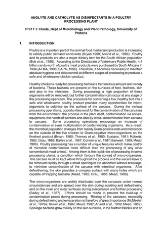
  • 11. Receiving Stunning Slaughter BleedingScaldingDefeathering Head and Neck Rem oval Spray W ashing Hock and Vent Cutting EviscerationSpray W ashingChilling Packaging Fig. 1.1. Flowsheet of stages in a typical poultry processing plant. 3. MATERIALS AND METHODS 3.1 Disinfectant solutions 3.1.1 Laboratory evaluation (Anolyte) Anolyte for minimum inhibitory concentration (MIC) determinations under laboratory conditions was supplied by Radical Waters. The MIC was determined on the same day the Anolyte solution was obtained. 3.1.2 Poultry Plant evaluation (Anolyte) Fresh Anolyte solution was prepared on the day of evaluation for a particular application. 3.1.3 Catholyte evaluation during scalding Catholyte was supplied by Radical Waters, for evaluation during scalding.
  • 12. NOTE: 0,01 % Sodium thioglycolate was added to all test solutions to neutralize the anolyte, before conducting microbiological analysis. 3.2 Applications, sample collection and preparation 3.2.1 Applications and sample collection - Pre-evisceration spray application: During this application, chicken carcases were sprayed with 100 ml of a 1:10 Anolyte solution. - Post-evisceration spray application: During this application, chicken carcases were sprayed with 1000 ml of a 1:10 Anolyte solution. - Spinchiller application: Chicken carcases were submerged in a 1:10 Anolyte solution at 10°C for 30 min. - Catholyte scalding application: Feathered chickens were submerged in a 100 % Catholyte solution for 7 min at a temperature of 54°C + 2°C. For each of the above applications, control samples of chickens were randomly taken off the processing line. 3.2.2 Sample preparation A composite meat and skin sample was prepared, by taking samples from the neck, back between the wings and under the wing 5g of sample was added to 45 ml of sterile Ringers solution to give a 1:10 dilution. From this a dilution series (10 -1 - 10-6 ) was prepared for plating. 3.3 Microbiological analysis This composite sample was placed into quarter strength Ringers solution in a 1+9 mass/volume ratio based on exact mass. Each composite sample was homogenised for 20 min (Seward Medical 400 Stomacher Lab Blender). Tenfold serial dilution’s in Ringers solution were plated out in duplicate on Nutrient Agar for the total bacteria count and on McConkey agar for the coliform bacteria count, using the spread-plate technique (ICMSF, 1978; Busta et al., 1984; von Holy et al., 1992) and incubated aerobically at 37 C for 48 h. Plates showing between 30 and 300 colony forming units (cfu) (or the highest number if below 30) were counted. Bacterial counts obtained from the plates of each duplicate set were meaned.
  • 13. 4. RESULTS Table 1 Microbiological quality of chicken carcass sprayed with 100 ml of anolyte immediately after defeathering, compared to the standard practise Anolyte Total bacteria cfu/g 1. 200 000 - 200 000 2. 60 000 80 000 70 000 3. 16 200 10 600 13 400 4. 10 800 7 600 9 200 5. 240 000 360 000 300 000 6. 220 000 130 000 17 5000 7. 20 000 15 000 17 500 8. 1 400 1 400 9. 820 000 820 000 10. 220 000 190 000 205 000 x Rang e 1,81 x 10⁵ 1,4 x 10 3 - 8,2 x 10 5 Control Total bacteria cfu/g 1. 590 000 800 000 695 000 2. 580 000 600 000 590 000 3. 90 000 150 000 120 000 4. 160 000 180 000 170 000 5. 230 000 260 000 245 000 6. 30 000 46 000 38 000 7. 38 600 54 600 46 600 8. 990 000 1150 000 1070 000 9. 10. 700 000 50 000 375 000 x Rang e 3,7 x 10⁵ 3,8 x 10 4 - 1,07 x 10 6 Spraying the chickens with anolyte immediately after defeathering, resulted in a 1 log difference in the bacterial number on average compared to the standard practise. This was considered a significant difference. Anolyte Coliform bacteria cfu/g 1. 11 100 9 300 10 200 2. 7 800 8 300 8 050 3. 30 500 30 000 32 500 4. 130 000 17 000 73 500 5. 12 500 12 200 12 350 6. 100 000 130 000 115 000 7. 7 100 6 300 6 700 8. 18 000 14 500 16 250 9. 840 000 770 000 805 000 10. 20 000 ND 20 000 x 1.09 x 10 5
  • 14. Rang e 8,05 x 10 3 - 8,05 x 10 5 Control Coliform bacteria cfu/g 1. 590 000 800 000 695 000 2. 11 900 8 900 10 400 3. 110 000 90 000 100 000 4. 40 600 38 900 39 750 5. 16 200 17 800 17 000 6. - - - 7. 90 000 280 000 185 000 8. - - - 9. 10 200 11 000 10 600 10. - 180 000 180 000 x s 1,54 x 10 5 1,0 x 10⁴ - 6,95 x 10 5 Figure 1 Microbiological quality of chicken carcass sprayed with 100 ml of anolyte immediately after defeathering, compared to the standard practise No significant difference in the number coliform bacteria was observed between the anolyte spray and the control (Figure 1). 4 5 6 Logcfu/g Total bacteria Coliforms Anolyte Control
  • 15.
  • 16. Table 2 Microbiological quality of chicken carcases using anolyte in a simulated spinchiller application compared to the standard practise Anolyte Total bacteria cfu/g 1. 250 000 340 000 295 000 2. 440 000 460 000 450 000 3. 140 000 - 140 000 4. 70 000 - 70 000 5. 16 200 - 16 200 6. 60 000 - 60 000 7. 20 800 18 200 19 500 8. 10 400 14 500 12 450 x Rang e 1,32 x 10⁵ 1,24 x 10 4 - 4,5 x 10 5 Control Total bacteria cfu/g 1. 15 500 - 15 500 2. 490 000 - 490 000 3. 23 800 - 23 800 4. 14 500 - 10 750 5. 8 000 7 000 7 500 6. 13 300 8 300 10 800 7. 9 900 - 9 600 8. 27 500 9 300 18 400 9. 5 100 19 300 12 200 x Rang e 6,66 x 10 4 9,6 x 10³ - 4,9 x 10⁵ During the simulated spinchiller application, the average total number of bacteria in the control was lower than the anolyte treatment. The anolyte treatment was nevertheless within the same range and hence not considered significantly different. Anolyte Coliform bacteria cfu/g 1. 2 000 2 000 2 000 2. 6 500 ND 6 500 3. 5 100 4 000 4 550 4. ND 8 000 8 000 5. ND 6 000 6 000 6. 9 600 8 800 9 200 X Rang e 6,04 x 10 3 2,5 x 10 3 - 8,0 x 10 3
  • 17. Control Coliform bacteria cfu/g 1. ND 5 800 5 800 2. - 4 200 4 200 3. 19 100 14 900 17 000 4. 11 600 - 11 600 5. 8 000 6 900 7 450 6. 4 500 7 500 6 000 7. 4 700 2 700 3 700 x Rang e 7,96 x 10 3 3,7 x 10 3 - 1,1 x 10 4 Figure 2 Microbiological quality of chicken carcases using anolyte in a simulated spinchiller application compared to the standard practise As with the total bacteria count, no significant difference was noted between the anolyte treatment and control (Figure 2). 3 4 5 6 Logcfu/g Total bacteria Coliforms Anolyte Control
  • 18. Table 3 Microbiologial quality of chicken carcases sprayed with 1 000ml of anolyte, compared to the standard practise Anolyte Total bacteria cfu/g 1. 28 400 ND 28 400 2. 300 000 130 000 215 000 3. 670 000 980 000 825 000 4. 920 000 1000 000 960 000 5. 39 000 49 800 44 400 6. 19 900 12 100 16 000 7. 840 000 910 000 875 000 8. 37 500 42 800 40 150 9. 24 600 36 200 30 400 10. 77 000 42 000 59 500 x Rang e 3,09 x 10 5 2,84 x 10 4 - 9,6 x 10 5 Control Total bacteria cfu/ml 1. 43 600 ND 43 600 2. 17 000 19 100 18 050 3. 39 000 25 600 32 300 4. 110 000 20 000 65 000 5. 270 000 280 000 275 000 6. 29 600 300 000 164 800 7. 2600 000 270 000 1435 000 8. 980 000 ND 980 000 9. 110 000 270 000 190 000 10. 24 000 27 300 25 650 x Rang e 3,22 x 10 5 1,80 x 10⁴ - 1.4 x 10⁶ Anolyte Coliform bacteria cfu/ml 1. 2 500 2 200 2 350 2. 72 000 72 000 3. 22 000 26 000 24 000 4. 4 000 4 200 4 100 5. 7 100 8 400 7 750 6. 1 600 3 300 2 450 7. 41 000 39 400 40 200 8. 22 100 17 900 20 000 9. 11 600 16 200 13 900 10. 14 300 12 700 13 500 x Rang e 2,00 x 10 4 4,1 x 10 3 - 7,0 x 10 4
  • 19. Control Coliform bacteria cfu/ml 1. 16 600 19 600 18 100 2. 7 000 1 900 4 450 3. 11 200 8 800 10 000 4. 6 200 6 100 6 150 5. 18 600 ND 18 600 6. 8 600 18 800 13 700 7. 25 500 20 300 22 900 8. 240 000 15 000 127 500 9. 11 700 12 000 11 850 10. 12 300 ND 12 300 x Rang e 2,45 x 10 4 4,45 x 10³ - 1,95 x 10 5 Figure 3 Microbiologial quality of chicken carcases sprayed with 1 000ml of anolyte, compared to the standard practise Neither the total bacteria number, or the coliform bacteria number differed significantly between the anolyte treatment and the control (Figure 3). 4 5 6 Logcfu/g Total bacteria Coliforms Anolyte Contol
  • 20. Table 4 Microbiological quality of chicken skin after scalding at 54°C in catholyte solution, compared to the standard practice Catholyte Total bacteria cfu/g 1. 200 000 150 000 175 000 2. 690 000 590 000 640 000 3. 460 000 580 000 520 000 x Rang e 4,45 x 10 5 1,75 x 10 5 - 6,4 x 10 5 Control Total bacteria cfu/g 1. 960 000 1280 000 1120 000 2. 350 000 220 000 285 000 3. 590 000 - 590 000 4. 240 000 - 240 000 5. 800 000 630 000 715 000 x Rang e 5,9 x 10 5 2,4 x 10⁵ - 1,2 x 10⁶ Catholyte Coliform bacteria cfu/g 1. 700 000 920 000 810 000 2. 9 700 910 500 460 100 3. 150 000 180 000 165 000 4. 15 000 90 000 52 500 x Rang e 3,7 x 10 5 5,3 x 10 4 - 8,1 x 10 5 Control Coliform bacteria cfu/g 1. 260 000 230 000 245 000 2. 510 000 760 000 635 000 3. 6 000 5 900 5 950 4. 20 000 40 000 30 000 5. 3 600 7 000 5 300 x Rang e 1,84 x 10 5 5,3 x 10 3 - 6,35 x 10 5
  • 21. Figure 4 Microbiological quality of chicken skin after scalding at 54°C in catholyte solution, compared to the standard practice Both the total bacteria and coliform bacteria numbers on the skin of chicken were concluded, that no significant difference existed between the two treatments (Figure 4). Table 5 Microbiological quality of feathers after scalding at 52°C in catholyte compared to the standard practice Catholyte Total bacteria cfu/g 1. 60 000 80 000 70 000 2. 1140 000 320 000 730 000 3. 1500 000 1500 000 1500 000 4. 15 000 20 000 17 500 x Rang e 5,79 x 10 5 1,75 x 10⁴ - 1,5 x 10⁶ Control Total bacteria cfu/g 1. 500 000 340 000 420 000 2. 330 000 780 000 550 000 3. 270 000 500 000 385 000 x Rang e 4,5 x 10 5 4,2 x 10 5 - 3,85 x 10 5 4 5 6 Logcfu/g Total bacteria Coliforms Catholyte Control
  • 22. The average number of bacteria for the catholyte treatment compared to the control was exactly the same. The range was also similar for the two treatments. Catholyte Coliform bacteria cfu/g 1. <10 <10 <10 2. <10 <10 <10 3. <10 <10 <10 4. <10 <10 <10 5. <10 <10 <10 x Rang e <10 <10 Control Coliform bacteria cfu/g 1. 15 000 15 000 15 000 2. 7 200 10 200 8 700 3. 8 900 13 400 11 150 4. 6 100 15 200 10 650 5. 15 000 60 000 37 500 x Rang e 1,66 x 10⁴ 8,7 x 10³ - 3,75 x 10⁴ Figure 5 Microbiological quality of feathers after scalding at 52°C in catholyte compared to the standard practice 0 1 2 3 4 5 6 Logcfu/ml Total bacteria Coliforms Catholyte Control
  • 23. No coliform bacteria were detected on the feathers, treated with the catholyte, in contrast to the control. This suggested, that the catholyte, either despersed all the coliforms, or killed all the coliforms on the feathers (Figure 5). 5. DISCUSSION During the laboratory evaluation of the anolyte solution, a 1:10 dilution gave the best result, using E.coli as test organism. The 1:20 and lower dilutions, did not result in a significant decrease of E.coli, using ca. 10 6 organisms as a challenge. Hence, it was decided to use the 1:10 dilution in our evaluation. The anolyte spray compared very favourably with the current practise in all applications. The most promising result was obtained immediately after defeathering, where the anolyte resulted in a significant decrease in the bacterial numbers compared to the control. It is highly recommended that this practise be followed, since it will reduce the bacterial numbers right at the outset and very early on during processing. Both the post-eviration spray application and the spinchiller application compared favorably with the current practice. Catholyte used during the scalding application, did not result in an improved result, with regards to the bacterial numbers on the skin, nor on the feathers. It should also be noted, that should the catholyte disperse the bacteria during scalding, without also killing the bacteria, it might result in a higher level of contamination, because bacteria attached to the feather will be dispersed, which may not be case with the current practise. 6. CONCLUSIONS It was concluded, that the anolyte solution (1:10 dilution) could replace the current practise and especially if it is introduced directly after defeathering it could have major benefits. The use of catholyte during the scalding application is not recommended. 7. RECOMMENDATIONS 1. The cost-effectivity of the anolyte solution (1:10 dilution) should be determined. If it is not comparable to the current practise, a comparable dilution of the anolyte should be evaluated. 2. A study should be conducted to determine the consistency of the anolyte product, with respect to its disinfection capability between different batches. 2. The antimicrobial properties of the catholyte solution should be determined.
  • 24. 8. REFERENCES 1. AFMA. 1996 Braaikuikenbedryf in Suid-Afrika. AFMA MATRIX 5(3): 27 2. Anand S.K., Mahapatra C.M., Pandey N.K. and Verma S.S. 1989 Microbial changes on chicken carcasses during processing. Indian J. Poult. Sci. 24(3): 203 - 209 3. Bailey J.S., Thomson J.E. and Cox N.A. 1987 Contamination of poultry during processing. [Edited by Cunningham F.E. and N.A. Cox] In The microbiology of poultry meat products. Academic Press, Orlando. 193 - 211. 4. Banwart George J. 1989 Basic Food Microbiology. 2nd ed. Published by Van Nostrand Reinhold. 5. Barnes Ella M., Impey C.S. and Parry R.T. 1973 The Sampling of Chickens, Turkeys, Ducks and Game Birds. [Edited by Board R.G. and D.W. Lovelock] In Sampling - Microbiological Monitoring of Environments, Society of Applied Bacteriology Technical Series No.7, Academic Press, London. 63 -73 6. Bok H.E., Holzapfel W.H., Odendaal E.S. and van der Linde H.J. 1986 Incidence of foodborne pathogens on retail broilers. International Journal of Food Microbiology 3: 273 - 285 7. Brenner, D.J. 1984 Family Enterobacteriaceae. [Edited by Murray R.G.E., Brenner D.J., Bryant M.P., Holt J.G., Krieg N.R., Moulder J.W., Pfennig N., Sneath P.H.A. and Staley J.T.] In Bergey's Manual of Systematic Bacteriology, Vol. I. Williams and Wilkins, Baltimore, USA. 409 - 498. 8. Brown M.H. and Baird-Parker A.C. 1982 The microbiological examination of meat.
  • 25. [Edited by Brown M.H.] In Meat Microbiology. Applied Science Publishers, London. 423 -520 9. Bryan F.L. 1980 Poultry and meat products. [Edited by Silliker J.H., Elliot R.P., Baird-Parker A.C., Bryan F.L., Christian J.H.B., Clark D.S., Olson J.C. and T.A. Roberts] In Microbial Ecology of Foods, Vol.2: Food Commodities. (ICMSF) 410 - 458 10. Busta F.F., Peterson E.H., Adams D.M. and Johnson M.G. 1984 Colony counts methods. [Edited by Speck M.L.] In Compedium of Methods for the Microbiological Examination of Foods, 2nd ed. American Public Health Association (APHA) Washington, DC. 62 - 83 11. Codex Alimentarius Commission (CAC) 1976 Recommended International Code of Hygiene Practice for Poultry Processing. CAC/RCP 14-1976, 2 nd edn. 12. Connor J.T., McMeekin T.A. and Patterson J.T. 1987 Prevention of microbial contamination in the poultry processing plant. [Edited by Smulders F.J.M.] In Elimination of Pathogenic Organisms from Meat and Poultry. Proceedings of the International Symposium: Prevention of Contamination, and Decontamination in the Meat Industry. Zeist, The Netherlands, 2 - 4 June 1986. Elsevier Science Publishers. 163 - 175 13. Cunningham F.E. 1987 Types of microorganisms associated with poultry carcasses. [Edited by Cunningham F.E. and N.A. Cox] In The microbiology of poultry meat products. Academic Press, Orlando. 29 - 42. 14. Dougherty T.J. 1976 A study of Salmonalla contamination in broiler flocks. Poultry Science 55: 1811 - 1815 15. Eley, 1992 Microbial Food Poisoning. Chapman and Hall, London 16. Eustace I.J. 1981 Control of bacterial contamination of meat during processing. Food Technology in Australia 33(1): 28 - 32 17. Evans J.B. 1986 Staphylococci. [Edited by Pearson A.M. and T.R. Dutson] In
  • 26. Advances in Meat Research, Vol.6: Meat and Health. Elsevier Science Publishers Ltd. 157 - 183 18. Farber J.M. and Idziak E.S. 1984 Attachment of psychrotrophic meat spoilage bacteria to muscle surfaces. Journal of Food Protection 47(2): 92 - 95 19. Firstenberg-Eden R. 1981 Attachment of bacteria to meat surfaces: A review. Journal of Food Protection 44(8): 602 - 607 20. Genigeorgis C., Hassuneh M. and Collins P. 1986 Campylobacter jejuni infection on poultry farms and its effect on poultry meat contamination during slaughter. Journal of Food Protection 49: 895 - 903 21. Geornaras I., Dykes G.A. and von Holy A. 1994 Microbial populations associated with refrigerated poultry. South African Journal of Science 90: 579 - 582 22. Goernaras I., de Jesus A.E., van Zyl E. and von Holy A. 1996 Bacterial populations associated with poultry processing in a South African abattoir. Food Microbiology 13: 457 - 465 23. Goernaras I., de Jesus A.E., van Zyl E. and von Holy A. 1997 Bacterial populations of different sample types from carcasses in the dirty area of a South African poultry abattoir. Journal of Food Protection 60: 551 - 554 24. Genstat 5 Committee of the Statistics Department. 1990 Rothamsted Experimental Station Genstat 5 Reference Manual, Release 2.2. Clarendon Press, Oxford. 25. Gibbs P.A., Patterson J.T. and Thompson J.K. 1978 The distribution of Staphylococcus aureus in a poultry processing plant. Journal of Applied Bacteriology 44: 401 - 410 26. Gill C.O. 1983 Meat spoilage and evaluation of the potential storage life of fresh meat. Journal of Food Protection 46(5): 444 - 452
  • 27. 27. Gill C.O. 1986 The control of microbial spoilage in fresh meats. [Edited by Pearson A.M. and T.R. Dutson] In Advances in Meat Research, Vol.2: Meat and Poultry Microbiology. Avi Publishing Company, Inc. 49 - 88 28. Government Gazette, No. 11881, May 1989, No. R. 1028. Animal Slaughter, Meat and Animal Products Hygiene Act, 1967 (Act. No. 87 of 1967) - Grades and Requirements for Abattoirs pp. 5 - 9. Republic of South Africa Government Printers. 29. Government Gazette, No. 2540, October 1969, No. R. 3505. Standing Regulations under the Animal Slaughter, Meat and Animal Products Hygiene Act, 1967 (Act No. 87 of 1967) Part XVIII pp. 34 - 44. Republic of South Africa Government Printers. 30. Grau F.H. 1986 Microbial ecology of meat and poultry. [Edited by Pearson A.M. and T.R. Dutson] In Advances in Meat Research, Vol.2: Meat and Poultry Microbiology. Avi Publishing Company, Inc. 1 - 36 31. Green S. 1974 Microbiology in the poultry processing industry. Process Biochemistry 9(1): 27 - 28 32. Hardie J.M. 1986 Genus Streptococcus. [Edited by Murray R.G.E., Brenner D.J., Bryant M.P., Holt J.G., Krieg N.R., Moulder J.W., Pfennig N., Sneath P.H.A. and Staley J.T.] In Bergey's Manual of Systematic Bacteriology, Vol. II. Williams and Wilkins, Baltimore, USA. 1043 - 1071. 33. Harrigan W.F. and McCane M.E. 1976 Laboratory methods in food and diary microbiology. Academic Press Inc. London 34. Holmes B., Owen R.J. and McMeekin T.J. 1984 Genus Flavobacterium. [Edited by Murray R.G.E., Brenner D.J., Bryant M.P., Holt J.G., Krieg N.R., Moulder J.W., Pfennig N., Sneath P.H.A. and Staley J.T.] In Bergey's Manual of Systematic Bacteriology, Vol. I. Williams and Wilkins, Baltimore, USA. 353 - 360.
  • 28. 35. International Commision on Microbiology Specifications for foods (ICMSF) 1978 Micro-organisms in Foods. 1 Their Significance and Methods of Enumeration, 2nd ed. University of Toronto Press, Canada 36. James W.O., Williams O.W., Prucha J.C., Johnston R. and Chritensen W. 1992 Profile of selected bacterial counts and Salmonella prevalence on raw poultry in a poultry slaughter establishment. JAVMA 200(1): 57 - 59 37. Jones F.T., Axtell R.C., Rives D.V., Scheideler S.E., Tarver F.R., Walker R.L. and Wineland M.J. 1991 A survey of Salmonella contamination in modern broiler production. Journal of Food Protection. 54(7): 502 - 513 38. Juni E. 1984 Genus Acinetobacter. [Edited by Murray R.G.E., Brenner D.J., Bryant M.P., Holt J.G., Krieg N.R., Moulder J.W., Pfennig N., Sneath P.H.A. and Staley J.T.] In Bergey's Manual of Systematic Bacteriology, Vol. I. Williams and Wilkins, Baltimore, USA. 303 - 307. 39. Kandler O. and Weiss N. 1986 Genus Lactobacillus. [Edited by Murray R.G.E., Brenner D.J., Bryant M.P., Holt J.G., Krieg N.R., Moulder J.W., Pfennig N., Sneath P.H.A. and Staley J.T.] In Bergey's Manual of Systematic Bacteriology, Vol. II. Williams and Wilkins, Baltimore, USA. 1209 - 1234 40. Keddie R.M., Collins M.D. and Jones D. 1986 Genus Arthrobacter. [Edited by Murray R.G.E., Brenner D.J., Bryant M.P., Holt J.G., Krieg N.R., Moulder J.W., Pfennig N., Sneath P.H.A. and Staley J.T.] In Bergey's Manual of Systematic Bacteriology, Vol. II. Williams and Wilkins, Baltimore, USA. 1288 - 1301. 41. Kersters K. and de Ley J. 1984 Genus Agrobacterium. [Edited by Murray R.G.E., Brenner D.J., Bryant M.P., Holt J.G., Krieg N.R., Moulder J.W., Pfennig N., Sneath P.H.A. and Staley J.T.] In Bergey's Manual of Systematic Bacteriology, Vol. Vol. I. Williams and Wilkins, Baltimore, USA. 244 - 254. 42. Kilsby D.C. 1982 Sampling schemes and limits. [Edited by Brown M.H.] In Meat
  • 29. Microbiology. Applied Science Publishers, London. 387 - 421 43. Kloos E.W. and Schleifer K.H. 1986 Genus Staphylococcus. [Edited by Murray R.G.E., Brenner D.J., Bryant M.P., Holt J.G., Krieg N.R., Moulder J.W., Pfennig N., Sneath P.H.A. and Staley J.T.] In Bergey's Manual of Systematic Bacteriology, Vol. II. Williams and Wilkins, Baltimore, USA. 1013 - 1035. 44. Kocur M. 1986 Genus Micrococcus. [Edited by Murray R.G.E., Brenner D.J., Bryant M.P., Holt J.G., Krieg N.R., Moulder J.W., Pfennig N., Sneath P.H.A. and Staley J.T.] In Bergey's Manual of Systematic Bacteriology, Vol. II. Williams and Wilkins, Baltimore, USA. 1004 - 1008. 45. Komagata K. and Suzuki K. 1986 Genus Aureobacterium. [Edited by Murray R.G.E., Brenner D.J., Bryant M.P., Holt J.G., Krieg N.R., Moulder J.W., Pfennig N., Sneath P.H.A. and Staley J.T.] In Bergey's Manual of Systematic Bacteriology, Vol. II. Williams and Wilkins, Baltimore, USA. 1323 - 1325. 46. Kraft A.A. 1986 Psycrotrophic organisms. [Edited by Pearson A.M. and T.R. Dutson] In Advances in Meat Research, Vol.2: Meat and Poultry Microbiology. Avi Publishing Company, Inc. 191 - 208 47. Lahellec C. and Colin P. 1981 Pseudomonas as spoilage agents in poultry: Observations concerning strains isolated at different stages of processing and during storage. [Edited by Roberts T.A., Hobbs G., Christian J.H.B. and N. Skovgaard] In Psychrotrophic Micro-organisms in Spoilage and Pathogenicity. Academic Press, London. 269 - 274 48. Lahellec C. and Colin P. 1979 Bacterial flora of poultry: Changes due to variations in ecological conditions during processing and storage. Archiv Für Lebensmittelhygiene 30 : 95 - 98 49. Lillard H.S. 1985 Bacterial cell characteristics and conditions influencing their
  • 30. adhesion to poultry skin. Journal of Food Protection 48(9): 803 - 807 50. McBride G.B., Skura B.J., Yada R.Y. and Bowmer E.J. 1980 Relationship between incidence of Salmonella contamination among pre-scalded, eviscerated and post- chilled chickens in a poultry processing plant. Journal of Food Protection 43(7): 538 - 542 51. McMeekin T.A. 1975 Spoilage association of chicken breast muscle. Applied Microbiology 29(1): 44 - 47 52a. McMeekin T.A. and Thomas C.J. 1979 Aspects of the microbial ecology of poultry processing and storage: A Review. Food Technology in Australia 31: 35 - 43 52b. McMeekin T.A., Thomas C.J. and McCall D. 1979 Scanning electron microscopy of micro-organisms on chicken skin. Journal of Applied Bacteriology 46:195 - 200 53. Microbiology and Food Safety Committee of the National Food Processors Association, 1993 Implementation of HACCP in a food processing plant. Journal of Food Protection 56 (6): 548 - 554 54. Mead G.C. 1989 Hygiene problems and control of process contamination. [Edited by Mead G.C.] In Processing of Poultry. Elsevier Science Publishers Ltd. 183 - 220 55. Mead G.C. 1982 Microbiology of poultry and game birds. [Edited by Brown M.H.] In Meat Microbiology. Applied Science Publishers Ltd. 67 - 101 56. Morris G.K. and Wells J.G. 1970 Salmonella contamination in a poultry-processing plant. Applied Microbiology 19 (15): 795 - 799 57. Mulder R.W.A.W. and Veerkamp C.H. 1974 Improvements in poultry slaughterhouse as a result of cleaning before cooling. Poultry Science 53: 1690 - 1974
  • 31. 58. Mulder R.W.A.W., Dorresteijn L.W.J. and Van Der Broek J. 1978 Cross- contamination during the scalding and plucking of broilers. Br. Poult. Sci. 19: 61 - 70 59. Notermans S., Dufrenne J. and Van Leeuwen W.J. 1982 Contamination of broiler chickens by Staphylococcus aureus during processing; incidence and origin. Journal of Applied Bacteriology 52: 275 -280 60. Notermans S., Terbijhe R.J. and Van Schothorst M. 1980 Removing faecal contamination of broilers by spray - cleaning during evisceration. Br. Poult. Sci. 21: 115 - 121 61. Notermans S. and Kampelmacher E.H. 1975 Further studies on the attachment of bacteria to skin. Br. Poult. Sci. 16: 487 - 496 62. Notermans S. and Kampelmacher E.H. 1974 Attachment of some bacterial strians to the skin of broiler chickens. Br. Poult. Sci. 15: 573 - 585 63. Olivier, M., Cloete, T.E., Veary, C.M and von Holy, A. 1996 Microbiological status of selected chicken carcases from a non-automated poultry processing plant. Journal of Basic Microbiology. 36(1): 41 - 49 64 Olivier, M., Cloete, T.E., Veary, C.M. and von Holy, A. 1995 Microbiological status of selected chicken carcases from a non-automated poultry processing plant. Proceedings of the Thirteenth Biennial SAAFoST International Congress. Durban, South Africa. p96. 65. Palleroni N.J. 1984 Genus Pseodomonas. [Edited by Murray R.G.E., Brenner D.J., Bryant M.P., Holt J.G., Krieg N.R., Moulder J.W., Pfennig N., Sneath P.H.A. and Staley J.T.] In Bergey's Manual of Systematic Bacteriology, Vol. I. Williams and Wilkins, Baltimore, USA. 141 - 199. 66. Parry R.T. 1989 Pre-slaughter handling and processing. [Edited by Mead G.C.] In Processing of Poultry. Elsevier Science Publishers Ltd. 65 - 101
  • 32. 67. Patterson J.T. 1972 Microbial sampling of poultry carcasses. Journal of Applied Bacteriology 35: 569 - 575 68. Popoff M. 1984 Genus Aeromonas. [Edited by Murray R.G.E., Brenner D.J., Bryant M.P., Holt J.G., Krieg N.R., Moulder J.W., Pfennig N., Sneath P.H.A. and Staley J.T.] In Bergey's Manual of Systematic Bacteriology, Vol. I. Williams and Wilkins, Baltimore, USA. 545 - 548. 69. Ralph W.J. and Tompkin R.B. 1984 Meat and poultry products. [Edited by Speck Marvin L.] In Compedium of Methods for the Microbiological Examination of Foods, 2nd ed. American Public Health Association (APHA) Washington, D.C. 611 - 623 70. Roberts D. 1982 Bacteria of public health significance. [Edited by Brown M.H.] In Meat Microbiology. Applied Science Publishers Ltd. 319 - 386 71. Silliker J.H. 1995 Microbiological testing and HACCP programs. Diary, Food and Environmental Sanitation 15 (10): 606 - 610 72. Silliker J.H. and Galois D.A. 1986 Salmonella. [Edited by Pearson A.M. and T.R. Dutson] In Advances in Meat Research, Vol.2: Meat and Poultry Microbiology. Avi Publishing Company, Inc. 209 - 229 73. Simonsen B. 1989 Microbiological critera for poultry products. [Edited by Mead G.C.] In Processing of Poultry. Elsevier Science Publishers Ltd. 221 - 250 74. Smeltzer T.I. 1981 Isolation of Campylobacter jejuni from poultry carcases. Australian Veterinary Journal 57: 511 - 512 75. South African Poultry Association (SAPA). 1996 Whats happening in the poultry and egg industry? Food Industries of South Africa (9): 9 - 15 76. South African Poultry Association (SAPA). 1993 Code of Practice for Broiler Processing Plant (Abattoirs) Annexure G 77. Statgraphics: Statistical graphics system by statistical graphics corporation, Plus
  • 33. Ware Software Products, STSC inc. USA 78. Thomas C.J. and McMeekin T.A. 1981 Attachment of Salmonella spp. to chicken muscle surfaces. Applied and Environmental Microbiology 42(1): 130 - 134 79. Thomas C.J. and McMeekin T.A. 1980 Contamination of broiler carcass skin during commercial processing procedures: an Electron microscopy Study. Applied and Environmental Microbiology 40(1): 133 - 144 80. Todd E.C.D. 1980 Poultry-associated foodborne disease - Its occurrence, cost, source and prevention. Journal of Food Protection 43(2): 129 - 139 81. Von Holy A., Holzapfel W.H. and Dykes G.A. 1992 Bacterial populations associated with Vienna sausage packaging. Food Microbiology 9: 45 - 53 82. WHO, 20 th Session ,1993 Codex Guidelines for the application of the Hazard Analysis Critical Control Point (HACCP) System. Codex Alimentarius Commission., Annex II, III and IV . 17 - 25 83. Zottola E.A. and Smith L.B. 1990 Pathogenic bacteria in meat and meat products. [Edited by Pearson A.M. and T.R. Dutson] In Advances in Meat Research, Vol.6: Meat and Health. Elsevier Science Publishers Ltd. 157 -183