

Recommended
More Related Content
What's hot
What's hot (20)
Similar to Mixed odontogenic tumor
Similar to Mixed odontogenic tumor (20)
More from IAU Dent
More from IAU Dent (20)
Recently uploaded
Recently uploaded (20)
Mixed odontogenic tumor
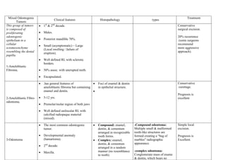
- 1. Mixed Odontogenic Tumors This group of tumors is composed of proliferating odontogenic epithelium in a cellular ectomesenchyme resembling the dental papilla. Clinical features Histopathology Posterior mandible 70%. Small (asymptomatic) – Large (Local swelling / failure of eruption). Well defined RL with sclerotic borders. 50% assoc. with unerupted teeth. Encapsulated. .has general features of ameloblastic fibroma but containing enamel and dentin. 5-12 yrs. Premolar/molar region of both jaws Well defined unilocular RL with calcified radiopaque material (mixed). The most common odontogenic tumor. Developmental anomaly (hamartoma). Treatment Males. 2-Ameloblastic Fibroodontoma. 1st & 2nd decade. 1-Ameloblastic Fibroma. types 3-Odontoma 2nd decade. Maxilla. Conservative surgical excision. 20% recurrence (some surgeons recommend more aggressive approach). Conservative curettage. Foci of enamel & dentin in epithelial structure. Prognosis is excellent Compound: enamel, dentin, & cementum arranged in recognizable tooth forms. Complex: enamel, dentin, & cementum arranged in a random manner (no resemblance to tooth). -Compound odontoma: Multiple small & malformed tooth-like structures are formed creating a “bag of marbles” radiographic appearance. -complex odontoma: Conglomerate mass of ename & dentin, which bears no Simple local excision. Prognosis is Excellent.
- 2. Compound (anterior maxilla). Complex (posterior of both jaws). Assoc. with unerupted tooth / block eruption. Odontogenic differentiation is fully expressed. Mostly radiodense. RL with well defined, smooth contours / later a well defined radiopaque appearance. Compound shows apparent tooth shapes while complex appears as a uniform opaque mass. Asymptomatic. Arise from altered fibroblast or myofibroblast that produce excess of mucopolysaccharides & incapable of producing mature collagen. 4-odontogenic myxoma. Benign, locally aggressive in bone or soft tissues. 20-40 yrs. Female. Posterior mandible 28% Unencapsulated, locally infiltrating tumor. Small (unilocular) anatomic resemblance of tooth. Stellate, spindle-shaped & round cells in myxoid stroma with few collagen bundles. It might be confused with chondromyxoid fibroma or with myxoid change in an enlarged dental follicle or papilla. Small: Curettage. Large: Surgical excision. 25% recurrence. Prognosis is good
- 3. Purely RL. Well circumscribed, expanding, detructive lesion. Maxillary lesion may extend to sinus, nasal cavity, orbit, or cranial cavity. 5-Cementoblastoma (true cementum) Large (multilocular). Teeth displacement / root resorption. 2nd & 3rd decades. Roots of mandibular posterior teeth. Slow growing / local expansion. Asymptomatic (may be pain). Radiographically: ball of dense material attached to the end of root. (opaque lesion). Opaque spicules radiate from the central mass. Mineralized materials. Multinucleated giant cells. This lesion resemble Osteoblastoma. Done by/ Mahdi 207 for ever دعواتكم شباب Surgical removal (of tooth together with mass). Prognosis is excellent