

Recommended
More Related Content
What's hot
What's hot (20)
Viewers also liked
Viewers also liked (20)
Similar to Papillary lesions
Similar to Papillary lesions (20)
More from IAU Dent
More from IAU Dent (20)
Recently uploaded
Recently uploaded (20)
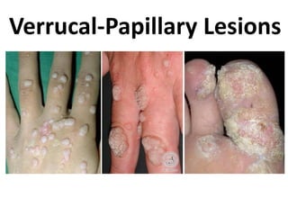
Papillary lesions
- 2. Verrucal-Papillary Lesions • Reactive lesions: • • • • • Papillary hyperplasia Condyloma Latum Squamous Papilloma Condyloma Acuminatum Focal epithelial hyperplasia • Neoplasms: • Keratoacanthoma • Verrucous carcinoma • Unknown etiology: • Pyostomatitis vegetans • Verruciform xanthoma Verrucous leukoplakia
- 3. Papillary hyperplasia • Etiology Removable dentures Hard palate and trauma Microorganisms (Candida A)
- 4. Papillary hyperplasia • Clinical features Site: Palate Erythematous, papillary cobblestone Ulceration is rare
- 5. Papillary hyperplasia Histopathology Small papillary projections covered with SSE Stroma is well vascularized Epithelium is hyperplastic with pseudoepitheliomatous features No evidence of dysplasia Pseudoepitheliomatous epithelium Epithelial dysplasia Mild, moderate and severe Malignant invasion of epithelium
- 6. Papillary hyperplasia Differential diagnosis: Nicotine stomatitis Multiple squamous papillomas Acanthosis nigricans Cowden syndrome Treatment and prognosis: mild: soft tissue agents and liners Surgical removal
- 7. Condyloma Latum Etiology: Secondary syphilis Clinically: Most common in the perianal area Orally: Smooth, lobulated or mushroom-like mass HP: Acanthosis, intra- and intercellular edema, inflammatory cell infiltration Treatment: It will regress after the treatment of syphilis Intracellular intercellular
- 8. Squamous Papilloma Oral Verruca Vulgaris Etiology HPV [2, 6, 11, 57] 2, 4 & 40 Cutaneous verruca 3 & 10 Flat warts 13 & 32 Focal epithelial hyperplasia Transmission might be through direct contact
- 9. Squamous Papilloma Clinical features Most common papillary lesion of the oral mucosa Palate, lip and vermilion and other parts of oral mucosa Generally single and less than 1 cm. Cauliflower-like surface
- 11. Squamous Papilloma Histopathology Growth of keratinized squamous epithelium Well vascularized stroma Inflammatory cell infiltration
- 12. Squamous Papilloma Differential diagnosis Verruciform xanthoma [gingiva, skin, Langerhans cells] Condyloma acuminatum [larger with broader base] Darier’s disease [multiple]
- 13. Squamous Papilloma Treatment Remove by surgery or Laser
- 14. Condyloma Acuminatum • Etiology Cauliflower HPV 6 & 11 Infectious lesion Frequent in HIV-infected persons
- 15. Condyloma Acuminatum • Clinical features Common in the anogenital region and may involve oral mucosa Early stage: multiple pink nodules Later it grows and coalesce to become: Soft, broad-base papillary growth
- 16. Condyloma Acuminatum • Histopathology Papillary projections covered by hyperplastic SSE Might be keratinized or non-keratinized Upper level of epithelium demonstrate perinuclear cellular vacuolization [koilocytic cells] Vascularized stroma with inflammatory infiltration Koilocytic cells
- 17. Condyloma Acuminatum • Treatment Surgical excision: Scalpel Laser CO2 Cryosurgery
- 18. Focal epithelial hyperplasia Heck’s disease Etiology: - Common among Americans - Irritation - Vitamin deficiency - HPV 13 and possibly 32 - Multi-focal epithelial hyperplasia
- 19. Focal epithelial hyperplasia Heck’s disease Clinical features: - Multiple nodular soft tissue masses - Buccal, labial mucosa and tongue - Whitish to pinky in color
- 20. Focal epithelial hyperplasia Heck’s disease Histopathology: - Acanthosis - Parakeratosis - Ballooning spinous cells
- 21. Focal epithelial hyperplasia Heck’s disease Treatment: - Regress spontaneously - Surgical removal
- 22. Keratoacanthoma Etiology Benign lesion Common at sun exposed skin and less on the vermilion Rarely on oral mucosa in such case it may originate from Fordyce’s granules Other etiologic agents: Viral infection trauma Chemical irritation …etc.
- 23. Keratoacanthoma Clinical features • • • Solitary or multiple Rapid enlargement of the papule within 4-8 weeks Asymptomatic nodules with central plug of keratin
- 24. Keratoacanthoma Histopathology Central keratin plug Marginal buttress of epithelium Pseudoepitheliomatous hyperplasia Intense mixed inflammatory infiltration No malignancy
- 25. Keratoacanthoma Differential diagnosis SCC Verruca vulgaris Treatment Careful follow-up It may regress spontaneously if no Surgical removal and HP examination
- 26. Verrucous carcinoma Etiology o The use of tobacco [smoking or smokeless] o HPV
- 27. Verrucous carcinoma Clinical features 5% of oral SCC Common sites: buccal mucosa and gingiva Early stage [verrucous hyperplasia] is benign or may arise from leukoplakia • Indurate firm with invasion to the subjacent tissue • • •
- 28. Verrucous carcinoma Histopathology Papillary surface, acanthotic and highly keratinized epithelium Submucosal invasion by bulbous well differentiated epithelium Minimal atypia
- 29. Verrucous carcinoma Differential diagnosis SCC Proliferative verrucous leukoplakia Treatment Surgery followed by radiotherapy
- 30. Pyostomatitis vegetans Etiology • Benign chronic mucocutaneous disease • Unknown etiology • Many cases are associated with gastrointestinal disturbances
- 31. Pyostomatitis vegetans Clinical features Erythematous and edematous oral mucosa Multiple yellow pustules 2-3 mm Papillary projections of oral mucosa male are more affected than female with mean age of 34 years
- 32. Pyostomatitis vegetans Histopathology Acanthosis Hyperkeratosis Pseudoepitheliomatous hyperplasia Inflammatory infiltration Ulceration and superficial epithelial necrosis
- 33. Pyostomatitis vegetans Treatment Controlling the associated diseases if present Topical corticosteroids may be used Additional antibiotic and multivitamins may be employed
- 34. Verruciform xanthoma Etiology • Unknown • Immunologic disorder
- 35. Verruciform xanthoma Clinical features • • • • • Well circumscribed Papillary surface Size: vary from 2mm. To 2cm. May appear on the skin Mean age 45 years
- 36. Verruciform xanthoma Histopathology Parakeratinized papillomatous epithelium Elongated rete ridges Presence of xanthoma [foam] cells in the lamina propria Langerhans cells
- 37. Verruciform xanthoma Treatment Conservative surgical excision
- 38. Differential diagnosis Papillary hyperplasia Condyloma latum Oral squamous papilloma Condyloma acuminatum Focal epithelial hyperplasia Keratoakanthoma Verrucous carcinoma Pyostomatitis vegetans Verruciform xanthoma
- 39. Differential diagnosis Papillary hyperplasia Condyloma latum Oral squamous papilloma Condyloma acuminatum Focal epithelial hyperplasia Keratoakanthoma Verrucous carcinoma
- 40. Differential diagnosis Papillary hyperplasia Condyloma latum Oral squamous papilloma Condyloma acuminatum Focal epithelial hyperplasia Keratoakanthoma Verrucous carcinoma
- 41. Differential diagnosis Papillary hyperplasia Condyloma latum Oral squamous papilloma Condyloma acuminatum Focal epithelial hyperplasia Keratoakanthoma Verrucous carcinoma
- 42. Differential diagnosis Papillary hyperplasia Condyloma latum Oral squamous papilloma Condyloma acuminatum Focal epithelial hyperplasia Keratoakanthoma Verrucous carcinoma
- 43. Differential diagnosis Papillary hyperplasia Condyloma latum Oral squamous papilloma Condyloma acuminatum Focal epithelial hyperplasia Keratoakanthoma Verrucous carcinoma
- 44. Differential diagnosis Papillary hyperplasia Condyloma latum Oral squamous papilloma Condyloma acuminatum Focal epithelial hyperplasia Keratoakanthoma Verrucous carcinoma
- 45. Differential diagnosis Papillary hyperplasia Condyloma latum Oral squamous papilloma Condyloma acuminatum Focal epithelial hyperplasia Keratoakanthoma Verrucous carcinoma
- 46. Differential diagnosis Papillary hyperplasia Condyloma latum Oral squamous papilloma Condyloma acuminatum Focal epithelial hyperplasia Keratoakanthoma Verrucous carcinoma
- 47. Differential diagnosis Papillary hyperplasia Condyloma latum Oral squamous papilloma Condyloma acuminatum Focal epithelial hyperplasia Keratoakanthoma Verrucous carcinoma
- 48. Differential diagnosis Papillary hyperplasia Condyloma latum Oral squamous papilloma Condyloma acuminatum Focal epithelial hyperplasia Keratoakanthoma Verrucous carcinoma
- 49. Differential diagnosis Papillary hyperplasia Condyloma latum Oral squamous papilloma Condyloma acuminatum Focal epithelial hyperplasia Keratoakanthoma Verrucous carcinoma
- 50. Differential diagnosis Papillary hyperplasia Condyloma latum Oral squamous papilloma Condyloma acuminatum Focal epithelial hyperplasia Keratoakanthoma Verrucous carcinoma
- 51. Differential diagnosis Papillary hyperplasia Condyloma latum Oral squamous papilloma Condyloma acuminatum Focal epithelial hyperplasia Keratoakanthoma Verrucous carcinoma
- 52. Differential diagnosis Papillary hyperplasia Condyloma latum Oral squamous papilloma Condyloma acuminatum Focal epithelial hyperplasia Keratoakanthoma Verrucous carcinoma Because there’s no histopathology