SlideShare une entreprise Scribd logo
1  sur  25
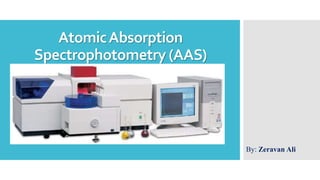
AtomicAbsorption
Spectrophotometry (AAS)
By: Zeravan Ali
Contents:
 Introduction.
 History.
 Principle.
 Instrumentation.
• Hollow Cathode Lamp.
• Electrodeless Discharge Lamp.
 Optical Considerations.
 AAS Detectors.
Applications of AAS.
Advantages of AAS.
Limitations of AAS.
References.
 Atomic absorption spectroscopy has become one of the most frequently
used tools in analytical chemistry.
 Atomic absorption methods measure the amount of energy (in the form of
photons of light, and thus a change in the wavelength) absorbed by the
sample. Specifically, a detector measures the wavelengths of light
transmitted by the sample (the "after" wavelengths), and compares them to
the wavelengths, which originally passed through the sample (the "before"
wavelengths).
 Atomic absorption spectrometry (AAS) is an easy, high-throughput, and
inexpensive technology used primarily to analyze compounds in solution.
As such, AAS is used in food and beverage, water, clinical, and
pharmaceutical analysis. It is also used in mining operations, such as to
determine the percentage of precious metal in rocks.
Introduction:
Introduction:
 Atomic absorption is so sensitive that it can measure down to parts per
billion of a grams (µg dm–3) in a sample. The technique makes use of the
wavelengths of light specifically absorbed by an element. They correspond
to the energies needed to promote electrons from one energy level to
another, higher, energy level.
 The process of atomic absorption is illustrated in Figure 1.
Figure 1: Atomic Absorption Process.
 The phenomenon of atomic absorption (AA) was first observed in 1802
with the discovery of the Fraunhofer lines in the sun's spectrum. It was not
until 1953 that Australian physicist Sir Alan Walsh demonstrated that
atomic absorption could be used as a quantitative analytical tool.
 In 1955 the modern era of atomic absorption spectroscopy began with the
work of Walsh and Alkemade and Milatz. The time since 1955 can be
divided into seven year periods. The first was an induction period (1955–
1962) when AA received attention from only a very few people. This was
followed by a growth period (1962–1969) when most of what we see today
was developed, and then by a period of relative stability (1969–1976) when
AA contributed greatly to other fields.
 We are now in a period of great change, which started in about 1976, due to
the impact of computer technology on individual laboratory instruments.
History:
 The absorption spectrum of an element in its gaseous atomic state consists of a series
of well defined and extremely narrow lines arising by the electronic transitions of outer
most electrons. In case of metals, most of these transitions belongs to visible and UV
regions.
 Most obvious wavelengths at which absorption or emission is observed are associated
with the transitions where minimal energy change occurs e.g., 3s-3p transition in Na
atom gives rise the emission of yellow light and this is referred as D-line transition.
 For the analysis, the sample (element) is converted into atomic vapour and then the
absorption, either in visible and UV region (at a selected wavelength), which is specific
for a given element is measured.
 For converting element into its gaseous atomic state initial step in the whole procedure
of estimation involves spraying a solution of the sample into the flame. This process is
accomplished by drawing the solution of the sample as a fine mist into a suitable flame.
In terms of Functionality, the flame serves as analogous to cell or cuvette containing
the solution in the conventional spectroscopy.
Principle:
 Spectrophotometric measurements are based on Beer’s Law (sometimes referred to as
the Beer-Lambert Law). When a monochromatic light beam (light with a particular
wavelength) is passed through a cell containing a specimen in a solution, part of the
light is absorbed and the rest is passed through the cell and reaches the detector.
 In spectrophotometry, transmittance is often measured as absorption (A) because there
is a linear relationship between absorbance and concentration of the analyte in the
solution
Lambert-beer’s Law
Principle:
Density C
l
I0 I
I = I0 e-k .l .C
Abs = -logI/I0 = k .l. C
k : proportional constant
l : path length
C : density (concentration)
 The major difference in the instrumentation of AAS and flame spectrophotometry is the
presence of a radiation source (a particular resonance wavelength cannot be isolated
from the continuous source using a prism or diffraction gratings). So, for this purpose, a
hollow cathode lamp is used (Figure 2).
Instrumentation:
Figure 2: Instrumentation of atomic absorption Spectrophotometer.
1. Light Sources
An atom absorbs light at discrete wavelengths. In order to measure this narrow light
absorption with maximum sensitivity, it is necessary to use a line source, which emits the
specific wavelengths which can be absorbed by the atom. Narrow line sources not only
provide high sensitivity, but also make atomic absorption a very specific analytical
technique with few spectral interferences. The two most common line sources used in
atomic absorption are the ‘‘hollow cathode lamp’’ and the ‘‘electrodeless discharge
lamp.’’
a. Hollow Cathode Lamp (HCL):
It contains a tungsten anode and cathode is a hollow cylindrical tube which is lined by the
element to be determined. These are sealed in the glass tube filled with an inert gas like
neon or argon at a low pressure. At the end of the cylinder is a window, made up of quartz
or pyrex, transparent to the emitted radiation. Each element in question will thus emit
monochromatic radiation characteristic of the emission spectrum of that particular element
involved. So, each element has its own unique lamp which must be used for the analysis.
Instrumentation:
 HCLs are available for most metallic elements. A schematic diagram of a HCL is shown
in Figure 3.
Instrumentation:
Figure 3: Hollow Cathode Lamp.
b. Electrodeless Discharge Lamp (EDL):
 Electrodeless discharge lamps are used less frequently than the HCLs except for analytes
such as arsenic and selenium. These lamps may be excited using either microwave energy
(although these tend to be less stable) or radiofrequency energy. The radiofrequency-
excited lamps are less intense than the microwave-excited ones, but are still 5–100 times
more intense than a standard HCL.
 In the electrodeless discharge lamp, a bulb contains the element of interest (or one of its
salts) in an argon atmosphere. The radiofrequency energy ionizes the argon and this, in
turn, excites the analyte element, causing it to produce its characteristic spectrum. For
analytes such as arsenic and selenium, these lamps
give a better signal-to-noise ratio than HCLs
and have a longer useful lifetime
Instrumentation:
Figure 4: Electrodeless Discharge Lamp.
2. Nebulizer
 It creates a fine spray of the sample for the introduction in the flame. The aerosol and
the fuel and oxidant are mixed thoroughly for the introduction into the flame.
 The standard nebulizer, which provides best performance with respect to minimizing
chemical interferences, is recommended for general-purpose applications. A High
Sensitivity Nebulizer is available for applications that require maximum sensitivity and
the lowest flame detection limits. The High Sensitivity Nebulizer utilizes an integral
ceramic impact bead to enhance atomization efficiency.
3. Atomizer:
 The elements which needs to be analysed needs to be in the atomic state. Here comes
the role of atomizer. It breaks down the molecules into the atoms by exposing the
analyte to high temperatures in a flame.
Instrumentation:
 There are two basic atom cells (a means of converting the sample, usually a liquid, into
free atoms) used in atomic absorption spectroscopy:
a. The Flame:
For flame systems, the sample is introduced via a nebulizer and spray chamber assembly.
Sample is drawn up the intake capillary by the Venturi effect. The liquid sample column
comes into contact with the fast moving flame gases and is shattered into small droplets. In
many systems it is further smashed into still smaller droplets by an impact bead. The flame
gases then carry the aerosol (the nebular) through a spray chamber containing a series of
baffles or flow spoilers. These act as a droplet size filter, passing the larger droplets to
waste (85–90% of the sample) and allowing the finer droplets to be transported to the
flame.
Instrumentation:
Instrumentation:
Figure 5: The flame process.
b. Electrothermal atomization:
 In electrothermal atomization the sample is introduced into the tube, which is then
heated in a series of steps at increasing temperature. The sample is dried at a
temperature just above the boiling point of the solvent (but not so hot as to cause
frothing and spitting of the sample); ashed (charred at an intermediate temperature to
remove as much of the concomitant matrix and potential interferences as possible
without losing any analyte); and then atomized at a high temperature. During the
atomization stage, the atoms leave the graphite surface and enter the light beam, where
they absorb the incident radiation. The ashing and atomizing temperatures used will
depend on the analyte of interest.
Instrumentation:
Figure 6: Electrothermal atomization.
Photometers
 The portion of an atomic absorption spectrometer’s optical system which conveys the
light from the source to the monochromator is referred to as the photometer. Three types
of photometers are typically used in atomic absorption instruments: single-beam,
double-beam and what might be called compensated single-beam or pseudo double-
beam.
1. Single-Beam Photometers:
 The instrument diagrammed in Figure 7 represents a fully functional ‘‘single-beam’’
atomic absorption spectrometer. It is called ‘‘single-beam’’ because all measurements
are based on the varying intensity of a single beam of light in a single optical path.
Optical Considerations:
Single-Beam Photometers:
Figure 7: Single-beam A.A Spectrometer.
2. Double-Beam Photometers:
 An alternate photometer configuration, known as ‘‘double-beam’’ (Figure 8) uses
additional optics to divide the light from the lamp into a ‘‘sample beam’’ (directed
through the sample cell) and a ‘‘reference beam’’ (directed around the sample cell). In
the double-beam system, the reference beam serves as a monitor of lamp intensity and
the response characteristics of common electronic circuitry. Therefore, the observed
absorbance, determined from a ratio of sample beam and reference beam readings, is
more free of effects due to drifting lamp intensities and other electronic anomalies
which similarly affect both sample and reference beams.
Optical Considerations:
Double-Beam Photometers:
Figure 8: Double-beam A.A Spectrometer.
3. Alternative Photometer Designs:
 There are several alternative system designs which provide advantages
similar to those of double-beam optical systems and the light throughput
characteristic of single-beam systems. Such systems can be described as
compensated single-beam or pseudo double-beam systems. One such
design uses two mechanically-adjusted mirrors to alternately direct the
entire output of the source through either the sample path (during sample
measurements) or through a reference path.
Optical Considerations:
 The photomultiplier tube is almost universally used as the detector type in AAS figure 9.
 Upon exiting the sample cell the beam of monochromatic light eventually strikes the
spectrophotometer’s detector.
 The role of the detector is to convert a light signal into an electrical signal. The
characteristics of an ideal detector are fast response times and a linear response over a
wide range of wavelengths with low noise and high sensitivity.
AAS Detectors:
Figure 9: photomultiplier tube.
Clinical analysis: Analyzing metals in biological fluids such as blood and urine.
Environmental analysis: Monitoring our environment – eg finding out the levels of
various elements in rivers, seawater, drinking water, air, petrol and drinks such as wine,
beer and fruit drinks.
Pharmaceuticals: In some pharmaceutical manufacturing processes, minute quantities of
a catalyst used in the process (usually a metal) are sometimes present in the final product.
By using AAS the amount of catalyst present can be determined.
Industry: Many raw materials are examined and AAS is widely used to check that the
major elements are present and that toxic impurities are lower than specified – eg in
concrete, where calcium is a major constituent, the lead level should be low because it is
toxic.
Mining: By using AAS the amount of metals such as gold in rocks can be determined to
see whether it is worth mining the rocks to extract the gold.
Applications of AAS:
• Minute amount of an element can be determined even in the
presence of high concentration of other elements.
• The technique is more sensitive than emission flame photometry.
• It is highly specific for a given element.
• It is rapid and requires only small amount of material.
Advantages of AAS:
• There is requirement of separate lamp for each of the element to be
determined.
• Elements such as Al, Ti, W, Mo, V, Si cannot be detected. This is due to the
fact that these elements give rise oxides of the atoms in the flame.
• The use of this technique is limited to metals only. With non-metals
difficulties arise from the strong absorption of light by the oxygen in the
light path and from the flame gases themselves.
• If aqueous solutions are used the prominent anion can affects the signal to a
noticeable degree.
Limitations of AAS:
1. Sneddon, J., Atomic absorption spectrometry: Atomic Absorption Spectrometry. Theory, Design and
Applications, edited by SJ Haswell, Elsevier Science Publishers, Amsterdam, 1992, Dfl. 345.00,(529
pages), ISBN: 0-444-88217-0, 1992, Elsevier.
2. Paul, V., et al., Atomic Absorption Spectroscopy (AAS) for Elemental Analysis of Plant Samples.
Manual of ICAR Sponsored Training Programme for Technical Staff of ICAR Institutes on
“Physiological Techniques to Analyze the Impact of Climate Change on Crop Plants”, 2017: p. 84.
3. Sommer, L., Analytical absorption spectrophotometry in the visible and ultraviolet: the principles2012:
Elsevier.
4. Koirtyohann, S., A history of atomic absorption spectroscopy. Spectrochimica Acta Part B: Atomic
Spectroscopy, 1980. 35(11-12): p. 663-670.
References:

Contenu connexe

Tendances

Atomic absorption spectroscopy
Atomic absorption spectroscopyAtomic absorption spectroscopy
Atomic absorption spectroscopymanishasoral
 
Atomic emission spectroscopy PPT
Atomic emission spectroscopy PPTAtomic emission spectroscopy PPT
Atomic emission spectroscopy PPTHumnaMehmood
 
Ion exchange chromatography
Ion exchange chromatography Ion exchange chromatography
Ion exchange chromatography Vharsha Haran
 
Flow injection analysis
Flow injection analysisFlow injection analysis
Flow injection analysisvedshree raole
 
atomic absorption spectroscopy
atomic absorption spectroscopyatomic absorption spectroscopy
atomic absorption spectroscopysharmasuriti
 
Atomic absorption spectroscopy
Atomic absorption spectroscopy Atomic absorption spectroscopy
Atomic absorption spectroscopy Zainab&Sons
 
INDUCTIVELY COUPLED PLASMA -ATOMIC EMISSION SPECTROSCOPY
INDUCTIVELY COUPLED PLASMA -ATOMIC EMISSION SPECTROSCOPYINDUCTIVELY COUPLED PLASMA -ATOMIC EMISSION SPECTROSCOPY
INDUCTIVELY COUPLED PLASMA -ATOMIC EMISSION SPECTROSCOPYParimi Anuradha
 
Atomic emission spectroscopy
Atomic emission spectroscopyAtomic emission spectroscopy
Atomic emission spectroscopyWasla Anum
 
Gas chromatography
Gas chromatographyGas chromatography
Gas chromatographypriyamr
 
Atomic absorption spectroscopy
Atomic absorption spectroscopyAtomic absorption spectroscopy
Atomic absorption spectroscopySeenam Iftikhar
 
Atomic absorption Spectrophotometry
Atomic absorption Spectrophotometry Atomic absorption Spectrophotometry
Atomic absorption Spectrophotometry Hari Sharan Makaju
 
Flame photometer (Atomic Emission Spectroscopy) Flame emission spectroscopy
Flame photometer (Atomic Emission Spectroscopy) Flame emission spectroscopyFlame photometer (Atomic Emission Spectroscopy) Flame emission spectroscopy
Flame photometer (Atomic Emission Spectroscopy) Flame emission spectroscopyMuhammad Asif Shaheeen
 
Flame Emission Spectroscopy
Flame Emission Spectroscopy Flame Emission Spectroscopy
Flame Emission Spectroscopy GauravMahalpure1
 

Tendances (20)

Atomic absorption spectroscopy
Atomic absorption spectroscopyAtomic absorption spectroscopy
Atomic absorption spectroscopy
 
Atomic emission spectroscopy PPT
Atomic emission spectroscopy PPTAtomic emission spectroscopy PPT
Atomic emission spectroscopy PPT
 
Ion exchange chromatography
Ion exchange chromatography Ion exchange chromatography
Ion exchange chromatography
 
Flow injection analysis
Flow injection analysisFlow injection analysis
Flow injection analysis
 
atomic absorption spectroscopy
atomic absorption spectroscopyatomic absorption spectroscopy
atomic absorption spectroscopy
 
Atomic absorption spectroscopy
Atomic absorption spectroscopy Atomic absorption spectroscopy
Atomic absorption spectroscopy
 
INDUCTIVELY COUPLED PLASMA -ATOMIC EMISSION SPECTROSCOPY
INDUCTIVELY COUPLED PLASMA -ATOMIC EMISSION SPECTROSCOPYINDUCTIVELY COUPLED PLASMA -ATOMIC EMISSION SPECTROSCOPY
INDUCTIVELY COUPLED PLASMA -ATOMIC EMISSION SPECTROSCOPY
 
Flame photometer
Flame  photometerFlame  photometer
Flame photometer
 
HPLC
HPLCHPLC
HPLC
 
Atomic emission spectroscopy
Atomic emission spectroscopyAtomic emission spectroscopy
Atomic emission spectroscopy
 
Radiochemical method
Radiochemical methodRadiochemical method
Radiochemical method
 
Gas chromatography
Gas chromatographyGas chromatography
Gas chromatography
 
FLAME EMISSION SPECTROSCOPY
FLAME EMISSION SPECTROSCOPY FLAME EMISSION SPECTROSCOPY
FLAME EMISSION SPECTROSCOPY
 
Flame photometry
Flame photometryFlame photometry
Flame photometry
 
Atomic absorption spectroscopy
Atomic absorption spectroscopyAtomic absorption spectroscopy
Atomic absorption spectroscopy
 
Aas and fes
Aas and fesAas and fes
Aas and fes
 
Atomic absorption Spectrophotometry
Atomic absorption Spectrophotometry Atomic absorption Spectrophotometry
Atomic absorption Spectrophotometry
 
Flame photometer (Atomic Emission Spectroscopy) Flame emission spectroscopy
Flame photometer (Atomic Emission Spectroscopy) Flame emission spectroscopyFlame photometer (Atomic Emission Spectroscopy) Flame emission spectroscopy
Flame photometer (Atomic Emission Spectroscopy) Flame emission spectroscopy
 
Flame Emission Spectroscopy
Flame Emission Spectroscopy Flame Emission Spectroscopy
Flame Emission Spectroscopy
 
Hyphenated techniques
Hyphenated techniquesHyphenated techniques
Hyphenated techniques
 

Similaire à Atomic absorption spectrophotometry

Atomic absorption-spectrometry
Atomic absorption-spectrometryAtomic absorption-spectrometry
Atomic absorption-spectrometryMuhammad Amien
 
Atomic absorption-spectrometry
Atomic absorption-spectrometryAtomic absorption-spectrometry
Atomic absorption-spectrometryMUsama10
 
Atomic absorption spectroscopy lec
Atomic absorption spectroscopy lecAtomic absorption spectroscopy lec
Atomic absorption spectroscopy lecZainab&Sons
 
Atomic Absorption Spectroscopy (AAS)
Atomic Absorption Spectroscopy (AAS)Atomic Absorption Spectroscopy (AAS)
Atomic Absorption Spectroscopy (AAS)Parijat Suryawanshi
 
Atomic_Absorption_Emission.ppt
Atomic_Absorption_Emission.pptAtomic_Absorption_Emission.ppt
Atomic_Absorption_Emission.pptZORAIZ HAIDER
 
Atomic absorption & flame emissionn.pptx
Atomic absorption & flame emissionn.pptxAtomic absorption & flame emissionn.pptx
Atomic absorption & flame emissionn.pptxDr. Vijaya Barge
 
Instru booklet (1)
Instru booklet (1)Instru booklet (1)
Instru booklet (1)Zainab&Sons
 
1606732404-atomic-absorption-emission-2.ppt
1606732404-atomic-absorption-emission-2.ppt1606732404-atomic-absorption-emission-2.ppt
1606732404-atomic-absorption-emission-2.pptRanaNoman21
 
Atomic absorption spectrophotometry
Atomic absorption spectrophotometryAtomic absorption spectrophotometry
Atomic absorption spectrophotometrySurabhi Pal
 
Atomic Absorption spectroscopy.pptx
Atomic Absorption spectroscopy.pptxAtomic Absorption spectroscopy.pptx
Atomic Absorption spectroscopy.pptxNeetuSoni21
 
1606732404-atomic-absorption-emission-2.ppt
1606732404-atomic-absorption-emission-2.ppt1606732404-atomic-absorption-emission-2.ppt
1606732404-atomic-absorption-emission-2.pptMuhammadJavedIqbal40
 

Similaire à Atomic absorption spectrophotometry (20)

Atomic absorption-spectrometry
Atomic absorption-spectrometryAtomic absorption-spectrometry
Atomic absorption-spectrometry
 
Atomic absorption-spectrometry
Atomic absorption-spectrometryAtomic absorption-spectrometry
Atomic absorption-spectrometry
 
Atomic absorption spectroscopy lec
Atomic absorption spectroscopy lecAtomic absorption spectroscopy lec
Atomic absorption spectroscopy lec
 
Atomic absorption spectroscopy
Atomic absorption spectroscopyAtomic absorption spectroscopy
Atomic absorption spectroscopy
 
Instru booklet
Instru bookletInstru booklet
Instru booklet
 
AAS interference.pptx
AAS interference.pptxAAS interference.pptx
AAS interference.pptx
 
Mridu- AAS.pptx
Mridu- AAS.pptxMridu- AAS.pptx
Mridu- AAS.pptx
 
APA seminar
APA seminarAPA seminar
APA seminar
 
Atomic Absorption Spectroscopy (AAS)
Atomic Absorption Spectroscopy (AAS)Atomic Absorption Spectroscopy (AAS)
Atomic Absorption Spectroscopy (AAS)
 
AAS.pptx
AAS.pptxAAS.pptx
AAS.pptx
 
Presentation1
Presentation1Presentation1
Presentation1
 
Atomic_Absorption_Emission.ppt
Atomic_Absorption_Emission.pptAtomic_Absorption_Emission.ppt
Atomic_Absorption_Emission.ppt
 
Atomic absorption & flame emissionn.pptx
Atomic absorption & flame emissionn.pptxAtomic absorption & flame emissionn.pptx
Atomic absorption & flame emissionn.pptx
 
Instru booklet (1)
Instru booklet (1)Instru booklet (1)
Instru booklet (1)
 
Atomic Absorption Spectroscopy (AAS)
Atomic Absorption Spectroscopy (AAS)Atomic Absorption Spectroscopy (AAS)
Atomic Absorption Spectroscopy (AAS)
 
1606732404-atomic-absorption-emission-2.ppt
1606732404-atomic-absorption-emission-2.ppt1606732404-atomic-absorption-emission-2.ppt
1606732404-atomic-absorption-emission-2.ppt
 
Atomic absorption spectrophotometry
Atomic absorption spectrophotometryAtomic absorption spectrophotometry
Atomic absorption spectrophotometry
 
Atomic Absorption spectroscopy.pptx
Atomic Absorption spectroscopy.pptxAtomic Absorption spectroscopy.pptx
Atomic Absorption spectroscopy.pptx
 
1606732404-atomic-absorption-emission-2.ppt
1606732404-atomic-absorption-emission-2.ppt1606732404-atomic-absorption-emission-2.ppt
1606732404-atomic-absorption-emission-2.ppt
 
Atomic Absorption Spectrophotometry - AAS
Atomic Absorption Spectrophotometry - AASAtomic Absorption Spectrophotometry - AAS
Atomic Absorption Spectrophotometry - AAS
 

Plus de ZeravanAli

Ghrelin and Leptin hormones
Ghrelin and Leptin hormonesGhrelin and Leptin hormones
Ghrelin and Leptin hormonesZeravanAli
 
Mutations of na
Mutations of naMutations of na
Mutations of naZeravanAli
 
A, b, and z forms of dna
A, b, and z forms of dnaA, b, and z forms of dna
A, b, and z forms of dnaZeravanAli
 
Biosensors in diagnostic purpose
Biosensors in diagnostic purposeBiosensors in diagnostic purpose
Biosensors in diagnostic purposeZeravanAli
 
Protein separation methods
Protein separation methods  Protein separation methods
Protein separation methods ZeravanAli
 
Essential fatty acids
Essential fatty acids Essential fatty acids
Essential fatty acids ZeravanAli
 

Plus de ZeravanAli (8)

Ghrelin and Leptin hormones
Ghrelin and Leptin hormonesGhrelin and Leptin hormones
Ghrelin and Leptin hormones
 
Mutations of na
Mutations of naMutations of na
Mutations of na
 
A, b, and z forms of dna
A, b, and z forms of dnaA, b, and z forms of dna
A, b, and z forms of dna
 
Biosensors in diagnostic purpose
Biosensors in diagnostic purposeBiosensors in diagnostic purpose
Biosensors in diagnostic purpose
 
Lipoproteins
LipoproteinsLipoproteins
Lipoproteins
 
Protein separation methods
Protein separation methods  Protein separation methods
Protein separation methods
 
Essential fatty acids
Essential fatty acids Essential fatty acids
Essential fatty acids
 
Fatty acids
Fatty acidsFatty acids
Fatty acids
 

Dernier

Harmful and Useful Microorganisms Presentation
Harmful and Useful Microorganisms PresentationHarmful and Useful Microorganisms Presentation
Harmful and Useful Microorganisms Presentationtahreemzahra82
 
Bioteknologi kelas 10 kumer smapsa .pptx
Bioteknologi kelas 10 kumer smapsa .pptxBioteknologi kelas 10 kumer smapsa .pptx
Bioteknologi kelas 10 kumer smapsa .pptx023NiWayanAnggiSriWa
 
Microphone- characteristics,carbon microphone, dynamic microphone.pptx
Microphone- characteristics,carbon microphone, dynamic microphone.pptxMicrophone- characteristics,carbon microphone, dynamic microphone.pptx
Microphone- characteristics,carbon microphone, dynamic microphone.pptxpriyankatabhane
 
Base editing, prime editing, Cas13 & RNA editing and organelle base editing
Base editing, prime editing, Cas13 & RNA editing and organelle base editingBase editing, prime editing, Cas13 & RNA editing and organelle base editing
Base editing, prime editing, Cas13 & RNA editing and organelle base editingNetHelix
 
Speech, hearing, noise, intelligibility.pptx
Speech, hearing, noise, intelligibility.pptxSpeech, hearing, noise, intelligibility.pptx
Speech, hearing, noise, intelligibility.pptxpriyankatabhane
 
Pests of Bengal gram_Identification_Dr.UPR.pdf
Pests of Bengal gram_Identification_Dr.UPR.pdfPests of Bengal gram_Identification_Dr.UPR.pdf
Pests of Bengal gram_Identification_Dr.UPR.pdfPirithiRaju
 
STOPPED FLOW METHOD & APPLICATION MURUGAVENI B.pptx
STOPPED FLOW METHOD & APPLICATION MURUGAVENI B.pptxSTOPPED FLOW METHOD & APPLICATION MURUGAVENI B.pptx
STOPPED FLOW METHOD & APPLICATION MURUGAVENI B.pptxMurugaveni B
 
Dubai Calls Girl Lisa O525547819 Lexi Call Girls In Dubai
Dubai Calls Girl Lisa O525547819 Lexi Call Girls In DubaiDubai Calls Girl Lisa O525547819 Lexi Call Girls In Dubai
Dubai Calls Girl Lisa O525547819 Lexi Call Girls In Dubaikojalkojal131
 
LIGHT-PHENOMENA-BY-CABUALDIONALDOPANOGANCADIENTE-CONDEZA (1).pptx
LIGHT-PHENOMENA-BY-CABUALDIONALDOPANOGANCADIENTE-CONDEZA (1).pptxLIGHT-PHENOMENA-BY-CABUALDIONALDOPANOGANCADIENTE-CONDEZA (1).pptx
LIGHT-PHENOMENA-BY-CABUALDIONALDOPANOGANCADIENTE-CONDEZA (1).pptxmalonesandreagweneth
 
BIOETHICS IN RECOMBINANT DNA TECHNOLOGY.
BIOETHICS IN RECOMBINANT DNA TECHNOLOGY.BIOETHICS IN RECOMBINANT DNA TECHNOLOGY.
BIOETHICS IN RECOMBINANT DNA TECHNOLOGY.PraveenaKalaiselvan1
 
FREE NURSING BUNDLE FOR NURSES.PDF by na
FREE NURSING BUNDLE FOR NURSES.PDF by naFREE NURSING BUNDLE FOR NURSES.PDF by na
FREE NURSING BUNDLE FOR NURSES.PDF by naJASISJULIANOELYNV
 
User Guide: Capricorn FLX™ Weather Station
User Guide: Capricorn FLX™ Weather StationUser Guide: Capricorn FLX™ Weather Station
User Guide: Capricorn FLX™ Weather StationColumbia Weather Systems
 
Call Girls in Munirka Delhi 💯Call Us 🔝8264348440🔝
Call Girls in Munirka Delhi 💯Call Us 🔝8264348440🔝Call Girls in Munirka Delhi 💯Call Us 🔝8264348440🔝
Call Girls in Munirka Delhi 💯Call Us 🔝8264348440🔝soniya singh
 
The dark energy paradox leads to a new structure of spacetime.pptx
The dark energy paradox leads to a new structure of spacetime.pptxThe dark energy paradox leads to a new structure of spacetime.pptx
The dark energy paradox leads to a new structure of spacetime.pptxEran Akiva Sinbar
 
Topic 9- General Principles of International Law.pptx
Topic 9- General Principles of International Law.pptxTopic 9- General Principles of International Law.pptx
Topic 9- General Principles of International Law.pptxJorenAcuavera1
 
Best Call Girls In Sector 29 Gurgaon❤️8860477959 EscorTs Service In 24/7 Delh...
Best Call Girls In Sector 29 Gurgaon❤️8860477959 EscorTs Service In 24/7 Delh...Best Call Girls In Sector 29 Gurgaon❤️8860477959 EscorTs Service In 24/7 Delh...
Best Call Girls In Sector 29 Gurgaon❤️8860477959 EscorTs Service In 24/7 Delh...lizamodels9
 
Pests of castor_Binomics_Identification_Dr.UPR.pdf
Pests of castor_Binomics_Identification_Dr.UPR.pdfPests of castor_Binomics_Identification_Dr.UPR.pdf
Pests of castor_Binomics_Identification_Dr.UPR.pdfPirithiRaju
 
User Guide: Magellan MX™ Weather Station
User Guide: Magellan MX™ Weather StationUser Guide: Magellan MX™ Weather Station
User Guide: Magellan MX™ Weather StationColumbia Weather Systems
 

Dernier (20)

Volatile Oils Pharmacognosy And Phytochemistry -I
Volatile Oils Pharmacognosy And Phytochemistry -IVolatile Oils Pharmacognosy And Phytochemistry -I
Volatile Oils Pharmacognosy And Phytochemistry -I
 
Harmful and Useful Microorganisms Presentation
Harmful and Useful Microorganisms PresentationHarmful and Useful Microorganisms Presentation
Harmful and Useful Microorganisms Presentation
 
Bioteknologi kelas 10 kumer smapsa .pptx
Bioteknologi kelas 10 kumer smapsa .pptxBioteknologi kelas 10 kumer smapsa .pptx
Bioteknologi kelas 10 kumer smapsa .pptx
 
Microphone- characteristics,carbon microphone, dynamic microphone.pptx
Microphone- characteristics,carbon microphone, dynamic microphone.pptxMicrophone- characteristics,carbon microphone, dynamic microphone.pptx
Microphone- characteristics,carbon microphone, dynamic microphone.pptx
 
Base editing, prime editing, Cas13 & RNA editing and organelle base editing
Base editing, prime editing, Cas13 & RNA editing and organelle base editingBase editing, prime editing, Cas13 & RNA editing and organelle base editing
Base editing, prime editing, Cas13 & RNA editing and organelle base editing
 
Speech, hearing, noise, intelligibility.pptx
Speech, hearing, noise, intelligibility.pptxSpeech, hearing, noise, intelligibility.pptx
Speech, hearing, noise, intelligibility.pptx
 
Pests of Bengal gram_Identification_Dr.UPR.pdf
Pests of Bengal gram_Identification_Dr.UPR.pdfPests of Bengal gram_Identification_Dr.UPR.pdf
Pests of Bengal gram_Identification_Dr.UPR.pdf
 
Hot Sexy call girls in Moti Nagar,🔝 9953056974 🔝 escort Service
Hot Sexy call girls in  Moti Nagar,🔝 9953056974 🔝 escort ServiceHot Sexy call girls in  Moti Nagar,🔝 9953056974 🔝 escort Service
Hot Sexy call girls in Moti Nagar,🔝 9953056974 🔝 escort Service
 
STOPPED FLOW METHOD & APPLICATION MURUGAVENI B.pptx
STOPPED FLOW METHOD & APPLICATION MURUGAVENI B.pptxSTOPPED FLOW METHOD & APPLICATION MURUGAVENI B.pptx
STOPPED FLOW METHOD & APPLICATION MURUGAVENI B.pptx
 
Dubai Calls Girl Lisa O525547819 Lexi Call Girls In Dubai
Dubai Calls Girl Lisa O525547819 Lexi Call Girls In DubaiDubai Calls Girl Lisa O525547819 Lexi Call Girls In Dubai
Dubai Calls Girl Lisa O525547819 Lexi Call Girls In Dubai
 
LIGHT-PHENOMENA-BY-CABUALDIONALDOPANOGANCADIENTE-CONDEZA (1).pptx
LIGHT-PHENOMENA-BY-CABUALDIONALDOPANOGANCADIENTE-CONDEZA (1).pptxLIGHT-PHENOMENA-BY-CABUALDIONALDOPANOGANCADIENTE-CONDEZA (1).pptx
LIGHT-PHENOMENA-BY-CABUALDIONALDOPANOGANCADIENTE-CONDEZA (1).pptx
 
BIOETHICS IN RECOMBINANT DNA TECHNOLOGY.
BIOETHICS IN RECOMBINANT DNA TECHNOLOGY.BIOETHICS IN RECOMBINANT DNA TECHNOLOGY.
BIOETHICS IN RECOMBINANT DNA TECHNOLOGY.
 
FREE NURSING BUNDLE FOR NURSES.PDF by na
FREE NURSING BUNDLE FOR NURSES.PDF by naFREE NURSING BUNDLE FOR NURSES.PDF by na
FREE NURSING BUNDLE FOR NURSES.PDF by na
 
User Guide: Capricorn FLX™ Weather Station
User Guide: Capricorn FLX™ Weather StationUser Guide: Capricorn FLX™ Weather Station
User Guide: Capricorn FLX™ Weather Station
 
Call Girls in Munirka Delhi 💯Call Us 🔝8264348440🔝
Call Girls in Munirka Delhi 💯Call Us 🔝8264348440🔝Call Girls in Munirka Delhi 💯Call Us 🔝8264348440🔝
Call Girls in Munirka Delhi 💯Call Us 🔝8264348440🔝
 
The dark energy paradox leads to a new structure of spacetime.pptx
The dark energy paradox leads to a new structure of spacetime.pptxThe dark energy paradox leads to a new structure of spacetime.pptx
The dark energy paradox leads to a new structure of spacetime.pptx
 
Topic 9- General Principles of International Law.pptx
Topic 9- General Principles of International Law.pptxTopic 9- General Principles of International Law.pptx
Topic 9- General Principles of International Law.pptx
 
Best Call Girls In Sector 29 Gurgaon❤️8860477959 EscorTs Service In 24/7 Delh...
Best Call Girls In Sector 29 Gurgaon❤️8860477959 EscorTs Service In 24/7 Delh...Best Call Girls In Sector 29 Gurgaon❤️8860477959 EscorTs Service In 24/7 Delh...
Best Call Girls In Sector 29 Gurgaon❤️8860477959 EscorTs Service In 24/7 Delh...
 
Pests of castor_Binomics_Identification_Dr.UPR.pdf
Pests of castor_Binomics_Identification_Dr.UPR.pdfPests of castor_Binomics_Identification_Dr.UPR.pdf
Pests of castor_Binomics_Identification_Dr.UPR.pdf
 
User Guide: Magellan MX™ Weather Station
User Guide: Magellan MX™ Weather StationUser Guide: Magellan MX™ Weather Station
User Guide: Magellan MX™ Weather Station
 

Atomic absorption spectrophotometry

  • 2. Contents:  Introduction.  History.  Principle.  Instrumentation. • Hollow Cathode Lamp. • Electrodeless Discharge Lamp.  Optical Considerations.  AAS Detectors. Applications of AAS. Advantages of AAS. Limitations of AAS. References.
  • 3.  Atomic absorption spectroscopy has become one of the most frequently used tools in analytical chemistry.  Atomic absorption methods measure the amount of energy (in the form of photons of light, and thus a change in the wavelength) absorbed by the sample. Specifically, a detector measures the wavelengths of light transmitted by the sample (the "after" wavelengths), and compares them to the wavelengths, which originally passed through the sample (the "before" wavelengths).  Atomic absorption spectrometry (AAS) is an easy, high-throughput, and inexpensive technology used primarily to analyze compounds in solution. As such, AAS is used in food and beverage, water, clinical, and pharmaceutical analysis. It is also used in mining operations, such as to determine the percentage of precious metal in rocks. Introduction:
  • 4. Introduction:  Atomic absorption is so sensitive that it can measure down to parts per billion of a grams (µg dm–3) in a sample. The technique makes use of the wavelengths of light specifically absorbed by an element. They correspond to the energies needed to promote electrons from one energy level to another, higher, energy level.  The process of atomic absorption is illustrated in Figure 1. Figure 1: Atomic Absorption Process.
  • 5.  The phenomenon of atomic absorption (AA) was first observed in 1802 with the discovery of the Fraunhofer lines in the sun's spectrum. It was not until 1953 that Australian physicist Sir Alan Walsh demonstrated that atomic absorption could be used as a quantitative analytical tool.  In 1955 the modern era of atomic absorption spectroscopy began with the work of Walsh and Alkemade and Milatz. The time since 1955 can be divided into seven year periods. The first was an induction period (1955– 1962) when AA received attention from only a very few people. This was followed by a growth period (1962–1969) when most of what we see today was developed, and then by a period of relative stability (1969–1976) when AA contributed greatly to other fields.  We are now in a period of great change, which started in about 1976, due to the impact of computer technology on individual laboratory instruments. History:
  • 6.  The absorption spectrum of an element in its gaseous atomic state consists of a series of well defined and extremely narrow lines arising by the electronic transitions of outer most electrons. In case of metals, most of these transitions belongs to visible and UV regions.  Most obvious wavelengths at which absorption or emission is observed are associated with the transitions where minimal energy change occurs e.g., 3s-3p transition in Na atom gives rise the emission of yellow light and this is referred as D-line transition.  For the analysis, the sample (element) is converted into atomic vapour and then the absorption, either in visible and UV region (at a selected wavelength), which is specific for a given element is measured.  For converting element into its gaseous atomic state initial step in the whole procedure of estimation involves spraying a solution of the sample into the flame. This process is accomplished by drawing the solution of the sample as a fine mist into a suitable flame. In terms of Functionality, the flame serves as analogous to cell or cuvette containing the solution in the conventional spectroscopy. Principle:
  • 7.  Spectrophotometric measurements are based on Beer’s Law (sometimes referred to as the Beer-Lambert Law). When a monochromatic light beam (light with a particular wavelength) is passed through a cell containing a specimen in a solution, part of the light is absorbed and the rest is passed through the cell and reaches the detector.  In spectrophotometry, transmittance is often measured as absorption (A) because there is a linear relationship between absorbance and concentration of the analyte in the solution Lambert-beer’s Law Principle: Density C l I0 I I = I0 e-k .l .C Abs = -logI/I0 = k .l. C k : proportional constant l : path length C : density (concentration)
  • 8.  The major difference in the instrumentation of AAS and flame spectrophotometry is the presence of a radiation source (a particular resonance wavelength cannot be isolated from the continuous source using a prism or diffraction gratings). So, for this purpose, a hollow cathode lamp is used (Figure 2). Instrumentation: Figure 2: Instrumentation of atomic absorption Spectrophotometer.
  • 9. 1. Light Sources An atom absorbs light at discrete wavelengths. In order to measure this narrow light absorption with maximum sensitivity, it is necessary to use a line source, which emits the specific wavelengths which can be absorbed by the atom. Narrow line sources not only provide high sensitivity, but also make atomic absorption a very specific analytical technique with few spectral interferences. The two most common line sources used in atomic absorption are the ‘‘hollow cathode lamp’’ and the ‘‘electrodeless discharge lamp.’’ a. Hollow Cathode Lamp (HCL): It contains a tungsten anode and cathode is a hollow cylindrical tube which is lined by the element to be determined. These are sealed in the glass tube filled with an inert gas like neon or argon at a low pressure. At the end of the cylinder is a window, made up of quartz or pyrex, transparent to the emitted radiation. Each element in question will thus emit monochromatic radiation characteristic of the emission spectrum of that particular element involved. So, each element has its own unique lamp which must be used for the analysis. Instrumentation:
  • 10.  HCLs are available for most metallic elements. A schematic diagram of a HCL is shown in Figure 3. Instrumentation: Figure 3: Hollow Cathode Lamp.
  • 11. b. Electrodeless Discharge Lamp (EDL):  Electrodeless discharge lamps are used less frequently than the HCLs except for analytes such as arsenic and selenium. These lamps may be excited using either microwave energy (although these tend to be less stable) or radiofrequency energy. The radiofrequency- excited lamps are less intense than the microwave-excited ones, but are still 5–100 times more intense than a standard HCL.  In the electrodeless discharge lamp, a bulb contains the element of interest (or one of its salts) in an argon atmosphere. The radiofrequency energy ionizes the argon and this, in turn, excites the analyte element, causing it to produce its characteristic spectrum. For analytes such as arsenic and selenium, these lamps give a better signal-to-noise ratio than HCLs and have a longer useful lifetime Instrumentation: Figure 4: Electrodeless Discharge Lamp.
  • 12. 2. Nebulizer  It creates a fine spray of the sample for the introduction in the flame. The aerosol and the fuel and oxidant are mixed thoroughly for the introduction into the flame.  The standard nebulizer, which provides best performance with respect to minimizing chemical interferences, is recommended for general-purpose applications. A High Sensitivity Nebulizer is available for applications that require maximum sensitivity and the lowest flame detection limits. The High Sensitivity Nebulizer utilizes an integral ceramic impact bead to enhance atomization efficiency. 3. Atomizer:  The elements which needs to be analysed needs to be in the atomic state. Here comes the role of atomizer. It breaks down the molecules into the atoms by exposing the analyte to high temperatures in a flame. Instrumentation:
  • 13.  There are two basic atom cells (a means of converting the sample, usually a liquid, into free atoms) used in atomic absorption spectroscopy: a. The Flame: For flame systems, the sample is introduced via a nebulizer and spray chamber assembly. Sample is drawn up the intake capillary by the Venturi effect. The liquid sample column comes into contact with the fast moving flame gases and is shattered into small droplets. In many systems it is further smashed into still smaller droplets by an impact bead. The flame gases then carry the aerosol (the nebular) through a spray chamber containing a series of baffles or flow spoilers. These act as a droplet size filter, passing the larger droplets to waste (85–90% of the sample) and allowing the finer droplets to be transported to the flame. Instrumentation:
  • 15. b. Electrothermal atomization:  In electrothermal atomization the sample is introduced into the tube, which is then heated in a series of steps at increasing temperature. The sample is dried at a temperature just above the boiling point of the solvent (but not so hot as to cause frothing and spitting of the sample); ashed (charred at an intermediate temperature to remove as much of the concomitant matrix and potential interferences as possible without losing any analyte); and then atomized at a high temperature. During the atomization stage, the atoms leave the graphite surface and enter the light beam, where they absorb the incident radiation. The ashing and atomizing temperatures used will depend on the analyte of interest. Instrumentation: Figure 6: Electrothermal atomization.
  • 16. Photometers  The portion of an atomic absorption spectrometer’s optical system which conveys the light from the source to the monochromator is referred to as the photometer. Three types of photometers are typically used in atomic absorption instruments: single-beam, double-beam and what might be called compensated single-beam or pseudo double- beam. 1. Single-Beam Photometers:  The instrument diagrammed in Figure 7 represents a fully functional ‘‘single-beam’’ atomic absorption spectrometer. It is called ‘‘single-beam’’ because all measurements are based on the varying intensity of a single beam of light in a single optical path. Optical Considerations:
  • 17. Single-Beam Photometers: Figure 7: Single-beam A.A Spectrometer.
  • 18. 2. Double-Beam Photometers:  An alternate photometer configuration, known as ‘‘double-beam’’ (Figure 8) uses additional optics to divide the light from the lamp into a ‘‘sample beam’’ (directed through the sample cell) and a ‘‘reference beam’’ (directed around the sample cell). In the double-beam system, the reference beam serves as a monitor of lamp intensity and the response characteristics of common electronic circuitry. Therefore, the observed absorbance, determined from a ratio of sample beam and reference beam readings, is more free of effects due to drifting lamp intensities and other electronic anomalies which similarly affect both sample and reference beams. Optical Considerations:
  • 19. Double-Beam Photometers: Figure 8: Double-beam A.A Spectrometer.
  • 20. 3. Alternative Photometer Designs:  There are several alternative system designs which provide advantages similar to those of double-beam optical systems and the light throughput characteristic of single-beam systems. Such systems can be described as compensated single-beam or pseudo double-beam systems. One such design uses two mechanically-adjusted mirrors to alternately direct the entire output of the source through either the sample path (during sample measurements) or through a reference path. Optical Considerations:
  • 21.  The photomultiplier tube is almost universally used as the detector type in AAS figure 9.  Upon exiting the sample cell the beam of monochromatic light eventually strikes the spectrophotometer’s detector.  The role of the detector is to convert a light signal into an electrical signal. The characteristics of an ideal detector are fast response times and a linear response over a wide range of wavelengths with low noise and high sensitivity. AAS Detectors: Figure 9: photomultiplier tube.
  • 22. Clinical analysis: Analyzing metals in biological fluids such as blood and urine. Environmental analysis: Monitoring our environment – eg finding out the levels of various elements in rivers, seawater, drinking water, air, petrol and drinks such as wine, beer and fruit drinks. Pharmaceuticals: In some pharmaceutical manufacturing processes, minute quantities of a catalyst used in the process (usually a metal) are sometimes present in the final product. By using AAS the amount of catalyst present can be determined. Industry: Many raw materials are examined and AAS is widely used to check that the major elements are present and that toxic impurities are lower than specified – eg in concrete, where calcium is a major constituent, the lead level should be low because it is toxic. Mining: By using AAS the amount of metals such as gold in rocks can be determined to see whether it is worth mining the rocks to extract the gold. Applications of AAS:
  • 23. • Minute amount of an element can be determined even in the presence of high concentration of other elements. • The technique is more sensitive than emission flame photometry. • It is highly specific for a given element. • It is rapid and requires only small amount of material. Advantages of AAS:
  • 24. • There is requirement of separate lamp for each of the element to be determined. • Elements such as Al, Ti, W, Mo, V, Si cannot be detected. This is due to the fact that these elements give rise oxides of the atoms in the flame. • The use of this technique is limited to metals only. With non-metals difficulties arise from the strong absorption of light by the oxygen in the light path and from the flame gases themselves. • If aqueous solutions are used the prominent anion can affects the signal to a noticeable degree. Limitations of AAS:
  • 25. 1. Sneddon, J., Atomic absorption spectrometry: Atomic Absorption Spectrometry. Theory, Design and Applications, edited by SJ Haswell, Elsevier Science Publishers, Amsterdam, 1992, Dfl. 345.00,(529 pages), ISBN: 0-444-88217-0, 1992, Elsevier. 2. Paul, V., et al., Atomic Absorption Spectroscopy (AAS) for Elemental Analysis of Plant Samples. Manual of ICAR Sponsored Training Programme for Technical Staff of ICAR Institutes on “Physiological Techniques to Analyze the Impact of Climate Change on Crop Plants”, 2017: p. 84. 3. Sommer, L., Analytical absorption spectrophotometry in the visible and ultraviolet: the principles2012: Elsevier. 4. Koirtyohann, S., A history of atomic absorption spectroscopy. Spectrochimica Acta Part B: Atomic Spectroscopy, 1980. 35(11-12): p. 663-670. References: