Presentation1.pptx, radiological imaging of congenital anomalies of the spine and spinal cord.
•Télécharger en tant que PPTX, PDF•
91 j'aime•9,141 vues


Recommandé
Recommandé
Contenu connexe
Tendances
Tendances (20)
En vedette
En vedette (20)
Similaire à Presentation1.pptx, radiological imaging of congenital anomalies of the spine and spinal cord.
Similaire à Presentation1.pptx, radiological imaging of congenital anomalies of the spine and spinal cord. (20)
Plus de Abdellah Nazeer
Plus de Abdellah Nazeer (20)
Dernier
Dernier (20)
Presentation1.pptx, radiological imaging of congenital anomalies of the spine and spinal cord.
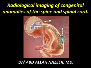
- 1. Radiological imaging of congenital anomalies of the spine and spinal cord. Dr/ ABD ALLAH NAZEER. MD.
- 2. Congenital abnormalities of the spine and spinal cord are referred to as spinal dysraphisms. Reviews normal embryological development of the spine and spinal cord and the imaging findings of congenital abnormalities of the spine and spinal cord with particular focus on MRI. Etiology: Multifactorial, Genetic, environmental factor and folic acid deficiency. The most common congenital anomalies is spinal dysraphism and the caudal spinal anomalies. The diagnosis is either prenatally by ultrasound, at birth, in the early childhood and in adulthood.
- 3. Radiological imaging techniques: Plain Radiography. Ultrasonography. Computed tomography(CT Scan). Magnetic resonance imaging(MRI). Nuclear Imaging.
- 4. Spinal Cord Development Spinal development can be summarized in three basic embryologic stages. The first stage is gastrulation and occurs during the second or third week of embryonic development. Gastrulation involves conversion of the embryonic disk from a bilaminar disk to a trilaminar disk composed of ectoderm, mesoderm, and endoderm. The second stage in spinal development is primary neurulation (weeks 3–4) in which the notochord and overlying ectoderm interact to form the neural plate. The neural plate bends and folds to form the neural tube, which then closes bidirectionally in a zipper like manner (Fig. 1). The final stage of spinal development is secondary neurulation (weeks 5–6). During this stage, a secondary neural tube is formed by the caudal cell mass. The secondary neural tube is initially solid and subsequently undergoes cavitation, eventually forming the tip of the conus medullaris and filum terminale by a process called retrogressive differentiation. Abnormalities in any of these steps can lead to spine or spinal cord malformations.
- 6. Neurulation and derangement of neurulation: Four stages of neurulation: 1- Formation of neural tube. 2- Shaping the neural plate. 3- Bending of the neural plate. 4- Fusion. Formation of neural tube. Shaping the neural plate. Bending of the neural plate. Fusion.
- 7. Categorization of Spinal Dysraphisms: Spinal dysraphisms can be broadly categorized into open and closed types. In an open spinal dysraphism, there is a defect in the overlying skin, and the neural tissue is exposed to the environment. In a closed spinal dysraphism, the neural tissue is covered by skin. Closed spinal dysraphisms can be further subcategorized on the basis of the presence or absence of a subcutaneous mass. Classification of spinal dysraphisms. Open Spinal Dysraphisms: not covered by intact skin: Myelocele Neural placode flush with skin surface Myelomeningocele Neural placode protrudes above skin surface Hemimyelocele Myelocele associated with diastematomyelia Hemimyelomeningocele Myelomeningocele associated with diastematomyelia
- 8. Closed Spinal Dysraphisms: covered by intact skin With a subcutaneous mass: Lipomyelocele Placode–lipoma interface within the spinal canal. Lipomyelomeningocele Placode–lipoma interface outside of the spinal canal. Meningocele Herniation of CSF-filled sac lined by dura. Terminal myelocystocele Terminal syrinx herniating into posterior meningocele. Myelocystocele Dilated central canal herniating through posterior spina bifida Without a subcutaneous mass Simple dysraphic states Intradural lipoma. Lipoma within the dural sac. Filar lipoma. Fibrolipomatous thickening of filum. Tight filum terminale. Hypertrophy and shortening of filum. Persistent terminal ventricle. Persistent cavity within conus medullaris. Dermal sinus. Epithelial lined fistula between neural tissue and skin surface. Complex dysraphic states Dorsal enteric fistula. Connection between bowel and skin surface Neurenteric cyst. More localized form of dorsal enteric fistula. Diastematomyelia . Separation of cord into two hemicords. Caudal agenesis. Total or partial agenesis of spinal column. Segmental spinal dysgenesis. Various segmentation anomalies.
- 9. Open Spinal Dysraphisms: Myelomeningocele and myelocele—Myelomeningoceles and myeloceles are caused by defective closure of the primary neural tube and are characterized clinically by exposure of the neural placode through a midline skin defect on the back. Myelomeningoceles account for more than 98% of open spinal dysraphisms. Myeloceles are rare. Open spinal dysraphisms are often diagnosed clinically, so imaging is not always performed. When imaging is performed, the main differentiating feature between a myelomeningocele and myelocele is the position of the neural placode relative to the skin surface. The neural placode protrudes above the skin surface with a myelomeningocele and is flush with the skin surface with a myelocele. Hemimyelomeningocele and hemimyelocele— Hemimyelomeningoceles and hemimyeloceles can also occur but are extremely rare. These conditions occur when a myelomeningocele or myelocele is associated with diastematomyelia (cord splitting) and one hemicord fails to neurulate.
- 10. Deranged neurulation: Spina Bifida aperta: Myelocele and myelomeningocele. Spina bifida aperta designates those forms of spinal dysraphism in which the neural tissue and meninges are exposed to environment because the skin, fascia, muscles and the bone are deficient in the midline of the back. Myelocele and Myelomeningocele Both myelocele and myelomeningocele are the result of nondisjunction of the cutaneous ectoderm from the neural ectoderm and failure of neural tube closure. The nonneurulated cord or placode is exposed through a dural, bony, and cutaneous defect (i.e., myelocele; The dorsal and ventral nerve roots exit from the ventral surface of the placode. With associated dorsal protrusion of the ventral subarachnoid space, the myelodysplasia is termed a myelomeningocele. These defects commonly occur at the lumbar or sacral level. Occasionally there is an associated diastematomyelia (hemimyelomeningocele) or dermal sinus. These defects are surgically closed shortly after birth. Hydrocephalus is often present; if so, shunting is established early. Subsequently, patients who have undergone such procedures may sac stenosis, progressive scoliosis, and cord ischemia or infarction.
- 11. A B C Myelomeningocele. A, Axial schematic of myelomeningocele shows neural placode (star) protruding above skin surface due to expansion of underlying subarachnoid space (arrow). B, Axial T2-weighted MR image in 1-day-old boy shows neural placode (black arrow) extending above skin surface due to expansion of underlying subarachnoid space (white arrow), which is characteristic of myelomeningocele. C, Sagittal T2-weighted MR image from same patient as in B with myelomeningocele shows neural placode (white arrow) protruding above skin surface due to expansion of underlying subarachnoid space (black arrow).
- 12. A B Myelocele. A, Axial schematic of myelocele shows neural placode (arrow) flush with skin surface. B, Axial T2-weighted MR image in 1-day-old girl shows exposed neural placode (arrow) that is flush with skin surface, consistent with myelocele. There is no expansion of underlying subarachnoid space.
- 13. Chiari malformation, also known as Arnold–Chiari malformation, is a malformation of the brain. It consists of a downward displacement of the cerebellar tonsils through the foramen magnum (the opening at the base of the skull), sometimes causing non-communicating hydrocephalus as a result of obstruction of cerebrospinal fluid (CSF) outflow. The cerebrospinal fluid outflow is caused by phase difference in outflow and influx of blood in the vasculature of the brain. It can cause headaches, fatigue, muscle weakness in the head and face, difficulty swallowing, dizziness, nausea, impaired coordination, and, in severe cases, paralysis. Classification: In the late 19th century, Austrian pathologist Hans Chiari described seemingly related anomalies of the hindbrain; the so-called Chiari malformations I, II and III. Later, other investigators added a fourth (Chiari IV) malformation. The scale of severity is rated I - IV, with IV being the most severe. Types III and IV are very rare.
- 14. Chiaric-1 malformation: A congenital malformation. Is generally asymptomatic during childhood, but often manifests with headaches and cerebellar symptoms. Herniation of cerebellar tonsils. Tonsillar ectopia of more than 3 mm below foramen magnum. Syringomyelia of cervical or cervicothoracic spinal cord can be seen. Sometimes the medullary kink and brainstem elongation can be seen. Syndrome of occipito atlantoaxial hypermobility is an acquired Chiari I malformation in patients with hereditary disorders of connective tissue. Patients who exhibit extreme joint hypermobility and connective tissue weakness as a result of Ehlers-Danlos syndrome or Marfan syndrome are susceptible to instabilities of the craniocervical junction; thus they are at risk for acquiring a Chiari malformation. This type is difficult to diagnose and treat.
- 16. Chiari Type 1 Malformation.
- 18. Chiari-11 malformation: Usually accompanied by a lumbar or lumbosacral myelomeningocele with tonsillar herniation below the foramen magnum. As opposed to the less pronounced tonsillar herniation seen with Chiari I, there is a larger cerebellar vermian displacement. Low lying torcular herophili, tectal beaking, and hydrocephalus with consequent clival hypoplasia are classic anatomic associations. The position of the torcular herophili is important for distinction from Dandy-Walker syndrome in which it is classically upturned. This is important because the hypoplastic cerebellum of Dandy-Walker may be difficult to distinguish from a Chiari malformation that has herniated or is ectopic on imaging. Colpocephaly may be seen due to the associated neural tube defect.
- 19. Fetal T2-weighted MR images. A, Chiari II malformation ( arrows ) on sagittal image. B, Lumbosacral myelocele with low-placed spinal cord ( arrow ) on axial image. C, Dysraphic defect plus placode ( arrow) on axial image.
- 20. Chiari II malformation ( arrows in A ) and lumbosacral myelomeningocele ( arrow in B ) on sagittal T2-weighted fetal MR images.
- 21. Chiari Type 11 Malformation.
- 22. Type II Chiari malformation in a 14-month-old child. Sagittal T1-weighted images demonstrating hydrocephalus (A) with a small posterior fossa and downward herniation of the cerebellum and medulla (B).
- 23. Chiari-111 malformation: It is associated with an occipital encephalocele containing a variety of abnormal neuroectodermal tissues. Syringomyelia and tethered cord as well as hydrocephalus is also seen. Chiari-111 malformation: Characterized by a lack of cerebellar development in which the cerebellum and brain stem lie within the posterior fossa with no relation to the foramen magnum. Associated with hypoplasia. Equivalent to primary cerebellar agenesis.
- 24. Type 3 Chiari malformation.
- 25. Chiari III.
- 26. Type IV Chiari malformation.
- 27. Type IV Chiari malformation.
- 28. Closed Spinal Dysraphisms With a Subcutaneous Mass Lipomas with a dural defect—Lipomas with a dural defect include both lipomyeloceles and Lipomyelomeningocele. These abnormalities result from a defect in primary neurulation whereby mesenchymal tissue enters the neural tube and forms lipomatous tissue. Lipomyeloceles and lipomyelomeningoceles are characterized clinically by the presence of a subcutaneous fatty mass above the intergluteal crease. The main differentiating feature between a lipomyelocele and lipomyelomeningocele is the position of the placode–lipoma interface . With a lipomyelocele, the placode–lipoma interface lies within the spinal canal. With a lipomyelomeningocele, the placode– lipoma interface lies outside of the spinal canal due to expansion of the subarachnoid space.
- 29. Meningocele—Herniation of a CSF-filled sac lined by dura and arachnoid mater is referred to as a meningocele. The spinal cord is not located within a meningocele but may be tethered to the neck of the CSF-filled sac. Posterior meningoceles herniate through a posterior spina bifida (osseous defect of posterior spinal elements) and are usually lumbar or sacral in location but also can occur in the occipital and cervical regions. Anterior meningoceles are usually presacral in location but also can occur elsewhere.
- 30. Lipomyelocele. A, Axial schematic of lipomyelocele shows placode–lipoma interface (arrow) lies within spinal canal. B, Axial T2-weighted MR image in 3-year-old girl shows placode–lipoma interface (arrow) within spinal canal, characteristic for lipomyelocele. C, Sagittal T1- weighted MR image in 3-year-old girl with lipomyelocele shows subcutaneous fatty mass (black arrow) and placode–lipoma interface (white arrow) within spinal canal.
- 31. Lipomyelocele with placode ( short arrows ), lipoma (L), and caudal hydromyelia ( long arrows ) on sagittal ( A ) and axial T1-weighted ( B ) MR images.
- 32. Lumbosacral lipomyelocele with caudal and presacral lipomas (L) and hydrosyringomyelia ( arrows ) on sagittal T1-weighted ( A ) and T2-weighted ( B ) MR images
- 33. Posterior meningocele. A, Sagittal T1-weighted MR image in in 12-month-old girl shows posterior herniation of CSF-filled sac (arrow) in occipital region, consistent with posterior meningocele. B, Sagittal T2-weighted MR image in 5-year-old boy shows large posterior meningocele (arrow) in cervical region. C, Sagittal T2-weighted MR image in 30-month-old girl shows small posterior meningocele (arrow) in lumbar region.
- 34. Meningocele. A and B, Sagittal (A) and axial (B) T2-weighted MR images in 6- month-old boy show small anterior meningocele (arrows).
- 35. Sagittal ( A ) and axial ( B and C ) T2-weighted MR images showing dural ectasia and intradural arachnoid cysts ( arrows in A ) with scoliosis, cord flattening ( short arrows in B and C ), and transforaminal meningoceles ( long arrows in B and C ).
- 37. Terminal myelocystocele—Herniation of large terminal syrinx (syringocele) into a posterior meningocele through a posterior spinal defect is referred to as a terminal myelocystocele. The terminal syrinx component communicates with the central canal, and the meningocele. component communicates with the subarachnoid space. The terminal syrinx and meningocele components do not usually communicate with each other. Myelocystocele—A non-terminal myelocystocele occurs when a dilated central canal herniates through a posterior spina bifida defect. Myelocystoceles are covered with skin and can occur anywhere but are most commonly seen in the cervical or cervicothoracic regions.
- 38. Terminal myelocystocele. A, Sagittal schematic of terminal myelocystocele shows terminal syrinx (star) herniating into large posterior meningocele (arrows). B and C, Sagittal (B) and axial (C) T2-weighted MR images in 1-month-old girl show terminal syrinx (white arrows) protruding through large posterior spina bifida defect and herniating into posterior meningocele component (black arrows). Sagittal image shows turbulent flow in more anterior meningocele component (star, B).
- 40. Closed Spinal Dysraphisms Without a Subcutaneous Mass Closed spinal dysraphisms without a subcutaneous mass can be subcategorized into simple and complex dysraphic states. Simple dysraphic states—Simple dysraphic states consist of intradural lipoma, filar lipoma, tight filum terminale, persistent terminal ventricle, and dermal sinus. An intradural lipoma refers to a lipoma located along the dorsal midline that is contained within the dural sac. No open spinal dysraphism is present. Intradural lipomas are most commonly lumbosacral in location and usually present with tethered-cord syndrome, a clinical syndrome of progressive neurologic abnormalities in the setting of traction on a low-lying conus medullaris. Fibrolipomatous thickening of the filum terminale is referred to as a filar lipoma. On imaging, a filar lipoma appears as a hyperintense strip of signal on T1-weighted MR images within a thickened filum terminale. Filar lipomas can be considered a normal variant if there is no clinical evidence of tethered-cord syndrome . Tight filum terminale is characterized by hypertrophy and shortening of the filum terminale.
- 41. The Tethered Cord Syndrome: Also known as “tight filum terminale syndrome,” tethered cord syndrome refers to low position of the conus medullaris (below the level of mid-L2) associated with a short and thickened filum terminale. The filar thickening (usually greater than 2 mm) is usually fibrous, fatty, or cystic, and may be associated with other dysraphic myelodysplasias, including lipomyelomeningocele and diastematomyelia. The thickened filum may also terminate in a lipoma or dermoid-epidermoid. The filar abnormality is usually best demonstrated on axial T 1 - weighted and T 2 -weighted MRI.
- 42. Filar lipoma. A and B, Sagittal (A) and axial (B) T1- weighted MR images in 2-year-old boy with filar lipoma (arrows), which has characteristic T1 hyperintensity and marked thickening of filum terminale.
- 43. Sagittal ( A ) and axial ( B and C ) T1-weighted MR images showing tethered cord with low conus ( short arrows in A and B ), dorsal lipoma ( long arrows in A and B ), and filar lipoma ( arrow heads in A and C ).
- 44. Sagittal ( A ) and axial ( B and C ) T1-weighted MR images showing lumbosacral dermal sinuses ( black arrows on A and C ) with tethered cord ( white arrows in A ) and lipoma ( white arrows in B and C ) in a newborn.
- 45. Tethered cord ( long arrow ), thick filum and lipoma ( short arrows ), and sacral extradural arachnoid cyst (A) on sagittal T1-weighted ( A ), axial T1-weighted ( B ), and axial T2-weighted ( C ) MR images.
- 48. Complex dysraphic states—Complex dysraphic states can be divided into two categories: disorders of midline notochordal integration, which include dorsal enteric fistula, neurenteric cyst, and diastematomyelia, and disorders of notochordal formation, which include caudal agenesis and segmental spinal dysgenesis. Disorders of midline notochordal integration: Dorsal enteric fistula and neurenteric cyst—A dorsal enteric fistula occurs when there is an abnormal connection between the skin surface and bowel. Neurenteric cysts represent a more localized form of dorsal enteric fistula. These cysts are lined with mucin-secreting epithelium similar to the gastrointestinal tract and are typically located in the cervicothoracic spine anterior to the spinal cord.
- 49. Neurenteric cyst in 3-year-old girl. A and B, Sagittal T2-weighted (A) and axial T1-weighted (B) MR images show bilobed neurenteric cyst (arrows) extending from central canal into posterior mediastinum. C, Three-dimensional CT reconstruction image shows osseous opening (arrow) through which neurenteric cyst passes. This opening is called the Kovalevsky canal.
- 50. Terminal ventricle: Persistence of a small, ependymal lined cavity within the conus medullaris is referred to as a persistent terminal ventricle. Key imaging features include location immediately above the filum terminale and lack of contrast enhancement, which differentiate this entity from other cystic lesions of the conus medullaris. A dermal sinus is an epithelial lined fistula that connects neural tissue or meninges to the skin surface. It occurs most frequently in the lumbosacral region and is often associated with a spinal dermoid at the level of the cauda equina or conus medullaris. Clinically, patients present with a midline dimple and may also have an associated hairy nevus, hyperpigmented patch, or capillary hemangioma. Surgical repair is of great importance because the fistulous connection between neural tissue and the skin surface can result in infectious complications such as meningitis and abscess.
- 51. Persistent terminal ventricle. A and B, Sagittal T2-weighted (A) and sagittal T1-weighted contrast-enhanced (B) MR images in 12-month-old boy show persistent terminal ventricle as cystic structure (arrows) at inferior aspect of conus medullaris, which does not enhance.
- 53. Dermal sinus. A and B, Sagittal schematic (A) and sagittal T2-weighted MR image (B) in 9- year-old girl show intradural dermoid (stars) with tract extending from central canal to skin surface (black arrows). Note tenting of dural sac at origin of dermal sinus (white arrows). C, Axial T2-weighted MR image from same patient as in B shows posterior location of hyperintense dermoid (arrow).
- 54. Thoracic dermal sinus ( black arrows ) and dermoid cyst ( white arrows ) on sagittal T1-weighted ( A ), sagittal T2- weighted ( B ), and axial T1-weighted ( C ) MR images.
- 55. Thoracic dermal sinus ( posterior black and white arrows in A, and B ) and cyst with enhancing abscess ( anterior white arrows in B ) with cord edema on sagittal T2-weighted ( A ) and gadolinium-enhanced T1- weighted ( B ) MR images.
- 56. Disorders of midline notochordal integration: Diastematomyelia—Separation of the spinal cord into two hemicords is referred to as diastematomyelia. The two hemicords are usually symmetric, although the length of separation is variable. There are two types of diastematomyelia. In type 1, the two hemicords are located within individual dural tubes separated by an osseous or cartilaginous septum. In type 2, there is a single dural tube containing two hemicords, sometimes with an intervening fibrous septum. Diastematomyelia can present clinically with scoliosis and tethered-cord syndrome. A hairy tuft on the patient’s back can be a distinctive finding on physical examination.
- 57. Type 1 diastematomyelia. A–C, Sagittal T2-weighted MR (A), axial T2-weighted MR (B), and axial CT with bone algorithm (C) images in 6-year-old boy show two dural tubes separated by osseous bridge (arrows), which is characteristic for type 1 diastematomyelia.
- 58. Lumbar diastematomyelia and split cord with two hemicords ( short arrows in A and B ), two dural sacs, and a bony septum ( long arrows in B and C ) on axial T2-weighted MR images ( A and B ) and an axial CT scan ( C ).
- 59. Type 2 diastematomyelia. A–C, Sagittal T1-weighted (A), coronal T1-weighted (B), and axial T2-weighted (C) MR images in 9-year-old girl show splitting of distal cord into two hemicords (white arrows, B and C) within single dural tube, which is characteristic for type 2 diastematomyelia. Incidental filum lipoma (black arrows, A and B) is present as well.
- 60. Lumbar diastematomyelia with tethered hemicords ( long arrows ), no septum, and a lipoma ( short arrow ) on sagittal T1-weighted ( A ) and axial T2-weighted ( B ) MR images.
- 61. Disorders of notochordal formation: Caudal agenesis—Caudal agenesis refers to total or partial agenesis of the spinal Column and may be associated with the following: anal imperforation, genital anomalies, renal dysplasia or aplasia, pulmonary hypoplasia, or limb abnormalities. Caudal agenesis can be categorized into two types. In type 1, there is a high position and abrupt termination of the conus medullaris. In type 2, there is a low position and tethering of the conus medullaris. Segmental spinal dysgenesis—The clinical–radiologic definition of segmental spinal dysgenesis includes several entities: segmental agenesis or dysgenesis of the thoracic or lumbar spine, segmental abnormality of the spinal cord or nerve roots, congenital paraparesis or paraplegia, and congenital lower limb deformities. Three-dimensional CT reconstructions can be helpful in showing various vertebral segmentation anomalies
- 62. Caudal agenesis. A and B, Sagittal T2-weighted (A) and sagittal T1-weighted (B) MR images in 6- month-old girl show agenesis of sacrum. Conus medullaris is high in position and wedge shaped (arrow) due to abrupt termination. These findings are characteristic of type 1 caudal agenesis. Distal cord syrinx (arrowhead) is present as well.
- 63. Caudal dysplasia with high imperforate anus, congenital lumbar kyphosis ( long arrow s), and dysplastic/hypoplastic conus medullaris ( short arrow ) on sagittal ( A ) and coronal ( B ) T1-weighted MR images.
- 64. MRI Sagittal T2w images of lumbo sacral spine shows: Hypoplastic S1 and S2 with failure of formation of S3 and onwards. Cord ending at a higher level at D12-L1 with 'wedge' shaped termination. No cord tethering. No terminal cord cyst or syrinx. Imaging diagnosis : Caudal Regression, Group I.
- 65. Teratoma - Genetics A teratoma is a disordered mass of cells which exhibit varying degrees of resemblance to normal tissues and structures. Disrupted or incomplete twinning versus uncontrolled stem cell? Some ovarian teratomas – parthenogenetic oocysts ? Fetus in fetus. Other teratomas - arise from stem cells
- 68. Congenital anomalies of the vertebral column.
- 69. Schematic drawing depicting the development of normal and abnormal vertebral bodies.
- 70. Asomia Congenital absence of the body of L2 Hypoplastic posterior elements are present (arrow). Left hemivertebra involving T11.
- 71. Butterfly vertebra. Coronal reformatted (A) and anterior coronal volume-rendered MDCT (B)images demonstrate a sagittal cleft within the L5 vertebral body.
- 73. Block vertebra. Sagittal reformatted (A) and anterior coronal volume rendered MDCT (B) images show L3-L4 block vertebra.
- 74. Dorsal hemivertebra and Metametric hemivertebrae Vertebrae with coronal clefts. in the lower dorsal and lumbar spine.
- 75. Hemi-vertebrae & Segmented Vertebrae.
- 76. Vertebral segmentation anomalies. A and B, Three-dimensional CT reconstruction image (A) in 4-year-old girl and schematic illustration (B) show multiple segmentation anomalies in lumbar spine (superior to inferior beginning at level of arrow): partial sagittal partition, butterfly vertebra, hemivertebra, tripedicular vertebra, and widely separated butterfly vertebra.
- 77. Limbus vertebra. Lateral radiograph (a) and sagittal thin slab reformatted MDCT images (b,c) show the defect at the anterosuperior border of L4.
- 78. Limbus vertebra. Axial MDCT image (a) and oblique volume rendered images (b) show a limbus vertebra.
- 79. L5 transitional vertebra in a 40-year-old woman who presented with left-sciatic pain. Axial thin-section (a) and coronal volume-rendered (b) MDCT images show A transitional lumbosacral vertebra, with a left hypertrophic transverse process (arrow) that articulates with the ilium and sacrum.
- 80. Lumbarization of L5 vertebra.
- 81. Spondylodysplasias: Spondylodysplasia refers to any developmental abnormality of the bony spinal column. This category includes idiopathic scoliosis, congenital scoliosis and kyphosis, Scheuermann disease, and the skeletal dysplasias (e.g., NF-1, mucopolysaccharidoses, spondyloepiphyseal dysplasia, Achondroplasia, Down syndrome). Spinal curvature abnormalities are common in this category. As defined by clinical examination and findings on standing plain films, they include scoliosis (lateral curvature), kyphosis (posterior angulation), and lordosis (increased anterior angulation). In the skeletal dysplasias, a major concern is mechanical compromise of the spinal neuraxis due to progressive canal/foraminal stenosis, kyphoscoliosis, or craniocervical instability. Evaluation usually requires both CT and MRI.
- 82. Spondylodysplasias Idiopathic scoliosis. Congenital scoliosis and kyphosis. Scheuermann/Schmorl disease. Neurofibromatosis. Achondroplasia. Mucopolysaccharidoses. Down syndrome. Spondyloepiphyseal dysplasia. Craniocervical anomalies. Klippel-Feil syndrome. Spinal vascular anomalies.
- 83. Idiopathic Scoliosis Idiopathic scoliosis is the most common curvature abnormality in childhood. The adolescent form, which is seen in children older than 10 years, is more common than the infantile or juvenile form. Adolescent idiopathic scoliosis is familial and most commonly seen in females. Characteristically, there is a right convex primary curve of the thoracic, thoracolumbar, or lumbar spine A rotatory component with pedicle asymmetry is characteristic, and a left convex secondary curve may present. Additional lordosis often occurs with curve progression. Complications of idiopathic scoliosis include curve progression, cardiopulmonary compromise, painful curves, cosmetic deformity, neurologic dysfunction, and degenerative joint disease. Congenital scoliosis and kyphosis result from formation or segmentation Anomalies . Failure of formation may result in vertebral aplasia or hypoplasia with wedge vertebra, hemivertebra, or butterfly vertebra. Anomalies of segmentation failure include pedicle fusion bars and block vertebra.
- 84. Idiopathic scoliosis with typical rightward thoracic lateral and rotatory curvature on a frontal plain film/computerized radiograph.
- 85. A and B, Congenital lumbar scoliosis with hemivertebrae ( arrows in A ) and hydrosyringomyelia ( arrows in B ) on coronal T2-weighted MR images. The hemivertebrae ( arrows ) and double curve are confirmed on coronal reformatted CT scan ( C ) and 3D reconstruction ( D ).
- 86. Scheuermann Disease An osteochondrodysplasia, Scheuermann disease is a common cause of juvenile and adolescent thoracic kyphosis. An abnormality of enchondral ossification is associated with degenerative and reactive changes of the vertebral end plates. Often there is multilevel end plate irregularity, disk space narrowing, vertebral body wedging, and Schmorles nodes . The kyphosis commonly occurs in the mid-thoracic spine. There may be progressive wedging with increasing thoracic kyphosis and lumbar lordosis. Scheuermann disease with multilevel, irregular thoracic disk space narrowing ( long arrows ) and Schmorl’s nodes ( short arrows ).
- 87. Neurofibromatosis Type 1: NF-1 is a common mesodermal dysplasia of childhood Characteristic anomalies include a short-segment kyphoscoliosis, posterior vertebral body scalloping, vertebral body wedging, apical vertebral rotation, dural ectasia, and meningoceles (e.g., thoracic, presacral). There may also be an associated plexiform neurofibroma and other nerve sheath tumors. Other anomalies are cervical kyphosis, hypoplasia of the spinous process, transverse process, or pedicle, and twisted-ribbon ribs. Dural ectasia and meningocele formation may also be seen with Marfan or Ehlers- Danlos syndrome and in familial cases.
- 88. A, Neurofibromatosis-1 with short-segment right lumbar scoliosis plus vertebral and rib deformities ( arrows ) on frontal plain film/computerized radiograph. B, Enhancing paraspinal plexiform neurofibroma ( arrows ) with foraminal extension is shown on coronal gadolinium-enhanced T1-weighted MR image along with other small intradural enhancing nerve sheath tumors.
- 89. Achondroplasia Achondroplasia is one of the osteochondrodysplasia (defective enchondral bone development) resulting in dwarfism. There is craniofacial dysmorphia with skull base constriction, including foramen magnum stenosis, short clivus, and small jugular Foramina. Macrocephaly and hydrocephalus (e.g., impaired venous outflow) occur, and there is enlargement of the subarachnoid spaces and ventricles. Other craniocervical abnormalities include odontoid hypoplasia, atlantoaxial (AA) instability, basilar impression, and occipitalization of the atlas. Scoliosis, kyphosis, or kyphoscoliosis may occur in about one third of patients with achondroplasia. Stenosis of the spinal canal may be cervical, thoracolumbar, lumbar, or may occur diffusely. There is platyspondyly with short pedicles, vertebral scalloping, and interpediculate narrowing. There may be associated spinal foraminal and canal compromise.
- 90. Achondroplasia, basilar invagination, and foramen magnum plus cervical spinal stenosis ( arrows ) on sagittal T1-weighted MR image ( A ), sagittal reformatted CT scan ( B ), and an axial CT scan ( C ). Also, there is characteristic macrocephaly and ventriculomegaly.
- 91. Mucopolysaccharidosis: Mucopolysaccharidosis may be associated with craniocervical abnormalities (e.g., Morquio syndrome; They include odontoid hypoplasia, occipital hypoplasia, ligamentous laxity, AA instability, and dural sac stenosis (mucopolysaccharide deposition with fibrosis; Foraminal and spinal canal encroachment may occur. Other vertebral anomalies are platyspondyly, beaking, wedging, gibbus deformity, and kyphoscoliosis. Down Syndrome: Down syndrome may be complicated by craniocervical instability, such as odontoid hypoplasia, os odontoideum, atlanto-occipital (AO) subluxation, or AA subluxation Scoliosis, degenerative cervical spine disease, tall vertebral bodies, vertebral fusions, and subluxations at other levels may also occur.
- 92. A, Morquio syndrome with craniocervical instability ( upper arrow ) plus thoracolumbar kyphoscoliosis ( lower arrow ) on a lateral plain film/computerized radiograph. B and C, Sagittal T2-weighted MR images show upper cervical dural sac stenosis with hyperintense cord injury ( arrow in B ) along with ventral cord deformity at the level of the kyphosis ( arrow in C ).
- 93. Lateral plain film/computerized radiograph ( A ), axial CT scan ( B ), and sagittal reformatted CT scan ( C ) showing Down syndrome with hypoplastic dens, os odontoideum (o), atlantoaxial instability, and post-dental canal stenosis ( short arrows ) with probable cord compression; C2, axis; a, anterior arch of atlas; p, posterior arch of atlas; b, basion; op, opisthion.
- 94. A, Sagittal T2-weighted MR image showing Down syndrome with hypoplastic dens, os odontoideum ( anterior arrow ), and atlantoaxial dislocation with high-intensity cervical spinal cord compressive injury ( posterior arrows ). B, Sagittal postoperative reformatted CT scan shows the stabilized anomaly, including the os odontoideum ( upper arrow ), decompressed canal, and metallic instrumentation plus bony fusion ( posterior arrows ).
- 95. Basilar Invagination: Basilar invagination refers to an occipital dysplasia with upward displacement of the margins of the foramen magnum anteriorly, posteriorly, laterally, or combined . The odontoid is superiorly displaced relative to McRae’s line. In addition the posterior fossa may be of small volume with an irregularly shaped foramen magnum and a short clivus. Basilar invagination may be (1) primary (i.e., developmental) and associated with other craniocervical anomalies or syndromes or (2) secondary (i.e., basilar impression) and associated with osteochondral dysplasias or metabolic disorders (e.g., rickets, fibrous dysplasia, achondroplasia, the mucopolysaccharidoses, osteogenesis imperfecta,osteomalacia, cleidocranial dysplasia). Basilar invagination may produce neural, CSF, or vascular compromise. There may be an associated Chiari I malformation, hydrocephalus, or hydrosyringomyelia.
- 96. Basilar invagination and Chiari I malformation on sagittal T2-weighted MR image with short clivus and high dens ( anterior arrows )plus low tonsils and low cervicomedullary junction( posterior arrows ).
- 97. Klippel-Feil Anomaly and Syndrome: The Klippel-Feil anomaly , which results from failure of segmentation, manifests as bony fusion of the cervical spine at one or more levels . The triad of low posterior hairline, short webbed neck, and limitation of neck motion represents the Klippel-Feil syndrome . Sprengel’s deformity of the scapula and a bridging omovertebral bone may also be present in Klippel-Feil syndrome. Other associated anomalies are genitourinary, cardiac, and musculoskeletal (e.g., limbs and digits). The spinal involvement may include posterior element abnormalities, occipitalization of the atlas, basilar impression, dens anomalies, and scoliosis. Chiari I malformation, hydrosyringomyelia, neurenteric cyst, or diastematomyelia may occur. Hypermobility and instability at unfused segments and early degenerative disease may lead to foraminal or spinal canal stenosis, osteophytic spurs, subluxation, facet arthropathy, or disk herniation. Occipitalization of the atlas refers to complete or partial fusion of the atlas to the occiput, which may be bony or fibrous. Usually, the anterior arch is assimilated into the anterior rim of the foramen magnum. The involvement may also include the posterior arch or lateral masses. Associated anomalies are odontoid hypoplasia, basilar invagination, AA instability, and Klippel-Feil anomaly.
- 98. Sagittal ( A ) and coronal ( B ) reformatted CT scans showing Klippel-Feil anomaly, occipitalization of the atlas, and cervical spinal stenosis, including fused cervical segments ( lower arrows ), fusion of atlas to the occiput ( upper black arrows ), and a small foramen magnum and C1 canal. Flexion ( C ) and extension ( D ) T2- weighted sagittal MR images show no translational craniocervical instability.
- 99. Odontoid Anomalies: Anomalies of the odontoid include aplasia, hypoplasia, and the os odontoideum. Aplasia is rare and causes severe AA dislocation. Hypoplasia leads to a short dens. Os odontoideum is a proatlas remnant or represents hypertrophy of the ossiculum terminale. Usually, there is odontoid hypoplasia, and the ossicle is located near the dens tip or the basion and lacks a normal dens fusion line. Hypertrophy of the anterior arch and hypoplasia or clefting of the posterior arch are often present. AA or occipitoaxial instability commonly occurs. Odontoid anomalies may be idiopathic or may be associated with skeletal dysplasias such as Morquio syndrome, Down syndrome, SED, and Klippel-Feil anomaly. Craniocervical Instability: Craniocervical instability includes translational or rotary AA instability and AO instability. Craniocervical instability often results from ligamentous deficiency or insufficiency (e.g. transverse ligament in AA instabilities) and is commonly associated with odontoid anomalies (as discussed earlier). AA and AO instabilities most commonly occur with Down syndrome. Other common causes are the skeletal dysplasias (e.g., Morquio syndrome, SED), rheumatoid arthritis, and trauma. Rotary AA instability includes displacement, subluxation, or dislocation and produces torticollis. It may be spontaneous or related to trauma, infection, or CCJ anomalies (e.g., odontoid anomalies, Klippel-Feil anomaly).
- 100. Craniocervical Instability secondary to tear of the transverse ligament.
- 101. Congenital spondylolisthesis Spondylolisthesis is a condition that involves a forward slip of a vertebra or vertebrae in relation to the rest of the spinal column. Herbinaux first described the condition in 1782 as a protruding sacral bone that complicated labor. Typically, this defect occurs between the fifth lumbar vertebra and the sacrum . However, spondylolisthesis has also been seen between the fourth and fifth lumbar vertebral levels and may even occur in the cervical region. The patients showed deficits in their neurological examinations and required stabilization and fusion. Studies showing an increase among family members with this defect suggest a genetic component. There are various types of spondylolisthesis. Thus, a classification system was established to differentiate the specific etiologies. Congenital spondylolisthesis, which is a Newman Type I defect. The cause of congenital or dysplastic spondylolisthesis, which is most commonly found at the lumbosacral junction, occurs as a result of an elongation of the pars interarticularis along with varying degrees of facet joint dysplasia . Patients may present with back pain, radicular complaints, tight hamstrings and various neurological symptoms.
- 103. Spinal Vascular Anomalies: Vascular anomalies of childhood may involve the paraspinal soft tissues, bony spinal column, spinal neuraxis, meninges, or multiple structures. Vascular anomalies of the CNS have traditionally been classified as arteriovenous malformations (AVMs), developmental venous anomalies, cavernous malformations, telangiectasias, and aneurysms. Intradural spinal vascular anomalies are usually AVMs or arteriovenous fistulae (AVFs) and are classifi d according to the anatomic site of origin or involvement. They may be classified as spinal cord AVMs (intramedullary), spinal dural AVFs, or metameric AVMs (Cobb syndrome), the last involving any or all layers of a spinal segment from the spinal cord to the skin. In spinal AVMs that are “high-flow” malformations, MRI may show nodular, serpiginous signal voids without tumor parenchyma, hemorrhage (subarachnoid or intramedullary), cord edema, infarct, myelomalacia, syrinx, or atrophy. Spinal AVFs may be “high-flow” or “low-flow” malformations. Spinal angiography is necessary for full evaluation of the vascular anomaly in anticipation of surgery or interventional therapy. Cavernous angiomas of the spinal cord are rare and may manifest as hemorrhage (subarachnoid or intramedullary) or myelopathy.
- 104. Cervical spinal cord arteriovenous malformation with high-flow vascular low intensities ( arrows ) on sagittal T1-weighted ( A ) and T2-weighted ( B ) MR images.
- 105. Spina bifida (Latin : "split spine") is a developmental congenital disorder caused by the incomplete closing of the embryonic neural tube . Spina bifida malformations fall into three categories: spina bifida occulta, spina bifida cystica with meningocele, and spina bifida cystica with myelomeningocele. The most common location of the malformations is the lumbar and sacral areas. Myelomeningocele is the most significant and common form, and this leads to disability in most affected individuals. The terms spina bifida and myelomeningocele are usually used interchangeably.
- 106. Spina bifida of lumbar and cervical spines.
- 108. Sagittal and axial MR images at the level of the L7-S1 disk of a young Bulldog with spina bifida.
- 109. Conclusion Congenital malformations of the spine and spinal cord can be complex and variable in imaging appearance. An organized approach to imaging findings with consideration of clinical and developmental factors allows greater ease in diagnosis. Spinal dysraphism or neural tube defect is a broad term encompassing a heterogenous group of congenital spinal anomalies which result from defective closure of neural tube early in the fetal life and anomalous development of the caudal cell mass. Neural tube defect exact emotional and economic toll on families and health care systems.
- 110. Thank You.