SlideShare une entreprise Scribd logo
1  sur  65
Dr/ ABD ALLAH NAZEER. MD.
Radiological imaging of the
nasopharyngeal diseases.
Adenoids
Nasopharyngeal abscess (MRI scan, sagittal view).
Nasopharyngeal abscess (MRI scan, axial view).
Latero and Retropharyngeal abscess (CT scan, axial view). B. Latero and
Retropharyngeal abscess (CT scan, sagittal view). C. Lateropharyngeal abscess
(CT scan, coronal view). D. Lateropharyngeal abscess (CT scan, axial view).
Inflammatory pseudotumor of the nasopharynx and skull base
is a benign, idiopathic disease that is often mistaken for a
neoplasm or infection owing to its aggressive behavior and
clinical presentation. It can present as a progressively
destructive mass and should be considered when repeated
tissue biopsies reveal acute or chronic inflammation without
evidence of malignant disease or infection. Nasopharyngeal
inflammatory pseudotumor with skull base invasion occurring
in patients with diabetes mellitus (DM). These patients had
repeated negative results from biopsies and cultures, and none
had associated cervical lymphadenopathy despite having an
aggressive destructive mass. We suggest that these findings,
coupled with clinical suspicion, will be helpful in making the
correct diagnosis of inflammatory pseudotumor. This is critical
in the management of these patients to institute the correct
treatment plan.
A 32-year-old man with right ptosis, trigeminal neuralgia and facial palsy 2 days after an
episode of acute otitis media treated with tympanocentesis. (a) Axial T1 weighted image
shows an intermediate signal intensity mass lesion involving the right nasopharyngeal
submucosal region (arrows). (b) Axial T2 weighted image shows low signal intensity
change of the lesion (arrows). Note the right mastoiditis change. (c) Coronal contrast-
enhanced T1 weighted image shows extension of the lesion to the right skull base,
cavernous sinus and Meckel’s cave (black arrows). Enhancement of the thickened
pachymeninges is also present (white arrows). (d) Axial T2 weighted and (e) contrast-
enhanced images obtained 2 months after a course of intravenous high-dose pulse
solumedrol therapy followed by oral corticosteroid medication show the mass lesion to be
significantly reduced in size. The pachymeningeal enhancement has nearly disappeared
A 32-year-old man with left hemicrania of 5 months’ duration and hemitongue atrophy of 1
months’ duration. (a) Axial T1 weighted image and (b) axial T2 weighted image show an ill-
defined hypointense lesion in the left nasopharyngeal submucosal region with extension to
the carotid space and skull base. The left internal carotid artery is encased and narrowed
(white arrow). Involvement of the hypoglossal canal (black arrow) is evident. The mass lesion
is distinguishable from the nasopharyngeal mucosa on T2 weighted images. (c) Axial contrast-
enhanced T1 weighted image with fat saturation shows moderate enhancement of the left
nasopharyngeal lesion, with extension around the left internal carotid artery into the skull
base, and involving the prevertebral muscles and hypoglossal canal (black arrow). (d) Axial T2
weighted and (e) contrast enhanced T1 weighted image obtained 2 months after a course of
oral corticosteroids show nearly complete resolution of the mass lesion.
A 37-year-old man with right hemicrania of 2 months’ duration. (a) Axial T2 weighted image
shows hypointensity and (b) axial contrast-enhanced T1 weighted image shows moderate
enhancement of the lesion in the right nasopharyngeal submucosa and carotid space (white
arrows), which encases the right internal carotid artery with extension to the skull base. There
is high signal change on T2 weighted images and abnormal contrast enhancement in the bone
marrow of the right occipital condyle (black arrows). (c) Contrast-enhanced MR angiography
shows segmental narrowing of the right upper cervical internal carotid artery (white arrows)
owing to the encasement by the carotid space lesion.
Thornwaldt cyst.
Thornwaldt cyst
Juvenile nasopharyngeal angiofibroma.
Neonatal teratomas are very rare congenital tumors which are formed from
cells derived from all three embryonic germ cell layers. The incidence of
neonatal teratomas is between 1:20,000 and 1:80,000 live births. Most occur in
the sacrococcygeal region and only 5% are found in the head and neck region,
specifically the cervical region followed by the nasopharynx. They can be
associated with a cleft palate, intracranial abnormalities including hemicrania
and anencephaly, other head and neck anomalies, diaphragmatic hernia and
renal and vertebral anomalies.
Nasopharyngeal teratomas most commonly present with difficulties in
swallowing and obstructive respiratory distress. The size and location serve as
the major causes of morbidity and mortality. Rapid airway stabilization,
including endotracheal intubation, cricothyrotomy or tracheotomy, is crucial in
the management of these patients. After securing the airway, imaging is very
useful to help differentiate this tumor from other masses. Ultimately surgical
excision is the mode of treatment. These tumors are often encapsulated or
pseudoencapsulated which yields excellent surgical prognosis.
The differential diagnosis for neonatal nasopharyngeal lesions is extensive and
includes: teratoma, dermoid, craniopharyngioma, encephalocele, cystic
hygroma, rhabdomyoscarcoma, hemangioma, lipoma, chordoma,
neurofibroma, Rathke’s pouch and sarcoma botryoides.
Nasopharyngeal teratoma.
(a) T2 weighted sequence in coronal plane showing the external portion of the tumour
consisting of lobulated well defined areas of fluid, solid tissue and fat, in keeping with
teratoma. There is involvement of the left temporal lobe of the brain (red arrow); (b) T1
weighted contrast enhanced sequence in sagittal plane. Internally, the tumour is occupying
the oropharynx, nasopharynx and oral cavity, effacing normal anatomy (red arrows).
Pleomorphic adenoma of the nasopharynx.
Nasopharyngeal hemangioma.
Chordoma is a low-grade malignant tumor that
arises from the remnants of notochord. They account
for 1-4% of malignant bone tumors and most
commonly present after the age of 30 years. Male to
female ratio is 1.8:1. Majority occur in sacrum or in
the clivus (a bony surface in the posterior cranial fossa
sloping upward from foramen magnum to dorsum
sella). Those located in the clivus are often associated
with a chronic headache and symptoms due to
compression of a cranial nerve. Lateral extension can
lead to a cerebellopontine angle tumor
symptomatology. In case of inferior spread, nasal
obstruction, bleeding, and even nasal mass appear.
Extraosseous Chordoma of the Nasopharynx.
Chordoma.
Two cases of cystic choristoma along the nasopharynx and the prestyloid spaces.
Paraganglioma of the nasopharynx.
Naso and parapharyngeal para-ganglioma.
Nasopharynx cancer or nasopharyngeal carcinoma (NPC) is the
most common cancer originating in the nasopharynx, the
uppermost region of the pharynx ("throat"), behind the nose
where the nasal passages and auditory tubes join the remainder
of the upper respiratory tract. NPC occurs in children and adults.
NPC differs significantly from other cancers of the head and
neck in its occurrence, causes, clinical behavior, and treatment.
It is vastly more common in certain regions of East Asia and
Africa than elsewhere, with viral, dietary and genetic factors
implicated in its causation. It is most common in males. It is a
squamous cell carcinoma or an undifferentiated type.
Squamous cells are a flat type of cell found in the skin and the
membranes that line some body cavities. Differentiation means
how different the cancer cells are from normal cells.
Undifferentiated is a word used to describe cells that do not
have their mature features or functions.
Classification:
Nasopharyngeal carcinoma, commonly known as
nasopharyngeal cancer, is classified as a malignant
neoplasm, or cancer, arising from the mucosal epithelium
of the nasopharynx, most often within the lateral
nasopharyngeal recess or fossa of Rosenmüller (a recess
behind the entrance of the Eustachian tube opening). The
World Health Organization classifies nasopharyngeal
carcinoma in three types. Type 1 (I) is squamous cell
carcinoma. Type 2a (II) is keratinizing undifferentiated
carcinoma. Type 2b (III) is nonkeratinizing undifferentiated
carcinoma. Type 2b (III) nonkeratinizing undifferentiated
form also known as lymphoepithelioma is most common,
and is most strongly associated with Epstein-Barr virus
infection of the cancerous cells.
Axial T2 weighted images shows a carcinoma that has infiltrated lateral to the
tensor and levator veli palatini. Solid arrow shows normal tensor veli palatini on
the right. Dashed arrow shows the abnormal left levator muscle which is being
displaced and the presence of tumor lateral to it.
Sagittal T1 image that shows NPC invasion into the clivus. There is a loss of
the inferior cortical margin of the clivus. The clivus is also of uniform lower signal
that reflects sclerotic change. 10B is as coronal T1 image in another patient that
shows NPC infiltration into the left aspect of the clivus (arrow).
Coronal T2 (left) and post gadolinium enhanced T1 (right) weighted images show
superior extension of NPC through the floor of the sphenoid sinus (arrows).
Two cases of nasopharyngeal tumour, extended laterally into the left masticator space and
subsequent perineural tumor tracking superiorly along V3 into the left cavernous sinus.
Carcinoma of the nasopharynx. Coronal T1 weighted, fat suppressed, contrast
enhanced MR image shows an enhancing lesion(T) in the nasopharynx with
extension into bilateral cavernous sinuses(white arrows) via foramen ovale (black
arrows). Both cavernous internal carotid arteries are encased by the lesion. There
is also extension into left infratemporal fossa(small open black arrow).
A case of locally advanced nasopharyngeal
carcinoma in a 44-year-old man with right-ear
tinnitus and hearing impairment of 1 year’s
duration. (a)AxialT1weighted image shows a
bulging right nasopharyngeal mass with
extension to the skull base, partially encasing
the right internal carotid artery. The right
parapharyngeal fat is obliterated. (b) Axial T2
weighted image shows mild hyperintensity of
the right nasopharyngeal mass. The signal
intensity of the mass is similar to that of the
nasopharyngeal mucosa, and the margin
between the normal mucosa and the mass
could not be delineated. Note the presence of
right mastoiditis change. (c) Axial and (d)
coronal contrast enhanced T1 weighted
images with fat saturation shows vivid
enhancement of the nasopharyngeal mass.
There is skull-base and cavernous sinus
invasion with encasement of the right internal
carotid artery, which is not narrowed (white
arrows). Bilateral neck lymphadenopathy
(black arrows) is prominent.
Burkett lymphoma of the nasopharynx.
Nasopharyngeal non-Hodgkin lymphoma.
Granulocytic sarcoma
in the right lateral
wall of the
nasopharynx (black
arrow), involving the
right pterygoid
muscles,
parapharyngeal space
Figure 1b: CT sagittal
image shows mass in
the nasopharynx with
extension into
sphenoid sinus Figure
1c: CT coronal image
shows that the mass is
confined to the
nasopharynx without
intracranial extension.
Leiomyosarcoma in the Nasopharynx:
Thank You.

Contenu connexe

Tendances

Presentation1.pptx, radiological anatomy of the neck.
Presentation1.pptx, radiological anatomy of the neck.Presentation1.pptx, radiological anatomy of the neck.
Presentation1.pptx, radiological anatomy of the neck.Abdellah Nazeer
 
Ct ANATOMY HEAD AND NECK
Ct ANATOMY HEAD AND NECKCt ANATOMY HEAD AND NECK
Ct ANATOMY HEAD AND NECKMUNEER khalam
 
Presentation1.pptx, radiological anatomy of the thigh and leg.
Presentation1.pptx, radiological anatomy of the thigh and leg.Presentation1.pptx, radiological anatomy of the thigh and leg.
Presentation1.pptx, radiological anatomy of the thigh and leg.Abdellah Nazeer
 
Presentation1, mri imaging of the prostate
Presentation1, mri imaging of the prostatePresentation1, mri imaging of the prostate
Presentation1, mri imaging of the prostateAbdellah Nazeer
 
Presentation1.pptx, radiological anatomy of the shoulder joint.
Presentation1.pptx, radiological anatomy of the shoulder joint.Presentation1.pptx, radiological anatomy of the shoulder joint.
Presentation1.pptx, radiological anatomy of the shoulder joint.Abdellah Nazeer
 
Presentation1.pptx, radiological imaging of spinal dysraphism.
Presentation1.pptx, radiological imaging of spinal dysraphism.Presentation1.pptx, radiological imaging of spinal dysraphism.
Presentation1.pptx, radiological imaging of spinal dysraphism.Abdellah Nazeer
 
CT perfusion physics and its application in Neuroimaging
CT perfusion physics and its application in NeuroimagingCT perfusion physics and its application in Neuroimaging
CT perfusion physics and its application in NeuroimagingDr.Suhas Basavaiah
 
Presentation1, radiological imaging of anal carcinoma.
Presentation1, radiological imaging of anal carcinoma.Presentation1, radiological imaging of anal carcinoma.
Presentation1, radiological imaging of anal carcinoma.Abdellah Nazeer
 
Radiology of the Elbow Joint. Dr. Sumit Sharma
Radiology of the Elbow Joint. Dr. Sumit SharmaRadiology of the Elbow Joint. Dr. Sumit Sharma
Radiology of the Elbow Joint. Dr. Sumit SharmaSumit Sharma
 
larynx Imaging 1st part laryngeal anatomy CT MRI Dr Ahmed Esawy
larynx Imaging 1st part laryngeal anatomy CT MRI Dr Ahmed Esawylarynx Imaging 1st part laryngeal anatomy CT MRI Dr Ahmed Esawy
larynx Imaging 1st part laryngeal anatomy CT MRI Dr Ahmed EsawyAHMED ESAWY
 
Vascular brain anatomy for Radiology by Dr Soumitra Halder
Vascular brain anatomy for Radiology by Dr Soumitra HalderVascular brain anatomy for Radiology by Dr Soumitra Halder
Vascular brain anatomy for Radiology by Dr Soumitra HalderSoumitra Halder
 
Imaging of white matter diseases
Imaging of white matter diseasesImaging of white matter diseases
Imaging of white matter diseasesNavni Garg
 
Diagnostic Imaging of Cerebellopontine Angle Masses
Diagnostic Imaging of Cerebellopontine Angle MassesDiagnostic Imaging of Cerebellopontine Angle Masses
Diagnostic Imaging of Cerebellopontine Angle MassesMohamed M.A. Zaitoun
 
Presentation2.pptx. posterior fossa tumour
Presentation2.pptx. posterior fossa tumourPresentation2.pptx. posterior fossa tumour
Presentation2.pptx. posterior fossa tumourAbdellah Nazeer
 

Tendances (20)

Presentation1.pptx, radiological anatomy of the neck.
Presentation1.pptx, radiological anatomy of the neck.Presentation1.pptx, radiological anatomy of the neck.
Presentation1.pptx, radiological anatomy of the neck.
 
Orbit imaging anatomy
Orbit imaging anatomyOrbit imaging anatomy
Orbit imaging anatomy
 
Ct ANATOMY HEAD AND NECK
Ct ANATOMY HEAD AND NECKCt ANATOMY HEAD AND NECK
Ct ANATOMY HEAD AND NECK
 
CSF cisterns
CSF cisternsCSF cisterns
CSF cisterns
 
Presentation1.pptx, radiological anatomy of the thigh and leg.
Presentation1.pptx, radiological anatomy of the thigh and leg.Presentation1.pptx, radiological anatomy of the thigh and leg.
Presentation1.pptx, radiological anatomy of the thigh and leg.
 
Presentation1, mri imaging of the prostate
Presentation1, mri imaging of the prostatePresentation1, mri imaging of the prostate
Presentation1, mri imaging of the prostate
 
Presentation1.pptx, radiological anatomy of the shoulder joint.
Presentation1.pptx, radiological anatomy of the shoulder joint.Presentation1.pptx, radiological anatomy of the shoulder joint.
Presentation1.pptx, radiological anatomy of the shoulder joint.
 
Presentation1.pptx, radiological imaging of spinal dysraphism.
Presentation1.pptx, radiological imaging of spinal dysraphism.Presentation1.pptx, radiological imaging of spinal dysraphism.
Presentation1.pptx, radiological imaging of spinal dysraphism.
 
CT perfusion physics and its application in Neuroimaging
CT perfusion physics and its application in NeuroimagingCT perfusion physics and its application in Neuroimaging
CT perfusion physics and its application in Neuroimaging
 
MRI SPINE ANATOMY
MRI SPINE ANATOMYMRI SPINE ANATOMY
MRI SPINE ANATOMY
 
Basic approach to brain tumor
Basic approach to brain tumorBasic approach to brain tumor
Basic approach to brain tumor
 
Presentation1, radiological imaging of anal carcinoma.
Presentation1, radiological imaging of anal carcinoma.Presentation1, radiological imaging of anal carcinoma.
Presentation1, radiological imaging of anal carcinoma.
 
Radiological anatomy of the brain
Radiological anatomy of the brainRadiological anatomy of the brain
Radiological anatomy of the brain
 
Radiology of the Elbow Joint. Dr. Sumit Sharma
Radiology of the Elbow Joint. Dr. Sumit SharmaRadiology of the Elbow Joint. Dr. Sumit Sharma
Radiology of the Elbow Joint. Dr. Sumit Sharma
 
Pre-FESS PNS CT
Pre-FESS PNS CTPre-FESS PNS CT
Pre-FESS PNS CT
 
larynx Imaging 1st part laryngeal anatomy CT MRI Dr Ahmed Esawy
larynx Imaging 1st part laryngeal anatomy CT MRI Dr Ahmed Esawylarynx Imaging 1st part laryngeal anatomy CT MRI Dr Ahmed Esawy
larynx Imaging 1st part laryngeal anatomy CT MRI Dr Ahmed Esawy
 
Vascular brain anatomy for Radiology by Dr Soumitra Halder
Vascular brain anatomy for Radiology by Dr Soumitra HalderVascular brain anatomy for Radiology by Dr Soumitra Halder
Vascular brain anatomy for Radiology by Dr Soumitra Halder
 
Imaging of white matter diseases
Imaging of white matter diseasesImaging of white matter diseases
Imaging of white matter diseases
 
Diagnostic Imaging of Cerebellopontine Angle Masses
Diagnostic Imaging of Cerebellopontine Angle MassesDiagnostic Imaging of Cerebellopontine Angle Masses
Diagnostic Imaging of Cerebellopontine Angle Masses
 
Presentation2.pptx. posterior fossa tumour
Presentation2.pptx. posterior fossa tumourPresentation2.pptx. posterior fossa tumour
Presentation2.pptx. posterior fossa tumour
 

En vedette

Nasopharynx and its diseases
Nasopharynx and its diseasesNasopharynx and its diseases
Nasopharynx and its diseasesVinay Bhat
 
Nasopharyngeal carcinoma
Nasopharyngeal carcinomaNasopharyngeal carcinoma
Nasopharyngeal carcinomasreelukp
 
Nose and paranasal sinuses according to new reference 1
Nose and paranasal sinuses according to new reference 1 Nose and paranasal sinuses according to new reference 1
Nose and paranasal sinuses according to new reference 1 Indian dental academy
 
Salivary glands
Salivary glandsSalivary glands
Salivary glandsddert
 
Pathology of pharynx
Pathology of pharynxPathology of pharynx
Pathology of pharynxDilnia Qader
 
Tumours of nasopharynx
Tumours of nasopharynxTumours of nasopharynx
Tumours of nasopharynxaaryaserin
 
Tumours of nasopharynx (2) itp class dr.davis - 03.06.16
Tumours of nasopharynx (2) itp class   dr.davis - 03.06.16Tumours of nasopharynx (2) itp class   dr.davis - 03.06.16
Tumours of nasopharynx (2) itp class dr.davis - 03.06.16ophthalmgmcri
 
Jna reg presentation
Jna reg presentationJna reg presentation
Jna reg presentationAngus Shao
 
Nasopharyngeal Airway (NPA): Banyak Manfaat namun Kurang Dikenal
Nasopharyngeal Airway (NPA): Banyak Manfaat namun Kurang DikenalNasopharyngeal Airway (NPA): Banyak Manfaat namun Kurang Dikenal
Nasopharyngeal Airway (NPA): Banyak Manfaat namun Kurang DikenalRobertus Arian Datusanantyo
 
Adenoiditis
AdenoiditisAdenoiditis
Adenoiditisfitango
 
Juvenile nasopharyngeal angiofibroma
Juvenile nasopharyngeal angiofibromaJuvenile nasopharyngeal angiofibroma
Juvenile nasopharyngeal angiofibromapraneeth koduru
 
Nasopharngeal angiofibroma
Nasopharngeal angiofibromaNasopharngeal angiofibroma
Nasopharngeal angiofibromaKomal Soomro
 
acute respiratory tract infection
acute respiratory tract infectionacute respiratory tract infection
acute respiratory tract infectionAnwar Ahmad
 

En vedette (20)

Nasopharynx and its diseases
Nasopharynx and its diseasesNasopharynx and its diseases
Nasopharynx and its diseases
 
Nasopharyngeal carcinoma
Nasopharyngeal carcinomaNasopharyngeal carcinoma
Nasopharyngeal carcinoma
 
Nasopharyngeal carcinoma
Nasopharyngeal carcinomaNasopharyngeal carcinoma
Nasopharyngeal carcinoma
 
Nose and paranasal sinuses according to new reference 1
Nose and paranasal sinuses according to new reference 1 Nose and paranasal sinuses according to new reference 1
Nose and paranasal sinuses according to new reference 1
 
Adenoiditis
AdenoiditisAdenoiditis
Adenoiditis
 
Salivary glands
Salivary glandsSalivary glands
Salivary glands
 
Pathology of pharynx
Pathology of pharynxPathology of pharynx
Pathology of pharynx
 
Tumours of nasopharynx
Tumours of nasopharynxTumours of nasopharynx
Tumours of nasopharynx
 
Tumours of nasopharynx (2) itp class dr.davis - 03.06.16
Tumours of nasopharynx (2) itp class   dr.davis - 03.06.16Tumours of nasopharynx (2) itp class   dr.davis - 03.06.16
Tumours of nasopharynx (2) itp class dr.davis - 03.06.16
 
Jna reg presentation
Jna reg presentationJna reg presentation
Jna reg presentation
 
Nasopharyngeal Airway (NPA): Banyak Manfaat namun Kurang Dikenal
Nasopharyngeal Airway (NPA): Banyak Manfaat namun Kurang DikenalNasopharyngeal Airway (NPA): Banyak Manfaat namun Kurang Dikenal
Nasopharyngeal Airway (NPA): Banyak Manfaat namun Kurang Dikenal
 
Neoplasms of nasopharynx
Neoplasms of nasopharynxNeoplasms of nasopharynx
Neoplasms of nasopharynx
 
Adenoids Hypertrophy
Adenoids HypertrophyAdenoids Hypertrophy
Adenoids Hypertrophy
 
Adenoiditis
AdenoiditisAdenoiditis
Adenoiditis
 
K vom
K vomK vom
K vom
 
Salivary gland
Salivary glandSalivary gland
Salivary gland
 
Juvenile nasopharyngeal angiofibroma
Juvenile nasopharyngeal angiofibromaJuvenile nasopharyngeal angiofibroma
Juvenile nasopharyngeal angiofibroma
 
Nasopharngeal angiofibroma
Nasopharngeal angiofibromaNasopharngeal angiofibroma
Nasopharngeal angiofibroma
 
Laryngeal Cancer
Laryngeal CancerLaryngeal Cancer
Laryngeal Cancer
 
acute respiratory tract infection
acute respiratory tract infectionacute respiratory tract infection
acute respiratory tract infection
 

Similaire à Presentation1.pptx, radiological imaging of the nasopharyngeal diseases.

Suprahyoid cysts on CT & MRI
Suprahyoid cysts on CT & MRISuprahyoid cysts on CT & MRI
Suprahyoid cysts on CT & MRIAdnan Rashid, MD
 
Orbital tumours
Orbital tumoursOrbital tumours
Orbital tumoursairwave12
 
Diagnostic Imaging of Pharynx & Larynx
Diagnostic Imaging of Pharynx & LarynxDiagnostic Imaging of Pharynx & Larynx
Diagnostic Imaging of Pharynx & LarynxMohamed M.A. Zaitoun
 
zfEuQE_114610.ppt
zfEuQE_114610.pptzfEuQE_114610.ppt
zfEuQE_114610.pptTyronBn
 
nasopharyngeal carcinoma an impportant cancer
nasopharyngeal carcinoma an impportant cancernasopharyngeal carcinoma an impportant cancer
nasopharyngeal carcinoma an impportant cancerMubasharullahjan
 
Nasopharyngeal carcinoma an importamnt cancer of nasopharynx
Nasopharyngeal carcinoma an importamnt cancer of nasopharynxNasopharyngeal carcinoma an importamnt cancer of nasopharynx
Nasopharyngeal carcinoma an importamnt cancer of nasopharynxMubasharullahjan
 
Larynx pathologies by dr avinash
Larynx pathologies  by dr avinashLarynx pathologies  by dr avinash
Larynx pathologies by dr avinashAnish Choudhary
 
Imaging of the neck part ii
Imaging of the neck part iiImaging of the neck part ii
Imaging of the neck part iiWafik Ebrahim
 
Case Report_2.pptx
Case Report_2.pptxCase Report_2.pptx
Case Report_2.pptxZahra1373
 
Presentation1.pptx, radiological imaging of salivary glands diseases.
Presentation1.pptx, radiological imaging of salivary glands diseases.Presentation1.pptx, radiological imaging of salivary glands diseases.
Presentation1.pptx, radiological imaging of salivary glands diseases.Abdellah Nazeer
 
Radiological imaging of salivary gland diseases.
Radiological imaging of salivary gland diseases.Radiological imaging of salivary gland diseases.
Radiological imaging of salivary gland diseases.Syed Yousaf Gilani
 
Skull base tumors & perineural spread radiology ppt
Skull base tumors & perineural spread radiology pptSkull base tumors & perineural spread radiology ppt
Skull base tumors & perineural spread radiology pptDr pradeep Kumar
 
Cystic masses of neck
Cystic masses of neckCystic masses of neck
Cystic masses of neckPRAMODG11
 
Presentation1.pptx, radiological imaging of extra nodal lymphoma.
Presentation1.pptx, radiological imaging of extra nodal lymphoma.Presentation1.pptx, radiological imaging of extra nodal lymphoma.
Presentation1.pptx, radiological imaging of extra nodal lymphoma.Abdellah Nazeer
 
Presentation1, radiological imaging of intra cranial dermoid tumours.
Presentation1, radiological imaging of intra cranial dermoid tumours.Presentation1, radiological imaging of intra cranial dermoid tumours.
Presentation1, radiological imaging of intra cranial dermoid tumours.Abdellah Nazeer
 
Presentation1, radiological imaging of tuberous sclerosis.
Presentation1, radiological imaging of tuberous sclerosis.Presentation1, radiological imaging of tuberous sclerosis.
Presentation1, radiological imaging of tuberous sclerosis.Abdellah Nazeer
 
MRI of Spine and very easy details of sp
MRI of Spine and very easy details of spMRI of Spine and very easy details of sp
MRI of Spine and very easy details of spssuserc66686
 

Similaire à Presentation1.pptx, radiological imaging of the nasopharyngeal diseases. (20)

Suprahyoid cysts on CT & MRI
Suprahyoid cysts on CT & MRISuprahyoid cysts on CT & MRI
Suprahyoid cysts on CT & MRI
 
Pns (1)
Pns (1)Pns (1)
Pns (1)
 
Carcinoma of Esophagus
Carcinoma of  EsophagusCarcinoma of  Esophagus
Carcinoma of Esophagus
 
Orbital tumours
Orbital tumoursOrbital tumours
Orbital tumours
 
Diagnostic Imaging of Pharynx & Larynx
Diagnostic Imaging of Pharynx & LarynxDiagnostic Imaging of Pharynx & Larynx
Diagnostic Imaging of Pharynx & Larynx
 
zfEuQE_114610.ppt
zfEuQE_114610.pptzfEuQE_114610.ppt
zfEuQE_114610.ppt
 
nasopharyngeal carcinoma an impportant cancer
nasopharyngeal carcinoma an impportant cancernasopharyngeal carcinoma an impportant cancer
nasopharyngeal carcinoma an impportant cancer
 
Nasopharyngeal carcinoma an importamnt cancer of nasopharynx
Nasopharyngeal carcinoma an importamnt cancer of nasopharynxNasopharyngeal carcinoma an importamnt cancer of nasopharynx
Nasopharyngeal carcinoma an importamnt cancer of nasopharynx
 
Larynx pathologies by dr avinash
Larynx pathologies  by dr avinashLarynx pathologies  by dr avinash
Larynx pathologies by dr avinash
 
Imaging of the neck part ii
Imaging of the neck part iiImaging of the neck part ii
Imaging of the neck part ii
 
Case Report_2.pptx
Case Report_2.pptxCase Report_2.pptx
Case Report_2.pptx
 
Presentation1.pptx, radiological imaging of salivary glands diseases.
Presentation1.pptx, radiological imaging of salivary glands diseases.Presentation1.pptx, radiological imaging of salivary glands diseases.
Presentation1.pptx, radiological imaging of salivary glands diseases.
 
Radiological imaging of salivary gland diseases.
Radiological imaging of salivary gland diseases.Radiological imaging of salivary gland diseases.
Radiological imaging of salivary gland diseases.
 
Skull base tumors & perineural spread radiology ppt
Skull base tumors & perineural spread radiology pptSkull base tumors & perineural spread radiology ppt
Skull base tumors & perineural spread radiology ppt
 
Cystic masses of neck
Cystic masses of neckCystic masses of neck
Cystic masses of neck
 
Presentation1.pptx, radiological imaging of extra nodal lymphoma.
Presentation1.pptx, radiological imaging of extra nodal lymphoma.Presentation1.pptx, radiological imaging of extra nodal lymphoma.
Presentation1.pptx, radiological imaging of extra nodal lymphoma.
 
Presentation1, radiological imaging of intra cranial dermoid tumours.
Presentation1, radiological imaging of intra cranial dermoid tumours.Presentation1, radiological imaging of intra cranial dermoid tumours.
Presentation1, radiological imaging of intra cranial dermoid tumours.
 
Lung ca & staging
Lung ca & stagingLung ca & staging
Lung ca & staging
 
Presentation1, radiological imaging of tuberous sclerosis.
Presentation1, radiological imaging of tuberous sclerosis.Presentation1, radiological imaging of tuberous sclerosis.
Presentation1, radiological imaging of tuberous sclerosis.
 
MRI of Spine and very easy details of sp
MRI of Spine and very easy details of spMRI of Spine and very easy details of sp
MRI of Spine and very easy details of sp
 

Plus de Abdellah Nazeer

Muculoskeletal Pediatic Imaging..pptx
Muculoskeletal Pediatic Imaging..pptxMuculoskeletal Pediatic Imaging..pptx
Muculoskeletal Pediatic Imaging..pptxAbdellah Nazeer
 
Presentation1, Ultrasound of the bowel loops and the lymph nodes..pptx
Presentation1, Ultrasound of the bowel loops and the lymph nodes..pptxPresentation1, Ultrasound of the bowel loops and the lymph nodes..pptx
Presentation1, Ultrasound of the bowel loops and the lymph nodes..pptxAbdellah Nazeer
 
Presentation1 Short cases MD..pptx
Presentation1 Short cases MD..pptxPresentation1 Short cases MD..pptx
Presentation1 Short cases MD..pptxAbdellah Nazeer
 
Presentation1, MD MCQ Cases..pptx
Presentation1, MD MCQ Cases..pptxPresentation1, MD MCQ Cases..pptx
Presentation1, MD MCQ Cases..pptxAbdellah Nazeer
 
Presentation1, Short Cases Quiz..pptx
Presentation1, Short Cases Quiz..pptxPresentation1, Short Cases Quiz..pptx
Presentation1, Short Cases Quiz..pptxAbdellah Nazeer
 
Presentation1, radiological imaging of lateral hindfoot impingement.
Presentation1, radiological imaging of lateral hindfoot impingement.Presentation1, radiological imaging of lateral hindfoot impingement.
Presentation1, radiological imaging of lateral hindfoot impingement.Abdellah Nazeer
 
Presentation2, radiological anatomy of the liver and spleen.
Presentation2, radiological anatomy of the liver and spleen.Presentation2, radiological anatomy of the liver and spleen.
Presentation2, radiological anatomy of the liver and spleen.Abdellah Nazeer
 
Presentation1, artifacts and pitfalls of the wrist and elbow joints.
Presentation1, artifacts and pitfalls of the wrist and elbow joints.Presentation1, artifacts and pitfalls of the wrist and elbow joints.
Presentation1, artifacts and pitfalls of the wrist and elbow joints.Abdellah Nazeer
 
Presentation1, artifact and pitfalls of the knee, hip and ankle joints.
Presentation1, artifact and pitfalls of the knee, hip and ankle joints.Presentation1, artifact and pitfalls of the knee, hip and ankle joints.
Presentation1, artifact and pitfalls of the knee, hip and ankle joints.Abdellah Nazeer
 
Presentation1, radiological imaging of artifact and pitfalls in shoulder join...
Presentation1, radiological imaging of artifact and pitfalls in shoulder join...Presentation1, radiological imaging of artifact and pitfalls in shoulder join...
Presentation1, radiological imaging of artifact and pitfalls in shoulder join...Abdellah Nazeer
 
Presentation1, radiological imaging of internal abdominal hernia.
Presentation1, radiological imaging of internal abdominal hernia.Presentation1, radiological imaging of internal abdominal hernia.
Presentation1, radiological imaging of internal abdominal hernia.Abdellah Nazeer
 
Presentation11, radiological imaging of ovarian torsion.
Presentation11, radiological imaging of ovarian torsion.Presentation11, radiological imaging of ovarian torsion.
Presentation11, radiological imaging of ovarian torsion.Abdellah Nazeer
 
Presentation1, musculoskeletal anatomy.
Presentation1, musculoskeletal anatomy.Presentation1, musculoskeletal anatomy.
Presentation1, musculoskeletal anatomy.Abdellah Nazeer
 
Presentation1, new mri techniques in the diagnosis and monitoring of multiple...
Presentation1, new mri techniques in the diagnosis and monitoring of multiple...Presentation1, new mri techniques in the diagnosis and monitoring of multiple...
Presentation1, new mri techniques in the diagnosis and monitoring of multiple...Abdellah Nazeer
 
Presentation1, radiological application of diffusion weighted mri in neck mas...
Presentation1, radiological application of diffusion weighted mri in neck mas...Presentation1, radiological application of diffusion weighted mri in neck mas...
Presentation1, radiological application of diffusion weighted mri in neck mas...Abdellah Nazeer
 
Presentation1, radiological application of diffusion weighted images in breas...
Presentation1, radiological application of diffusion weighted images in breas...Presentation1, radiological application of diffusion weighted images in breas...
Presentation1, radiological application of diffusion weighted images in breas...Abdellah Nazeer
 
Presentation1, radiological application of diffusion weighted images in abdom...
Presentation1, radiological application of diffusion weighted images in abdom...Presentation1, radiological application of diffusion weighted images in abdom...
Presentation1, radiological application of diffusion weighted images in abdom...Abdellah Nazeer
 
Presentation1, radiological application of diffusion weighted imges in neuror...
Presentation1, radiological application of diffusion weighted imges in neuror...Presentation1, radiological application of diffusion weighted imges in neuror...
Presentation1, radiological application of diffusion weighted imges in neuror...Abdellah Nazeer
 
Presentation1, mr physics.
Presentation1, mr physics.Presentation1, mr physics.
Presentation1, mr physics.Abdellah Nazeer
 
Presentation1. ct physics.
Presentation1. ct physics.Presentation1. ct physics.
Presentation1. ct physics.Abdellah Nazeer
 

Plus de Abdellah Nazeer (20)

Muculoskeletal Pediatic Imaging..pptx
Muculoskeletal Pediatic Imaging..pptxMuculoskeletal Pediatic Imaging..pptx
Muculoskeletal Pediatic Imaging..pptx
 
Presentation1, Ultrasound of the bowel loops and the lymph nodes..pptx
Presentation1, Ultrasound of the bowel loops and the lymph nodes..pptxPresentation1, Ultrasound of the bowel loops and the lymph nodes..pptx
Presentation1, Ultrasound of the bowel loops and the lymph nodes..pptx
 
Presentation1 Short cases MD..pptx
Presentation1 Short cases MD..pptxPresentation1 Short cases MD..pptx
Presentation1 Short cases MD..pptx
 
Presentation1, MD MCQ Cases..pptx
Presentation1, MD MCQ Cases..pptxPresentation1, MD MCQ Cases..pptx
Presentation1, MD MCQ Cases..pptx
 
Presentation1, Short Cases Quiz..pptx
Presentation1, Short Cases Quiz..pptxPresentation1, Short Cases Quiz..pptx
Presentation1, Short Cases Quiz..pptx
 
Presentation1, radiological imaging of lateral hindfoot impingement.
Presentation1, radiological imaging of lateral hindfoot impingement.Presentation1, radiological imaging of lateral hindfoot impingement.
Presentation1, radiological imaging of lateral hindfoot impingement.
 
Presentation2, radiological anatomy of the liver and spleen.
Presentation2, radiological anatomy of the liver and spleen.Presentation2, radiological anatomy of the liver and spleen.
Presentation2, radiological anatomy of the liver and spleen.
 
Presentation1, artifacts and pitfalls of the wrist and elbow joints.
Presentation1, artifacts and pitfalls of the wrist and elbow joints.Presentation1, artifacts and pitfalls of the wrist and elbow joints.
Presentation1, artifacts and pitfalls of the wrist and elbow joints.
 
Presentation1, artifact and pitfalls of the knee, hip and ankle joints.
Presentation1, artifact and pitfalls of the knee, hip and ankle joints.Presentation1, artifact and pitfalls of the knee, hip and ankle joints.
Presentation1, artifact and pitfalls of the knee, hip and ankle joints.
 
Presentation1, radiological imaging of artifact and pitfalls in shoulder join...
Presentation1, radiological imaging of artifact and pitfalls in shoulder join...Presentation1, radiological imaging of artifact and pitfalls in shoulder join...
Presentation1, radiological imaging of artifact and pitfalls in shoulder join...
 
Presentation1, radiological imaging of internal abdominal hernia.
Presentation1, radiological imaging of internal abdominal hernia.Presentation1, radiological imaging of internal abdominal hernia.
Presentation1, radiological imaging of internal abdominal hernia.
 
Presentation11, radiological imaging of ovarian torsion.
Presentation11, radiological imaging of ovarian torsion.Presentation11, radiological imaging of ovarian torsion.
Presentation11, radiological imaging of ovarian torsion.
 
Presentation1, musculoskeletal anatomy.
Presentation1, musculoskeletal anatomy.Presentation1, musculoskeletal anatomy.
Presentation1, musculoskeletal anatomy.
 
Presentation1, new mri techniques in the diagnosis and monitoring of multiple...
Presentation1, new mri techniques in the diagnosis and monitoring of multiple...Presentation1, new mri techniques in the diagnosis and monitoring of multiple...
Presentation1, new mri techniques in the diagnosis and monitoring of multiple...
 
Presentation1, radiological application of diffusion weighted mri in neck mas...
Presentation1, radiological application of diffusion weighted mri in neck mas...Presentation1, radiological application of diffusion weighted mri in neck mas...
Presentation1, radiological application of diffusion weighted mri in neck mas...
 
Presentation1, radiological application of diffusion weighted images in breas...
Presentation1, radiological application of diffusion weighted images in breas...Presentation1, radiological application of diffusion weighted images in breas...
Presentation1, radiological application of diffusion weighted images in breas...
 
Presentation1, radiological application of diffusion weighted images in abdom...
Presentation1, radiological application of diffusion weighted images in abdom...Presentation1, radiological application of diffusion weighted images in abdom...
Presentation1, radiological application of diffusion weighted images in abdom...
 
Presentation1, radiological application of diffusion weighted imges in neuror...
Presentation1, radiological application of diffusion weighted imges in neuror...Presentation1, radiological application of diffusion weighted imges in neuror...
Presentation1, radiological application of diffusion weighted imges in neuror...
 
Presentation1, mr physics.
Presentation1, mr physics.Presentation1, mr physics.
Presentation1, mr physics.
 
Presentation1. ct physics.
Presentation1. ct physics.Presentation1. ct physics.
Presentation1. ct physics.
 

Presentation1.pptx, radiological imaging of the nasopharyngeal diseases.

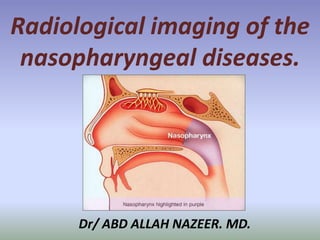
  • 1. Dr/ ABD ALLAH NAZEER. MD. Radiological imaging of the nasopharyngeal diseases.
  • 2.
  • 3.
  • 4.
  • 5.
  • 6.
  • 7.
  • 8.
  • 9.
  • 11.
  • 12.
  • 13.
  • 14.
  • 15. Nasopharyngeal abscess (MRI scan, sagittal view). Nasopharyngeal abscess (MRI scan, axial view).
  • 16. Latero and Retropharyngeal abscess (CT scan, axial view). B. Latero and Retropharyngeal abscess (CT scan, sagittal view). C. Lateropharyngeal abscess (CT scan, coronal view). D. Lateropharyngeal abscess (CT scan, axial view).
  • 17. Inflammatory pseudotumor of the nasopharynx and skull base is a benign, idiopathic disease that is often mistaken for a neoplasm or infection owing to its aggressive behavior and clinical presentation. It can present as a progressively destructive mass and should be considered when repeated tissue biopsies reveal acute or chronic inflammation without evidence of malignant disease or infection. Nasopharyngeal inflammatory pseudotumor with skull base invasion occurring in patients with diabetes mellitus (DM). These patients had repeated negative results from biopsies and cultures, and none had associated cervical lymphadenopathy despite having an aggressive destructive mass. We suggest that these findings, coupled with clinical suspicion, will be helpful in making the correct diagnosis of inflammatory pseudotumor. This is critical in the management of these patients to institute the correct treatment plan.
  • 18. A 32-year-old man with right ptosis, trigeminal neuralgia and facial palsy 2 days after an episode of acute otitis media treated with tympanocentesis. (a) Axial T1 weighted image shows an intermediate signal intensity mass lesion involving the right nasopharyngeal submucosal region (arrows). (b) Axial T2 weighted image shows low signal intensity change of the lesion (arrows). Note the right mastoiditis change. (c) Coronal contrast- enhanced T1 weighted image shows extension of the lesion to the right skull base, cavernous sinus and Meckel’s cave (black arrows). Enhancement of the thickened pachymeninges is also present (white arrows). (d) Axial T2 weighted and (e) contrast- enhanced images obtained 2 months after a course of intravenous high-dose pulse solumedrol therapy followed by oral corticosteroid medication show the mass lesion to be significantly reduced in size. The pachymeningeal enhancement has nearly disappeared
  • 19. A 32-year-old man with left hemicrania of 5 months’ duration and hemitongue atrophy of 1 months’ duration. (a) Axial T1 weighted image and (b) axial T2 weighted image show an ill- defined hypointense lesion in the left nasopharyngeal submucosal region with extension to the carotid space and skull base. The left internal carotid artery is encased and narrowed (white arrow). Involvement of the hypoglossal canal (black arrow) is evident. The mass lesion is distinguishable from the nasopharyngeal mucosa on T2 weighted images. (c) Axial contrast- enhanced T1 weighted image with fat saturation shows moderate enhancement of the left nasopharyngeal lesion, with extension around the left internal carotid artery into the skull base, and involving the prevertebral muscles and hypoglossal canal (black arrow). (d) Axial T2 weighted and (e) contrast enhanced T1 weighted image obtained 2 months after a course of oral corticosteroids show nearly complete resolution of the mass lesion.
  • 20. A 37-year-old man with right hemicrania of 2 months’ duration. (a) Axial T2 weighted image shows hypointensity and (b) axial contrast-enhanced T1 weighted image shows moderate enhancement of the lesion in the right nasopharyngeal submucosa and carotid space (white arrows), which encases the right internal carotid artery with extension to the skull base. There is high signal change on T2 weighted images and abnormal contrast enhancement in the bone marrow of the right occipital condyle (black arrows). (c) Contrast-enhanced MR angiography shows segmental narrowing of the right upper cervical internal carotid artery (white arrows) owing to the encasement by the carotid space lesion.
  • 21.
  • 22.
  • 25.
  • 26.
  • 27.
  • 28.
  • 31.
  • 32. Neonatal teratomas are very rare congenital tumors which are formed from cells derived from all three embryonic germ cell layers. The incidence of neonatal teratomas is between 1:20,000 and 1:80,000 live births. Most occur in the sacrococcygeal region and only 5% are found in the head and neck region, specifically the cervical region followed by the nasopharynx. They can be associated with a cleft palate, intracranial abnormalities including hemicrania and anencephaly, other head and neck anomalies, diaphragmatic hernia and renal and vertebral anomalies. Nasopharyngeal teratomas most commonly present with difficulties in swallowing and obstructive respiratory distress. The size and location serve as the major causes of morbidity and mortality. Rapid airway stabilization, including endotracheal intubation, cricothyrotomy or tracheotomy, is crucial in the management of these patients. After securing the airway, imaging is very useful to help differentiate this tumor from other masses. Ultimately surgical excision is the mode of treatment. These tumors are often encapsulated or pseudoencapsulated which yields excellent surgical prognosis. The differential diagnosis for neonatal nasopharyngeal lesions is extensive and includes: teratoma, dermoid, craniopharyngioma, encephalocele, cystic hygroma, rhabdomyoscarcoma, hemangioma, lipoma, chordoma, neurofibroma, Rathke’s pouch and sarcoma botryoides.
  • 35. (a) T2 weighted sequence in coronal plane showing the external portion of the tumour consisting of lobulated well defined areas of fluid, solid tissue and fat, in keeping with teratoma. There is involvement of the left temporal lobe of the brain (red arrow); (b) T1 weighted contrast enhanced sequence in sagittal plane. Internally, the tumour is occupying the oropharynx, nasopharynx and oral cavity, effacing normal anatomy (red arrows).
  • 36. Pleomorphic adenoma of the nasopharynx.
  • 38. Chordoma is a low-grade malignant tumor that arises from the remnants of notochord. They account for 1-4% of malignant bone tumors and most commonly present after the age of 30 years. Male to female ratio is 1.8:1. Majority occur in sacrum or in the clivus (a bony surface in the posterior cranial fossa sloping upward from foramen magnum to dorsum sella). Those located in the clivus are often associated with a chronic headache and symptoms due to compression of a cranial nerve. Lateral extension can lead to a cerebellopontine angle tumor symptomatology. In case of inferior spread, nasal obstruction, bleeding, and even nasal mass appear.
  • 39. Extraosseous Chordoma of the Nasopharynx.
  • 41. Two cases of cystic choristoma along the nasopharynx and the prestyloid spaces.
  • 42. Paraganglioma of the nasopharynx.
  • 43. Naso and parapharyngeal para-ganglioma.
  • 44. Nasopharynx cancer or nasopharyngeal carcinoma (NPC) is the most common cancer originating in the nasopharynx, the uppermost region of the pharynx ("throat"), behind the nose where the nasal passages and auditory tubes join the remainder of the upper respiratory tract. NPC occurs in children and adults. NPC differs significantly from other cancers of the head and neck in its occurrence, causes, clinical behavior, and treatment. It is vastly more common in certain regions of East Asia and Africa than elsewhere, with viral, dietary and genetic factors implicated in its causation. It is most common in males. It is a squamous cell carcinoma or an undifferentiated type. Squamous cells are a flat type of cell found in the skin and the membranes that line some body cavities. Differentiation means how different the cancer cells are from normal cells. Undifferentiated is a word used to describe cells that do not have their mature features or functions.
  • 45. Classification: Nasopharyngeal carcinoma, commonly known as nasopharyngeal cancer, is classified as a malignant neoplasm, or cancer, arising from the mucosal epithelium of the nasopharynx, most often within the lateral nasopharyngeal recess or fossa of Rosenmüller (a recess behind the entrance of the Eustachian tube opening). The World Health Organization classifies nasopharyngeal carcinoma in three types. Type 1 (I) is squamous cell carcinoma. Type 2a (II) is keratinizing undifferentiated carcinoma. Type 2b (III) is nonkeratinizing undifferentiated carcinoma. Type 2b (III) nonkeratinizing undifferentiated form also known as lymphoepithelioma is most common, and is most strongly associated with Epstein-Barr virus infection of the cancerous cells.
  • 46.
  • 47.
  • 48.
  • 49.
  • 50. Axial T2 weighted images shows a carcinoma that has infiltrated lateral to the tensor and levator veli palatini. Solid arrow shows normal tensor veli palatini on the right. Dashed arrow shows the abnormal left levator muscle which is being displaced and the presence of tumor lateral to it.
  • 51.
  • 52.
  • 53. Sagittal T1 image that shows NPC invasion into the clivus. There is a loss of the inferior cortical margin of the clivus. The clivus is also of uniform lower signal that reflects sclerotic change. 10B is as coronal T1 image in another patient that shows NPC infiltration into the left aspect of the clivus (arrow).
  • 54. Coronal T2 (left) and post gadolinium enhanced T1 (right) weighted images show superior extension of NPC through the floor of the sphenoid sinus (arrows).
  • 55.
  • 56.
  • 57. Two cases of nasopharyngeal tumour, extended laterally into the left masticator space and subsequent perineural tumor tracking superiorly along V3 into the left cavernous sinus.
  • 58. Carcinoma of the nasopharynx. Coronal T1 weighted, fat suppressed, contrast enhanced MR image shows an enhancing lesion(T) in the nasopharynx with extension into bilateral cavernous sinuses(white arrows) via foramen ovale (black arrows). Both cavernous internal carotid arteries are encased by the lesion. There is also extension into left infratemporal fossa(small open black arrow).
  • 59.
  • 60. A case of locally advanced nasopharyngeal carcinoma in a 44-year-old man with right-ear tinnitus and hearing impairment of 1 year’s duration. (a)AxialT1weighted image shows a bulging right nasopharyngeal mass with extension to the skull base, partially encasing the right internal carotid artery. The right parapharyngeal fat is obliterated. (b) Axial T2 weighted image shows mild hyperintensity of the right nasopharyngeal mass. The signal intensity of the mass is similar to that of the nasopharyngeal mucosa, and the margin between the normal mucosa and the mass could not be delineated. Note the presence of right mastoiditis change. (c) Axial and (d) coronal contrast enhanced T1 weighted images with fat saturation shows vivid enhancement of the nasopharyngeal mass. There is skull-base and cavernous sinus invasion with encasement of the right internal carotid artery, which is not narrowed (white arrows). Bilateral neck lymphadenopathy (black arrows) is prominent.
  • 61. Burkett lymphoma of the nasopharynx.
  • 63. Granulocytic sarcoma in the right lateral wall of the nasopharynx (black arrow), involving the right pterygoid muscles, parapharyngeal space Figure 1b: CT sagittal image shows mass in the nasopharynx with extension into sphenoid sinus Figure 1c: CT coronal image shows that the mass is confined to the nasopharynx without intracranial extension.
  • 64. Leiomyosarcoma in the Nasopharynx: