PTPM008B PTM of Oncology and Palliative Care-related Medic…
•
2 likes•2,008 views

PTPM008B PTM of Oncology and Palliative Care-related Medic…

Recommended
Recommended
More Related Content
Viewers also liked
Viewers also liked (19)
More from Abdul Rehman S Mulla
More from Abdul Rehman S Mulla (12)
Recently uploaded
Recently uploaded (20)
PTPM008B PTM of Oncology and Palliative Care-related Medic…
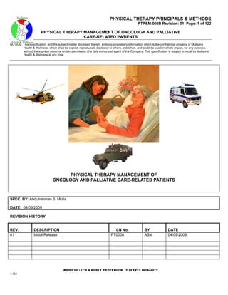
- 1. PHYSICAL THERAPY PRINCIPALS & METHODS PTP&M:008B Revision: 01 Page: 1 of 122 PHYSICAL THERAPY MANAGEMENT OF ONCOLOGY AND PALLIATIVE CARE-RELATED PATIENTS NOTICE: This specification, and the subject matter disclosed therein, embody proprietary information which is the confidential property of Mullsons Health & Wellness, which shall be copied, reproduced, disclosed to others, published, and could be used in whole or part, for any purpose, without the express advance written permission of a duly authorized agent of the Company. This specification is subject to recall by Mullsons Health & Wellness at any time. PHYSICAL THERAPY MANAGEMENT OF ONCOLOGY AND PALLIATIVE CARE-RELATED PATIENTS SPEC. BY: Abdulrehman S. Mulla DATE: 04/09/2009 REVISION HISTORY REV. DESCRIPTION CN No. BY DATE 01 Initial Release PT0008 ASM 04/09/2009 Medicine: it’s a noble profession, it serves humanity 1/122
- 2. PHYSICAL THERAPY PRINCIPALS & METHODS PTP&M:008B Revision: 01 Page: 2 of 122 PHYSICAL THERAPY MANAGEMENT OF ONCOLOGY AND PALLIATIVE CARE-RELATED PATIENTS NOTICE: This specification, and the subject matter disclosed therein, embody proprietary information which is the confidential property of Mullsons Health & Wellness, which shall be copied, reproduced, disclosed to others, published, and could be used in whole or part, for any purpose, without the express advance written permission of a duly authorized agent of the Company. This specification is subject to recall by Mullsons Health & Wellness at any time. TABLE OF CONTENTS PAGE 5.0 BREAST CANCER: 7 5.1 NONINVASIVE BREAST CANCER: 8 5.2 INVASIVE BREAST CANCER: 8 5.3 RISK FACTORS: 8 5.3.1 RISK FACTORS FOR BREAST CANCER INCLUDE: 9 A. AGE: 9 B. RACE AND ETHNICITY: 9 C. FAMILY AND PERSONAL HISTORY: 10 D. GENETIC FACTORS: 10 E. EXPOSURE TO ESTROGEN: 10 F. BREAST ABNORMALITIES: 11 5.3.3 PHYSICAL CHARACTERISTICS: 13 A. ENVIRONMENTAL FACTORS: 13 I. EXPOSURE TO ESTROGEN-LIKE INDUSTRIAL CHEMICALS. 13 II. EXPOSURE TO DIETHYLSTILBESTROL. 13 III. RADIATION EXPOSURE. 13 IV. DISPROVEN RISK FACTORS: 13 5.4 SYMPTOMS: 13 5.5 DIAGNOSIS: 14 5.5.1 MONTHLY SELF-EXAMINATION: 14 5.6 MAMMOGRAMS: 15 5.6.1 CURRENT RECOMMENDATIONS FOR SCREENING: 15 5.6.2 OTHER IMAGING TECHNIQUES: 16 A. MAGNETIC RESONANCE IMAGING AND ULTRASOUND: 16 B. SCINTIMAMMOGRAPHY: 18 5.6.3 BIOPSY: 18 5.7 LYMPHADENECTOMY: 20 5.7.1 SENTINEL NODE BIOPSY: 20 5.8 PROGNOSIS: 21 5.8.1 LOCATION OF THE TUMOR: 21 5.8.2 HORMONE RECEPTOR-POSITIVE OR –NEGATIVE: 22 5.8.3 TUMOR MARKERS: 22 5.8.4 GENE EXPRESSION PROFILING: 23 5.8.5 OTHER FACTORS FOR PREDICTING OUTLOOK: 23 A. TUMOR SIZE AND SHAPE: 23 B. RATE OF CELL DIVISION: 23 5.9 EFFECT OF EMOTIONS AND PSYCHOLOGICAL SUPPORT: 23 5.9.1 TREATMENT: 23 A. LOCAL TREATMENT: 24 B. SYSTEMIC TREATMENT: 24 C. CANCER STAGE AND TREATMENT OPTIONS: 24 D. TREATMENT OPTIONS FOR DCIS INCLUDE: 25 E. TREATMENT OPTIONS FOR LCIS INCLUDE: 25 I. STAGE I AND II (EARLY-STAGE INVASIVE): 26 II. STAGE II CANCER IS CLASSIFIED AS EITHER STAGE IIA OR STAGE IIB: 26 1. Treatment options for stage i and stage ii breast cancer may include: 26 2. Stage iii (locally advanced): 26 a. In stage iiia breast cancer, the tumor is either: 26 3. Treatment options for stage iiia breast cancer are the same as those for stages i and ii. 26 a. Stage iiib: 26 b. Stage iiic: 27 c. Treatment options for operable stage iii breast cancer: 27 5. Stage iv (advanced cancer): 27 a. Treatment options for stage iv cancer include: 28 5.10 POST-TREATMENT CARE: 28 5.10.1 GENETIC COUNSELING MAY BE HELPFUL IF YOU HAVE: 29 5.10.2 PREGNANCY AFTER BREAST CANCER TREATMENT: 29 Medicine: it’s a noble profession, it serves humanity 2/122
- 3. PHYSICAL THERAPY PRINCIPALS & METHODS PTP&M:008B Revision: 01 Page: 3 of 122 PHYSICAL THERAPY MANAGEMENT OF ONCOLOGY AND PALLIATIVE CARE-RELATED PATIENTS NOTICE: This specification, and the subject matter disclosed therein, embody proprietary information which is the confidential property of Mullsons Health & Wellness, which shall be copied, reproduced, disclosed to others, published, and could be used in whole or part, for any purpose, without the express advance written permission of a duly authorized agent of the Company. This specification is subject to recall by Mullsons Health & Wellness at any time. 5.11 RECURRENT BREAST CANCER: 29 5.12 SURGERY: 30 5.12.1 BREAST-CONSERVING PROCEDURES: 30 A. LUMPECTOMY: 30 B. BREAST-CONSERVING SURGERY: 30 5.12.2 MASTECTOMY: 30 5.12.3 COMPLICATIONS AND SIDE EFFECTS OF SURGERY: 31 5.13 RADIATION: 32 5.13.1 ADMINISTRATION OF RADIATION THERAPY: 32 A. EXTERNAL BEAM RADIATION: 32 B. BRACHYTHERAPY: 32 C. SIDE EFFECTS OF RADIATION THERAPY: 33 D. LONG-TERM COMPLICATIONS: 33 5.14 CHEMOTHERAPY: 33 5.14.1 SIDE EFFECTS OF CHEMOTHERAPY: 34 5.14.2 HIGH-DOSE CHEMOTHERAPY WITH BONE MARROW OR PERIPHERAL-BLOOD STEM CELL TRANSPLANTATION: 35 A. HORMONE THERAPY: 35 B. TAMOXIFEN AND SELECTIVE ESTROGEN RECEPTOR MODULATORS (SERMS): 35 5.15 AROMATASE INHIBITORS: 36 5.16 OVARIAN ABLATION: 36 5.16.1 CHEMICAL OVARIAN ABLATION: 36 5.16.2 BILATERAL OOPHORECTOMY: 37 5.17 AFTER IMPLANT SURGERY: 37 5.18 AFTER YOU LEAVE: 38 5.19 CALL YOUR DOCTOR IF: 39 5.20 CANCER REHABILITATION: 39 5.20.1 LYMPHEDEMA REHABILITATION: 40 5.20.2 EXERCISES AFTER BREAST SURGERY: 40 6.0 LUNG CANCER: 43 6.1 SYMPTOMS: 43 6.2 CAUSES: 43 6.2.1 SMOKING CAUSES LUNG CANCER: 44 6.3 TYPES OF LUNG CANCER: 44 6.4 RISK FACTORS 44 6.5 TESTS AND DIAGNOSIS: 45 6.5.1 SCREENING: 45 6.5.2 DIAGNOSIS: 45 6.5.3 STAGING: 47 A. STAGES OF NON-SMALL CELL LUNG CANCER: 47 I. Stage I. 47 II. Stage II. 47 III. Stage IIIA. 47 IV. Stage IIIB. 47 V. Stage IV. 48 B. STAGES OF SMALL CELL LUNG CANCER: 49 6.6 COMPLICATIONS: 49 6.7 TREATMENTS AND DRUGS: 50 6.7.1 SURGERY: 51 6.7.2 CHEMOTHERAPY: 51 6.7.3 RADIATION THERAPY: 52 6.7.4 TARGETED DRUG THERAPY: 52 6.7.5 CLINICAL TRIALS: 53 6.7.6 SUPPORTIVE (PALLIATIVE) CARE: 53 6.8 PREVENTION: 53 6.9 ALTERNATIVE MEDICINE: 54 6.10 PHYSICAL THERAPY AFTER LUNG CANCER: 55 6.10.1 AEROBIC EXERCISE AS THERAPY FOR CANCER FATIGUE: 55 A. TRAINING PROGRAM: 56 7.0 PROSTATE CANCER: 58 7.1 SYMPTOMS: 59 Medicine: it’s a noble profession, it serves humanity 3/122
- 4. PHYSICAL THERAPY PRINCIPALS & METHODS PTP&M:008B Revision: 01 Page: 4 of 122 PHYSICAL THERAPY MANAGEMENT OF ONCOLOGY AND PALLIATIVE CARE-RELATED PATIENTS NOTICE: This specification, and the subject matter disclosed therein, embody proprietary information which is the confidential property of Mullsons Health & Wellness, which shall be copied, reproduced, disclosed to others, published, and could be used in whole or part, for any purpose, without the express advance written permission of a duly authorized agent of the Company. This specification is subject to recall by Mullsons Health & Wellness at any time. 7.2 CAUSES: 60 7.3 RISK FACTORS: 60 7.4 WHEN TO SEEK MEDICAL ADVICE: 61 7.5 TESTS AND DIAGNOSIS: 61 7.5.1 DETERMINING HOW FAR THE CANCER HAS SPREAD: 61 7.5.2 GRADING: 63 7.5.3 STAGING: 63 A. STAGE I. 63 B. STAGE II. 63 C. STAGE III. 63 D. STAGE IV. 63 7.6 COMPLICATIONS: 64 7.6.1 THE TYPICAL COMPLICATIONS OF PROSTATE CANCER AND ITS TREATMENTS INCLUDE: 64 A. SPREAD OF CANCER: 64 B. PAIN: 64 C. DIFFICULTY URINATING (URINARY INCONTINENCE): 64 D. ERECTILE DYSFUNCTION (ED) OR IMPOTENCE: 64 E. DEPRESSION: 64 7.7 TREATMENTS AND DRUGS: 65 7.6.1 EXTERNAL BEAM RADIATION THERAPY (EBRT): 65 A. SIDE EFFECTS OF EBRT: 65 7.6.2 RADIOACTIVE SEED IMPLANTS: 66 A. SIDE EFFECTS OF RADIOACTIVE SEED IMPLANTS: 66 7.6.3 HORMONE THERAPY: 68 A. SIDE EFFECTS OF HORMONE THERAPY: 68 B. CERTAIN HORMONE THERAPY MEDICATIONS CAN ALSO CAUSE: 68 7.6.4 RADICAL PROSTATECTOMY: 69 A. SIDE EFFECTS OF RADICAL PROSTATECTOMY: 70 7.6.5 ROBOT-ASSISTED LAPAROSCOPIC RADICAL PROSTATECTOMY (RALRP): 70 7.6.6 WATCHFUL WAITING: 70 7.6.7 CHEMOTHERAPY: 71 7.6.8 CRYOTHERAPY: 71 7.6.9 GENE THERAPY AND IMMUNE THERAPY: 72 7.7 PREVENTION: 72 7.8 COPING AND SUPPORT: 73 7.9 ALTERNATIVE MEDICINE: 73 7.10 PHYSICAL THERAPY AND EXERCISE BEFORE AND AFTER PROSTATE CANCER: 75 7.10.1 PHYSICAL THERAPY / EXERCISE PROGRAM: 75 7.10.2 MANUAL THERAPY: 76 7.10.3 SEVEN BENEFITS OF REGULAR PHYSICAL ACTIVITY: 77 7.10.4 BLADDER CANCER REHABILITATION: 79 A. PHYSICAL THERAPY / EXERCISE PROGRAM: 79 I. BORG RATING OF PERCEIVED EXERTION (RPE): 79 II. FLEXIBILITY PROGRAM: 80 III. THE BENEFITS OF STRETCHING: 80 IV. PELVIC FLOOR EXERCISES FOR WOMEN: 80 1. PELVIC FLOOR EXERCISE ONE: 80 2. PELVIC FLOOR EXERCISE TWO: 80 V. PELVIC FLOOR EXERCISES FOR MEN: 81 1. HOW TO DO THE EXERCISES: 81 2. HOW LONG TO CONTINUE WITH THE EXERCISES: 82 8.0 LEUKEMIA: 84 8.1 TYPES OF LEUKEMIA: 84 8.1.1 COMMON TYPES OF LEUKEMIA: 85 8.2 LEUKEMIA RISKS 86 8.2.1 RISK FACTORS FOR LEUKEMIA: 86 8.3 SYMPTOMS OF LEUKEMIA: 86 8.3.1 COMMON SYMPTOMS OF LEUKEMIA: 86 8.4 LEUKEMIA DIAGNOSES? 87 8.4.1 DIFFERENTIAL DIAGNOSIS 88 Medicine: it’s a noble profession, it serves humanity 4/122
- 5. PHYSICAL THERAPY PRINCIPALS & METHODS PTP&M:008B Revision: 01 Page: 5 of 122 PHYSICAL THERAPY MANAGEMENT OF ONCOLOGY AND PALLIATIVE CARE-RELATED PATIENTS NOTICE: This specification, and the subject matter disclosed therein, embody proprietary information which is the confidential property of Mullsons Health & Wellness, which shall be copied, reproduced, disclosed to others, published, and could be used in whole or part, for any purpose, without the express advance written permission of a duly authorized agent of the Company. This specification is subject to recall by Mullsons Health & Wellness at any time. 8.5 LEUKEMIA TREATMENT: 89 8.5.1 GETTING A SECOND OPINION: 89 8.5.2 PREPARING FOR TREATMENT: 89 8.5.3 METHODS OF TREATMENT: 90 A. CHEMOTHERAPY: 90 B. BIOLOGICAL THERAPY: 92 C. RADIATION THERAPY: 93 D. STEM CELL TRANSPLANTATION: 93 8.5.4 SIDE EFFECTS OF TREATMENT FOR LEUKEMIA: 97 A. CHEMOTHERAPY: 97 B. BIOLOGICAL THERAPY: 98 C. RADIATION THERAPY: 98 D. STEM CELL TRANSPLANTATION: 98 8.5.5 WHAT HAPPENS AFTER TREATMENT FOR LEUKEMIA? 98 A. SUPPORTIVE CARE: 98 B. NUTRITION: 99 C. FOLLOWUP CARE: 99 D. SUPPORT FOR PEOPLE WITH LEUKEMIA: 99 8.5.6 WHAT DOES THE FUTURE HOLD FOR PATIENTS WITH LEUKEMIA?: 100 8.6 LEUKEMIA AT A GLANCE: 100 8.7 PHYSICAL THERAPY INTERVENTIONS: 100 8.7.1 EVIDENCE: 100 A. MAKING THERAPY FUN: 101 I. RECOMMENDED INTERVENTION: 101 II. PHYSICAL THERAPY PRECAUTIONS: 101 III. PSYCHOSOCIAL MANIFESTATIONS: 101 IV. OTHER INFORMATION: 102 9.0 LYMPHOMA: 103 9.1 LYMPHOMA CAUSES: 106 9.2 LYMPHOMA SYMPTOMS: 107 9.2.1 SYMPTOMS OF LYMPHOMA MAY INCLUDE THE FOLLOWING: 107 9.3 WHEN TO SEEK MEDICAL CARE: 107 9.4 EXAMS AND TESTS: 107 9.4.1 BLOOD TESTS: 108 A. BIOPSY: 108 B. IMAGING STUDIES: 109 C. BONE MARROW EXAMINATION: 109 D. OTHER TESTS: 109 9.4.2 STAGING: 110 9.4.3 PROGNOSTIC FACTORS: 111 9.5 LYMPHOMA TREATMENT: 111 9.5.1 MEDICAL TREATMENT: 113 A. RADIATION THERAPY: 113 B. CHEMOTHERAPY: 113 I. CHEMOTHERAPY PILLS: 113 II. DRUG COMBINATIONS; 114 III. CHEMOTHERAPY CYCLES. 114 C. BIOLOGICAL THERAPY: 114 I. MONOCLONAL ANTIBODIES: 114 II. CYTOKINES: 114 III. VACCINES: 115 9.5.2 MEDICATIONS: 115 9.5.3 OTHER THERAPIES: 115 A. WATCHFUL WAITING: 115 B. CLINICAL TRIALS: 116 C. COMPLEMENTARY/ALTERNATIVE THERAPIES: 116 9.6 NEXT STEPS: 116 9.7 PREVENTION: 116 9.8 OUTLOOK: 117 9.9 SUPPORT GROUPS AND COUNSELLING: 117 Medicine: it’s a noble profession, it serves humanity 5/122
- 6. PHYSICAL THERAPY PRINCIPALS & METHODS PTP&M:008B Revision: 01 Page: 6 of 122 PHYSICAL THERAPY MANAGEMENT OF ONCOLOGY AND PALLIATIVE CARE-RELATED PATIENTS NOTICE: This specification, and the subject matter disclosed therein, embody proprietary information which is the confidential property of Mullsons Health & Wellness, which shall be copied, reproduced, disclosed to others, published, and could be used in whole or part, for any purpose, without the express advance written permission of a duly authorized agent of the Company. This specification is subject to recall by Mullsons Health & Wellness at any time. 9.10 PHYSICAL THERAPY INTERVENTIONS FOR LYMPHOMA: 117 9.10.1 PHYSICAL EVALUATION: 117 A. PHYSICAL THERAPY / EXERCISE PROGRAM: 118 B. BORG RATING OF PERCEIVED EXERTION (RPE) - 118 C. FLEXIBILITY PROGRAM: 118 D. MANUAL THERAPY: 118 10.0 PALLIATIVE CARE: 119 10.1 THE ROLE OF PHYSIOTHERAPY IN PALLIATIVE CARE: 120 10.1.1 CONDITIONS SEEN IN PALLIATIVE CARE: 120 10.1.2 AIMS OF PHYSIOTHERAPY: 121 Medicine: it’s a noble profession, it serves humanity 6/122
- 7. PHYSICAL THERAPY PRINCIPALS & METHODS PTP&M:008B Revision: 01 Page: 7 of 122 PHYSICAL THERAPY MANAGEMENT OF ONCOLOGY AND PALLIATIVE CARE-RELATED PATIENTS NOTICE: This specification, and the subject matter disclosed therein, embody proprietary information which is the confidential property of Mullsons Health & Wellness, which shall be copied, reproduced, disclosed to others, published, and could be used in whole or part, for any purpose, without the express advance written permission of a duly authorized agent of the Company. This specification is subject to recall by Mullsons Health & Wellness at any time. 5.0 BREAST CANCER: Breast cancers are potentially life-threatening malignancies that develop in one or both breasts. The structure of the female breast is important in understanding this cancer: The interior of the female breast consists mostly of fatty and fibrous connective tissues. It is divided into about 20 sections called lobes. Each lobe is further subdivided into a collection of lobules, structures that contain small milk-producing glands. These glands secrete milk into a complex system of tiny ducts. The ducts carry the milk through the breast and converge in a collecting chamber located just below the nipple. Breast cancer is either noninvasive (referred to as in situ, confined to the site of origin) or invasive (spreading). Medicine: it’s a noble profession, it serves humanity 7/122
- 8. PHYSICAL THERAPY PRINCIPALS & METHODS PTP&M:008B Revision: 01 Page: 8 of 122 PHYSICAL THERAPY MANAGEMENT OF ONCOLOGY AND PALLIATIVE CARE-RELATED PATIENTS NOTICE: This specification, and the subject matter disclosed therein, embody proprietary information which is the confidential property of Mullsons Health & Wellness, which shall be copied, reproduced, disclosed to others, published, and could be used in whole or part, for any purpose, without the express advance written permission of a duly authorized agent of the Company. This specification is subject to recall by Mullsons Health & Wellness at any time. 5.1 NONINVASIVE BREAST CANCER: Noninvasive breast cancers include: Ductal carcinoma in situ (also called intraductal carcinoma or DCIS). DCIS consist of cancer cells in the lining of the duct. DCIS is a non-invasive, early cancer, but if left untreated, it may sometimes progress to an invasive, infiltrating ductal breast cancer. DCIS is the most common type of noninvasive breast cancer. Lobular carcinoma in situ, or LCIS. Although it is technically not a cancer, lobular carcinoma in situ is a marker for an increased risk of invasive cancer the same or both breasts. A diagnosis of these early cancers (DCIS and LCIS) is made when there is no evidence of invasion. 5.2 INVASIVE BREAST CANCER: Invasive cancer occurs when cancer cells spread beyond the basement membrane, which covers the underlying connective tissue in the breast. This tissue is rich in blood vessels and lymphatic channels that are capable of carrying cancer cells beyond the breast. Invasive breast cancers include the following: Invasive (also called infiltrating) ductal carcinoma. This is invasive breast cancer that penetrates the wall of a milk-passage duct. It comprises between 70 - 80% of all breast cancer cases. Invasive (also called infiltrating) lobular carcinoma. This invasive cancer has spread through the wall of a milk-producing lobule. It accounts for 10 - 15% of all breast cancers. It may sometimes appear in both breasts, sometimes in several separate locations. 5.3 RISK FACTORS: Breast cancer is the second leading cause of cancer deaths in women today (after lung cancer) and is the most common cancer among women, excluding nonmelanoma skin cancers. According to the American Cancer Society, about 1.3 million women will be diagnosed with breast cancer annually worldwide about 465,000 will Medicine: it’s a noble profession, it serves humanity 8/122
- 9. PHYSICAL THERAPY PRINCIPALS & METHODS PTP&M:008B Revision: 01 Page: 9 of 122 PHYSICAL THERAPY MANAGEMENT OF ONCOLOGY AND PALLIATIVE CARE-RELATED PATIENTS NOTICE: This specification, and the subject matter disclosed therein, embody proprietary information which is the confidential property of Mullsons Health & Wellness, which shall be copied, reproduced, disclosed to others, published, and could be used in whole or part, for any purpose, without the express advance written permission of a duly authorized agent of the Company. This specification is subject to recall by Mullsons Health & Wellness at any time. die from the disease. Breast cancer death rates have been dropping steadily since 1990, according to the Society, because of earlier detection and better treatments. About 40,910 breast cancer deaths are expected in 2007. According to the American Cancer Society, in general, breast cancer rates have risen about 30% in the past 25 years in western countries, due in part to increased screening which detects the cancer in earlier stages. In the United States, though, breast cancer rates decreased by 10% between 2000-2004, due in part to a reduction in the use of hormone replacement therapy. Although breast cancer rates are rising in many western countries, deaths from the disease have decreased in some countries as a result of improved screening and treatment. The lifetime probability of developing breast cancer in developed countries is about 4.8%, according to the American Cancer Society (the probability is about 13% for any type of cancer). In developing countries, the lifetime probability of developing breast cancer is about 1.8%. Breast Cancer Worldwide Breast (All ages) Incidence Deaths China Zimbabwe India Japan Brazil Singapore Italy Switzerland Australia Canada Netherlands UK Sweden Denmark France United States 5.3.1 RISK FACTORS FOR BREAST CANCER INCLUDE: A. AGE: Most cases of breast cancer occur in women older than age 60. According to the American Cancer Society, about 1 in 8 cases of invasive breast cancer are found in women younger than age 45, while 2 in 3 cases of invasive breast cancer occur in women age 55 and older. B. RACE AND ETHNICITY: Breast cancer is slightly more common among white woman than African-American, Asian, Latina, or Native American women. However, African-American women tend to have more aggressive types of breast cancer tumors and are more likely to die from breast cancer than women of other races. It is unclear whether this is mainly due to biologic or socioeconomic reasons. Social and economic factors make it less likely that African-American women will be screened, so they are Medicine: it’s a noble profession, it serves humanity 9/122
- 10. PHYSICAL THERAPY PRINCIPALS & METHODS PTP&M:008B Revision: 01 Page: 10 of 122 PHYSICAL THERAPY MANAGEMENT OF ONCOLOGY AND PALLIATIVE CARE-RELATED PATIENTS NOTICE: This specification, and the subject matter disclosed therein, embody proprietary information which is the confidential property of Mullsons Health & Wellness, which shall be copied, reproduced, disclosed to others, published, and could be used in whole or part, for any purpose, without the express advance written permission of a duly authorized agent of the Company. This specification is subject to recall by Mullsons Health & Wellness at any time. more likely to be diagnosed at a later stage. They are also less likely to have access to effective treatments. Breast cancer is also more prevalent among Jewish women of Eastern European (Ashkenazi) descent (see Genetic Factors). C. FAMILY AND PERSONAL HISTORY: Women who have a family history of breast cancer are at increased risk for developing breast cancer themselves. Having a first-degree relative (mother, sister, or daughter) who has been diagnosed with breast cancer doubles the risk for developing breast cancer. Women who have had ovarian cancer are at increased risk for developing breast cancer. And, a personal history of breast cancer increases the risk of developing a new cancer in the same or other breast. D. GENETIC FACTORS: About 5 - 10% of breast cancer cases are due to inherited genetic mutations. BRCA Genes. Inherited mutations in genes known as BRCA1 or BRCA2 are responsible for most cases of hereditary breast cancers, ovarian cancers, or both in families with a history of these cancers. BRCA gene mutations are present in only about 0.5% of the overall population. However, certain ethnic groups -- such as Jewish women of Eastern European (Ashkenazi) descent -- have a higher prevalence (2.5%) of BRCA gene mutations. BRCA gene mutations are also seen in some African-American and Hispanic women. Screening Guidelines for BRCA Genes. The U.S. Preventive Services Task Force (USPSTF) recommends that women at high risk should be tested for BRCA genes, but does not recommend routine genetic counseling or testing in low-risk women (no family history of BRCA 1 or 2 genetic mutations). Risk assessment is based on a womans family history of breast and ovarian cancer (on both the maternal and paternal sides). The relevance of the inherited BRCA1 or BRCA2 mutations to survival is controversial. Some studies have suggested that these mutations are linked to less lethal breast cancer. Others suggest that they do not change prognosis or may worsen it. Women with these genetic mutations do have a greater risk for a new cancer to develop. Patients with BRCA1 mutations tend to develop tumors that are hormone receptor negative, which can behave more aggressively. Other Genetic Mutations. Other genes associated with increased hereditary breast cancer risk include p53, CHEK2, ATM, and PTEN. E. EXPOSURE TO ESTROGEN: Because growth of breast tissue is highly sensitive to estrogens, the more estrogen a woman is exposed to over her lifetime, the higher her risk for breast cancer. Duration of Estrogen Exposure. Early age at menarche (first menstrual period) or later age at menopause may slightly increase a womans risk for breast cancer. Pregnancy. Women who have never had children or who had their first child after age 30 may have a slightly increased breast cancer risk. Having children at an early age, and having multiple pregnancies, reduces breast cancer risk. Evidence does not show an association between abortion and increased breast cancer risk. Studies have been mixed on whether breast-feeding decreases breast cancer risk. Breast- feeding reduces a womans total number of menstrual cycles, and thereby estrogen exposure, Medicine: it’s a noble profession, it serves humanity 10/122
- 11. PHYSICAL THERAPY PRINCIPALS & METHODS PTP&M:008B Revision: 01 Page: 11 of 122 PHYSICAL THERAPY MANAGEMENT OF ONCOLOGY AND PALLIATIVE CARE-RELATED PATIENTS NOTICE: This specification, and the subject matter disclosed therein, embody proprietary information which is the confidential property of Mullsons Health & Wellness, which shall be copied, reproduced, disclosed to others, published, and could be used in whole or part, for any purpose, without the express advance written permission of a duly authorized agent of the Company. This specification is subject to recall by Mullsons Health & Wellness at any time. which may account for its possible protective effects. Some studies suggest that the longer a woman breast-feeds, the lower her risk. Oral Contraception. Although studies have been conflicting about whether estrogen in oral contraception increases the chances for breast cancer, the most recent research indicates that current or former oral contraceptive use does not significantly increase breast cancer risk. Women who have used oral contraceptives may have slightly more risk for breast cancer than women who have never used them, but this risk declines once a woman stops using birth control pills. Hormone Replacement Therapy. Many studies have reported a higher risk for breast cancer in postmenopausal women who take combination hormone replacement therapy (HRT), which contains both estrogen and progestin. A combination of estrogen and testosterone also increases breast cancer risk. Several studies of women who had a hysterectomy indicate that estrogen alone does not increase overall breast cancer risk when the drug is used for 7 years or less. However, women who take estrogen for 10 - 15 years or more do have an increased risk, especially women who are already at higher risk for breast cancer. In addition, HRT increases breast cancer density, making mammograms more difficult to read. This can cause cancer to be diagnosed at a later stage. Women who take estrogen HRT should be aware that they need frequent mammogram screenings. The North American Menopause Society recommends that women who are at risk for breast cancer should avoid hormone therapy and try other options to manage menopausal symptoms such as hot flashes. Most doctors recommend that women use HRT only for short-term relief of menopausal symptoms. [For more information, see In-Depth Report #40: Menopause.] Infertility and Infertility Treatments. Despite some concerns that infertility treatments using the drug clomiphene may increase the risk for breast cancer, most studies do not show an association. Some studies indicate that ovulation induction with clomiphene may actually decrease breast cancer risk. (Clomphine is related to tamoxifen, a drug that is used for breast cancer prevention in high-risk women.) F. BREAST ABNORMALITIES: Abnormalities or Breast Conditions Suggesting a Higher Risk. Certain factors and breast conditions may increase the risk for breast cancer: Dense breast tissue is associated with a higher risk for breast cancer. Studies suggest that women with highly dense tissue have 2 - 6 times the risk of women with the least dense tissue. Genetic factors play a large role in breast density. Hormone replacement therapy also increases breast density. In addition, dense breasts make mammograms more difficult to read, which increases the likelihood of missing early signs of cancer. Benign proliferative breast disease, or unusual cell growth known as atypical hyperplasia, is a significant risk factor for breast cancer. Some common benign breast abnormalities that pose few or no risks include the following: Cysts. These mostly occur in women in their middle-to-late reproductive years and can be eliminated simply by aspirating fluid from them. Medicine: it’s a noble profession, it serves humanity 11/122
- 12. PHYSICAL THERAPY PRINCIPALS & METHODS PTP&M:008B Revision: 01 Page: 12 of 122 PHYSICAL THERAPY MANAGEMENT OF ONCOLOGY AND PALLIATIVE CARE-RELATED PATIENTS NOTICE: This specification, and the subject matter disclosed therein, embody proprietary information which is the confidential property of Mullsons Health & Wellness, which shall be copied, reproduced, disclosed to others, published, and could be used in whole or part, for any purpose, without the express advance written permission of a duly authorized agent of the Company. This specification is subject to recall by Mullsons Health & Wellness at any time. Fibroadenoma. These are solid benign lumps that occur in women ages 15 - 30. Breast abscesses during breast-feeding. Nipple discharge. Discharge from the nipple is worrisome to patients, but is unlikely to be a sign of cancer. Unexplained discharge still warrants evaluation, however. Mastalgia. This is breast pain that occurs in association with, or independently from, the menstrual cycle. About 8 - 10% of women experience moderate-to-severe breast pain associated with their menstrual cycle. In general, breast pain does not need assessment unless it is severe and prolonged. Medicine: it’s a noble profession, it serves humanity 12/122
- 13. PHYSICAL THERAPY PRINCIPALS & METHODS PTP&M:008B Revision: 01 Page: 13 of 122 PHYSICAL THERAPY MANAGEMENT OF ONCOLOGY AND PALLIATIVE CARE-RELATED PATIENTS NOTICE: This specification, and the subject matter disclosed therein, embody proprietary information which is the confidential property of Mullsons Health & Wellness, which shall be copied, reproduced, disclosed to others, published, and could be used in whole or part, for any purpose, without the express advance written permission of a duly authorized agent of the Company. This specification is subject to recall by Mullsons Health & Wellness at any time. 5.3.3 PHYSICAL CHARACTERISTICS: The following physical characteristics have been associated with increased risk: Obesity increases the risk for all types of estrogen receptor-positive breast cancers. Women who gain weight after menopause are most at risk. (On a positive note, losing weight after menopause decreases breast cancer risk.) In postmenopausal women, estrogen is produced in fat tissue. High amounts of fatty tissue increase levels of estrogen in the body, leading to faster growth of estrogen- sensitive cancers. Estrogen is involved in building bone mass. Therefore, women with heavy, dense bones are likely to have higher estrogen levels and to be at greater risk for breast cancer. Some studies have found a greater risk for breast cancer in taller women, possibly due to the higher estrogen levels associated with greater bone growth. A. ENVIRONMENTAL FACTORS: I. EXPOSURE TO ESTROGEN-LIKE INDUSTRIAL CHEMICALS. Chemicals with estrogen-like effects, called xenoestrogens, have been under suspicion for years. There has been particular concern with pesticides containing organochlorines (DDT and its metabolites, such as dieldrin) and pyrethroids (permethrin), but at this time evidence of any causal association is very weak. II. EXPOSURE TO DIETHYLSTILBESTROL. Women who took diethylstilbestrol (DES) to prevent miscarriage have a slightly increased risk for breast cancer. There may also be a slightly increased risk for their daughters (commonly called quot;DES daughtersquot;), who were exposed to the drug when their mothers took it during pregnancy. III. RADIATION EXPOSURE. Heavy exposure to radiation is a significant risk factor for breast cancer. Girls who received high-dose radiation therapy for cancer face an increased risk for breast cancer in adulthood. Low-dose radiation exposure before age 20 may increase the risk for women with BRCA genetic mutations. IV. DISPROVEN RISK FACTORS: Antiperspirants or use of deodorants after shaving have not been linked with any higher risk for breast cancer. There is also no evidence that bras increase breast risk. Abortion does not increase risk. 5.4 SYMPTOMS: Breast cancers in their early stages are usually painless. Often the first symptom is the discovery of a hard lump. Fifty percent of such masses are found in the upper outer quarter of the breast. The lump may make the affected breast appear elevated or asymmetric. The nipple may be retracted or scaly. Sometimes the skin of the breast is dimpled like the skin of an orange. In some cases there is a bloody or clear discharge from the nipple. Many cancers, however, produce no symptoms and cannot be felt on examination. They can be detected only with a mammogram. Breast self-exam Medicine: it’s a noble profession, it serves humanity 13/122
- 14. PHYSICAL THERAPY PRINCIPALS & METHODS PTP&M:008B Revision: 01 Page: 14 of 122 PHYSICAL THERAPY MANAGEMENT OF ONCOLOGY AND PALLIATIVE CARE-RELATED PATIENTS NOTICE: This specification, and the subject matter disclosed therein, embody proprietary information which is the confidential property of Mullsons Health & Wellness, which shall be copied, reproduced, disclosed to others, published, and could be used in whole or part, for any purpose, without the express advance written permission of a duly authorized agent of the Company. This specification is subject to recall by Mullsons Health & Wellness at any time. 5.5 DIAGNOSIS: Breast Examination by a Health Professional. Women ages 20 - 49 should have a physical examination by a health professional every 1 - 2 years. Those over age 50 should be examined annually. Self-Examinations. Women have been encouraged to perform a self-examination each month, but some studies have reported no difference in mortality rates between women who do self-examination and those who do not. This does not mean women should stop attempting self-examinations, but they should not replace the annual examination done by a health professional. 5.5.1 MONTHLY SELF-EXAMINATION: 1. Pick a time of the month that is easy to remember and perform self-examination at that time each month. The breast has normal patterns of thickness and lumpiness that change within a monthly period, and a consistently scheduled examination will help differentiate between what is normal from abnormal. Many doctors recommend breast awareness rather than formal monthly self- examinations. 2. Stand in front of a mirror. Breasts should be basically the same size (one may be slightly larger than the other). Check for changes or redness in the nipple area. Look for changes in the appearance of the skin. With hands on the hips, push the pelvis forward and pull the shoulders back and observe the breasts for irregularities. Repeat the observation with hands behind the head. Move each arm and shoulder forward. 3. Lie down on the back with a rolled towel under one shoulder. Apply lotion or bath oil over the breast area. The finger action should be as follows: Using the 2nd, 3rd, and 4th finger pads (not tips) held together, make dime-sized circles. Press lightly first to feel the breast area, then press harder using a circular motion. Medicine: it’s a noble profession, it serves humanity 14/122
- 15. PHYSICAL THERAPY PRINCIPALS & METHODS PTP&M:008B Revision: 01 Page: 15 of 122 PHYSICAL THERAPY MANAGEMENT OF ONCOLOGY AND PALLIATIVE CARE-RELATED PATIENTS NOTICE: This specification, and the subject matter disclosed therein, embody proprietary information which is the confidential property of Mullsons Health & Wellness, which shall be copied, reproduced, disclosed to others, published, and could be used in whole or part, for any purpose, without the express advance written permission of a duly authorized agent of the Company. This specification is subject to recall by Mullsons Health & Wellness at any time. Using this motion, start from the collarbone and move downward to underneath the breast. Shift the fingers slightly over, slightly overlapping the previously checked region, and work upward back to the collarbone. Repeat this up-and-down examination until the entire breast area has been examined. Be sure to cover the entire area from the collarbone to the bottom of the breast area and from the middle of the chest to the armpits. Move the towel under the other shoulder and repeat the procedure. Examine the nipple area, by gently lifting and squeezing it and checking for discharge. 4. Repeat step 3 in an upright position. (The shower is the best place for this, using plenty of soap.) Note: A lump can be any size or shape and can move around or remain fixed. Of special concern are specific or unusual lumps that appear to be different from the normal varying thicknesses in the breast. 5.6 MAMMOGRAMS: 5.6.1 CURRENT RECOMMENDATIONS FOR SCREENING: Mammograms are very effective low-radiation screening methods for breast cancer. At this time, the U.S. Preventive Services Task Force recommends screening mammograms, with or without breast examination, every 1 - 2 years for all women over age 40. Guidelines from the American College of Physicians (ACP), however, debate whether women with a low risk for breast cancer should begin mammogram screening at age 40. The 2007 guidelines, instead, recommend that women in their 40s ask their doctor when they should begin having the test. In contrast, the American Cancer Society and the U.S. National Cancer Institute continue to endorse annual screening for women age 40 and older. Supporters of the ACP guidelines believe that their recommendations reflect some of the risks involved in screening younger women. These risks include radiation exposure and unnecessary biopsies. Mammographies in younger women produce a relatively high rate of false-positive results (when the test falsely indicates breast cancer). Scientists are working on new technologies to improve mammography's accuracy, but more work is needed. Medicine: it’s a noble profession, it serves humanity 15/122
- 16. PHYSICAL THERAPY PRINCIPALS & METHODS PTP&M:008B Revision: 01 Page: 16 of 122 PHYSICAL THERAPY MANAGEMENT OF ONCOLOGY AND PALLIATIVE CARE-RELATED PATIENTS NOTICE: This specification, and the subject matter disclosed therein, embody proprietary information which is the confidential property of Mullsons Health & Wellness, which shall be copied, reproduced, disclosed to others, published, and could be used in whole or part, for any purpose, without the express advance written permission of a duly authorized agent of the Company. This specification is subject to recall by Mullsons Health & Wellness at any time. Opponents of the ACP guidelines argue that mammograms help catch tumors while they are in their earliest and most treatable stages, and that the most deadly types of breast cancer tend to occur in women in their 40s. After age 50, all guidelines recommend annual screenings. As a woman ages, her risk for developing breast cancer increases. (Women over age 65 account for most new cases of breast cancer.) Women with risk factors for breast cancer, including a close family member with the disease, should consider having annual mammograms starting 10 years earlier than the age at which the relative was diagnosed. 5.6.2 OTHER IMAGING TECHNIQUES: A. MAGNETIC RESONANCE IMAGING AND ULTRASOUND: Magnetic resonance imaging (MRI) and ultrasound techniques can detect very small tumors (less than half an inch). However, they are expensive and time-consuming procedures, and ultrasound may yield more false-positive results. Nevertheless, some doctors believe they are important in identifying small tumors missed on mammography in women who are receiving lumpectomy or breast-conserving surgeries. Such findings allow surgeons to remove the optimal amount of abnormal tissue. Ultrasound may be particularly helpful for women with dense breast tissue who show signs of breast cancer. In 2007, the American Cancer Society recommended that high-risk women have an MRI of their breast with their annual mammogram, including those who have: Medicine: it’s a noble profession, it serves humanity 16/122
- 17. PHYSICAL THERAPY PRINCIPALS & METHODS PTP&M:008B Revision: 01 Page: 17 of 122 PHYSICAL THERAPY MANAGEMENT OF ONCOLOGY AND PALLIATIVE CARE-RELATED PATIENTS NOTICE: This specification, and the subject matter disclosed therein, embody proprietary information which is the confidential property of Mullsons Health & Wellness, which shall be copied, reproduced, disclosed to others, published, and could be used in whole or part, for any purpose, without the express advance written permission of a duly authorized agent of the Company. This specification is subject to recall by Mullsons Health & Wellness at any time. A BRCA1 or BRCA2 mutation A first-degree relative (parent, sibling, child) with a BRCA1 or BRCA2 mutation, even if they have yet to be tested themselves A lifetime risk of breast cancer that has been scored at 20 - 25% or greater based on various risk assessment tools that evaluate family history and other factors Had radiation to the chest between ages 10 - 30 Li-Fraumeni syndrome, Cowden syndrome, or Bannayan-Riley-Ruvalcaba syndrome, or may have one of these genetic syndromes based on a history in a first-degree relative For women who have had cancer diagnosed in one breast, MRIs can also be very helpful for detecting hidden tumors in the other breast. An important study reported that MRI scans of women who were diagnosed with cancer in one breast detected over 90% of cancers in the other breast that had been previously missed by mammography or clinical breast exam. Currently, few women who are diagnosed with cancer in one breast are offered an MRI of the other breast. Some doctors advocate MRIs for all women newly diagnosed with breast cancer; others oppose this view. MRI Medicine: it’s a noble profession, it serves humanity 17/122
- 18. PHYSICAL THERAPY PRINCIPALS & METHODS PTP&M:008B Revision: 01 Page: 18 of 122 PHYSICAL THERAPY MANAGEMENT OF ONCOLOGY AND PALLIATIVE CARE-RELATED PATIENTS NOTICE: This specification, and the subject matter disclosed therein, embody proprietary information which is the confidential property of Mullsons Health & Wellness, which shall be copied, reproduced, disclosed to others, published, and could be used in whole or part, for any purpose, without the express advance written permission of a duly authorized agent of the Company. This specification is subject to recall by Mullsons Health & Wellness at any time. scans may be most useful for younger women with breast cancer who have dense breast tissue that may obscure tumors from mammography readings. MRIs are less likely to be helpful for older women with early tumors in one breast and clear mammography readings in the other. It is very important that women have MRIs at qualified centers that perform many of these procedures each year. MRI is a complicated procedure and requires special equipment and experienced radiologists. MRI facilities should also be able to offer biopsies when suspicious findings are detected. B. SCINTIMAMMOGRAPHY: In scintimammography, a radioactive chemical is injected into the circulatory system, which is then selectively taken up by the tumor and revealed on mammograms. This method is used for women who have had abnormal mammograms or for women who have dense breast tissue. It is not used for regular screening or as an alternative to mammography. 5.6.3 BIOPSY: A definitive diagnosis of breast cancer can be made only by a biopsy (a microscopic examination of a tissue sample of the suspicious area). When a lump can be felt and is suspicious for cancer on mammography, an excisional biopsy may be recommended. This biopsy is a surgical procedure for removing the suspicious tissue and typically requires general anesthetic. Medicine: it’s a noble profession, it serves humanity 18/122
- 19. PHYSICAL THERAPY PRINCIPALS & METHODS PTP&M:008B Revision: 01 Page: 19 of 122 PHYSICAL THERAPY MANAGEMENT OF ONCOLOGY AND PALLIATIVE CARE-RELATED PATIENTS NOTICE: This specification, and the subject matter disclosed therein, embody proprietary information which is the confidential property of Mullsons Health & Wellness, which shall be copied, reproduced, disclosed to others, published, and could be used in whole or part, for any purpose, without the express advance written permission of a duly authorized agent of the Company. This specification is subject to recall by Mullsons Health & Wellness at any time. A core biopsy involves a small incision and the insertion of a spring-loaded hollow needle that removes several samples. The patient only requires local anesthetic. A wire localization biopsy may be performed if mammography detects abnormalities but there is no lump. With this procedure, using mammography as a guide, the doctor inserts a small wire hook through a hollow needle and into the suspicious tissue. The needle is withdrawn, and the hook is used by the surgeon to locate and remove the lesion. The patient may receive local or general anesthetic. A vacuum-assisted device may be used for some biopsies. This uses a single probe through which a vacuum is used to draw out tissue. It allows several samples to be taken without having to remove and re-insert the probe. Final analysis of the breast tissue may take several days. Medicine: it’s a noble profession, it serves humanity 19/122
- 20. PHYSICAL THERAPY PRINCIPALS & METHODS PTP&M:008B Revision: 01 Page: 20 of 122 PHYSICAL THERAPY MANAGEMENT OF ONCOLOGY AND PALLIATIVE CARE-RELATED PATIENTS NOTICE: This specification, and the subject matter disclosed therein, embody proprietary information which is the confidential property of Mullsons Health & Wellness, which shall be copied, reproduced, disclosed to others, published, and could be used in whole or part, for any purpose, without the express advance written permission of a duly authorized agent of the Company. This specification is subject to recall by Mullsons Health & Wellness at any time. 5.7 LYMPHADENECTOMY: If breast cancer has been determined, the next diagnostic step is to find out how far it has spread. To do this, the doctor performs a procedure called an axillary lymphadenectomy, which partially or completely removes the lymph nodes in the armpit beside the affected breast (called axillary lymph nodes). It may require a hospital stay of 1 - 2 days. Once the lymph nodes are removed, they are analyzed to determine whether subsequent treatment needs to be more or less aggressive: If no cancer is found in the lymph nodes, the condition is referred to as node negative breast cancer. The chances are good that the cancer has not spread and is still local. If cancer cells are present in the lymph nodes, the cancer is called node positive. Their presence increases the possibility that the cancer has spread microscopically to other areas of the body. In such cases, however, it is still not known if the cancer has metastasized beyond the lymph nodes or, if so, to what extent. The doctor may perform further tests to see if the cancer has spread to the bone (bone scan), lungs (x-ray or CT scan) or brain (MRI or CT scan). Side effects of the procedure may include increased risk for infection and pain, swelling in the arm from fluid build-up, and impaired sensation and restricted movement in the affected arm. 5.7.1 SENTINEL NODE BIOPSY: Sentinel node biopsy is a less invasive alternative to axillary lymph node dissection. This procedure can help determine if cancer has spread beyond the nodes. If the doctor finds no evidence of cancer, the patient may not need to have a complete axillary lymphadenectomy. Sentinel node biopsy involves: The procedure uses an injection of a tiny amount of a tracer, either a radioactively-labeled substance (radioisotope) or a blue dye, into the tumor site. The tracer or dye then flows through the lymphatic system into the sentinel node. This is the first lymph node to which any cancer would spread. The sentinel lymph node and possibly one or two others are then removed. If they do not show any signs of cancer, it is highly likely that the remaining lymph nodes will be cancer free, making further surgery unnecessary. Patients who have a sentinel node biopsy tend to have better arm function and a shorter hospital stay than those who have an axillary node biopsy. The American Society of Clinical Oncology's guidelines recommend sentinel node biopsy instead of axillary lymph node dissection for women with early stage breast cancer who do not have nodes that can be felt during a physical exam. It is still not known if the Medicine: it’s a noble profession, it serves humanity 20/122
- 21. PHYSICAL THERAPY PRINCIPALS & METHODS PTP&M:008B Revision: 01 Page: 21 of 122 PHYSICAL THERAPY MANAGEMENT OF ONCOLOGY AND PALLIATIVE CARE-RELATED PATIENTS NOTICE: This specification, and the subject matter disclosed therein, embody proprietary information which is the confidential property of Mullsons Health & Wellness, which shall be copied, reproduced, disclosed to others, published, and could be used in whole or part, for any purpose, without the express advance written permission of a duly authorized agent of the Company. This specification is subject to recall by Mullsons Health & Wellness at any time. sentinel node biopsy has any survival advantages compared to standard lymph node removal procedures. 5.8 PROGNOSIS: Breast cancer is the second most lethal cancer in women. (Lung cancer is the leading cancer killer in women.) The good news is that early detection and new treatments have improved survival rates. The 5-year survival rate for women diagnosed with cancer is 80%. About 88% of women diagnosed with breast cancer will survive at least 10 years. Unfortunately, women in lower social and economic groups still have significantly lower survival rates than women in higher groups. Several factors are used to determine the risk for recurrence and the likelihood of successful treatment. They include: Location of the tumor and how far it has spread Whether the tumor is hormone receptor-positive or -negative Tumor markers Gene expression Tumor size and shape Rate of cell division The good news is that women are living longer with breast cancer. Due to better treatment options, breast cancer mortality rates declined by about 25% since 1990. However, survivors must live with the uncertainties of possible recurrent cancer and some risk for complications from the treatment itself. Recurrences of cancer usually develop within 5 years of treatment. About 25% of recurrences and half of new cancers in the opposite breast occur after 5 years. 5.8.1 LOCATION OF THE TUMOR: The location of the tumor is a major factor in outlook: If the cancer is ductal carcinoma in situ (DCIS) or has not spread to the lymph nodes (node negative), the 5-year survival rates with treatment are up to 98%. If the cancer has spread to the lymph nodes or beyond the primary site (node positive), the 5-year survival rate is about 84%. Medicine: it’s a noble profession, it serves humanity 21/122