
Recommended
More Related Content
What's hot
What's hot (20)
Similar to Retina drwaing
Similar to Retina drwaing (20)
More from abhishek ghelani
More from abhishek ghelani (20)
Recently uploaded
Recently uploaded (20)
Retina drwaing
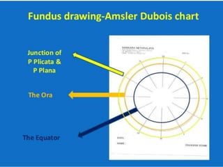
- 2. Requisites for Fundus drawing: examination table indirect ophthalmoscope 20 D lens a scleral depressor coloured pencils (mainly red, blue, green, yellow, black and brown)
- 3. •Retinal arterioles •Neovascularization •Vascular abnormalities •Vortex veins •Attached retina •Haemorrhages (pre- retinal and intra- retinal) Vascular tumor Vortex vein Pre retinal h’ge arteriole
- 4. •Open interior of conventionalretinal breaks (tears, holes) •Open interior of outer layer holes in retinoschisis •Normal macula is drawn as a red dot Schisis outer layer hole hole HST Pre retinal h’ge Retinal h’ge Vortex vein Vascular tumor
- 5. •Open portion of GRT or large dialyses •Inner portion of thin areas of retina •Open portion of retinal holes in inner layer of retinoschisis Schisis outer layer hole HST hole Vortex vein Pre retinal h’ge Retinal h’ge Vascular tumor Retinal dialysis Schisis inner layer hole Open portion of GRT
- 6. •Detached retina •Retinal viens •Outlines of retinal breaks •Outlines of ora serrata •Meridional,radial,fixed & circumferential folds Detached retina Retinal vein Outline of ora
- 7. •VR traction tufts •Retinal granular tags & tufts •Outline of flat neo- vascularization •Outline of lattice degeneration [inner ‘x’] •Outline of thin areas of retina •Intra-retinal cysts [with overlying curvilinear stripes to show configuration] lattice Thin area of retina Flat neovascularization Detached retina Outline of ora Retinal vein Shifting fluid of RD
- 8. •Inner layer of retinoschisis •White with or without pressure •Detached parsplana epithelium anterior to seperation of ora serrata Inner layer retinoschisis Detached pars epi. Rolled edge of tear Flat neovascularization Retinal vein Detached retina
- 11. asteroid Frosting or snowflakes on schisis or lattice membra nes in vitreous Elevated NVE FVP Weiss ring Vitreous substitute
- 12. •Uveal tissue •Pars plana cysts •Ciliary processess •Striae ciliaris •Pigment beneath detached retina Pars plana cyst Pigment beneath detached retina
- 13. •Outline of CRA beneath detached retina •Pigment Epithelial Detachment •Outline of posterior staphyloma PED Pigment beneath detached retina
- 14. •Malignant choroidal melanomas •Edge of buckle beneath detached retina •Choroidal detachment CD
- 15. •Intraretinal oedema •Intraretinal subretinal hard yellow exudate •Deposits in the RPE •Coloboma hard exudates
- 16. •MNF •Disk pallor •Post-PHC retinal edema •Fresh laser spots •drusen edema Cilliary nerve Fresh LASER spot Fresh LASER spot
- 17. •Edge of buckle beneath attached retina •Outline of CRA •Hyperpigmentation as a result of previous tt with CryoPHCDiathermy hyperpigmentation Sheathed vessel
- 18. •Naevi •Sheathed vessels •Outline of Long & Short Post Ciliary vein & nerve •Pigment in choroid, or RPEhyperpigmentation in attached retina Sheathed vessel hyperpigmentation Demarcation line
- 19. •Pigmented demarcation lines at attached margin of detached retina or within detached retina Edge of buckle beneath attached retina Demarcation line Sheathed vessel Outline of CRA
- 20. •Remember: •Choroidal tumours are drawn in brown colour and retinoblastomas are outlined in blue and coloured yellow. •While documenting a retinoschisis, the inner layer is generally outlined and cross lined in blue. While the open retinal holes within inner layer is outlined in blue with inner portion cross lined in red, the holes in the outer layer are outlined in blue and inner layer is coloured red.