Thyroid dysfunction dr. mohammed ibrahim youssef (1)(1)
•Download as PPTX, PDF•
6 likes•2,057 views

This document discusses guidelines for managing thyroid dysfunction during pregnancy and postpartum. It covers several topics: 1. Hypothyroidism - Recommends adjusting levothyroxine doses preconception and during pregnancy to maintain TSH below 2.5 mIU/L. Women with thyroid antibodies should be monitored. 2. Hyperthyroidism - Distinguishes gestational transient hyperthyroidism from Graves' disease. For Graves', antithyroid drugs aim to keep FT4 at upper limit of normal. Surgery may be considered in some cases. 3. Autoimmune thyroid disease - Recommends measuring TSH monthly in first half and once in third trimester for women with thyroid antibodies

Recommended
Recommended
More Related Content
What's hot
What's hot (20)
Similar to Thyroid dysfunction dr. mohammed ibrahim youssef (1)(1)
Similar to Thyroid dysfunction dr. mohammed ibrahim youssef (1)(1) (20)
Recently uploaded
Recently uploaded (20)
Thyroid dysfunction dr. mohammed ibrahim youssef (1)(1)
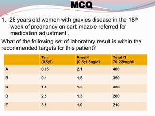
- 1. MCQ 1. 28 years old women with gravies disease in the 18th week of pregnancy on carbimazole referred for medication adjustment . What of the following set of laboratory result is within the recommended targets for this patient? Total t3 70:220ng/dl Freet4 (0.8;1.8ng/dl Tsh (0.5;5) 4002.10.05A 3501.80.1B 3301.51.5C 2801.32.5D 2101.03.5E
- 2. 2. A 24 years old woman in the first trimester of pregnancy present with heat intolerance , palpitation and failure to achieve expected weight gain. she has no prior history of thyroid disease and takes only prenatal vitamins. on examination , her pulse is 112beat /min. she has no proptosis or periorbital soft tissue changes her thyroid is slightly enlarged , and she has resting hand tremors . free t4 is 25.7(normal 10.3-23.2 pmol/l)and serum TSH is 0.05 MIU/l Which one of the following laboratory stuties is the most useful in determining the etiology of this patient’s thyrotoxicosis? A. TSH receptor antibody testing B. Thyroid perioxidase antibodies. C. Serum HCG level. D. Free t3:freet4 ratio E. RAIU usin I131 23/04/1436 2 video
- 3. Dr. Mohammed Ibrahim Youssef Family Medicine Specialist Albassam Diabetic Center .KSH
- 4. Objective: Give an overview on updated clinical practice guidelines (Published august.2012) for the management of thyroid dysfunction during pregnancy and postpartum . Will be reviewed august 2015. 23/04/1436 4
- 5. Objective: What are the normal physiological changes during pregnancy? How can we make interpretation of TFT in the context of these changes? hypothyroidism, or hyperthyroidism, how managed what's our target? Thyroid autoimmunity ! is it risky ? Thyroid nodules & caner , how to manage? Screening of thyroid dysfunctions ! with or against?
- 6. Thyroid problems during pregnancy. 1. Hypothyroidism. 2. Hyperthyroidism. 3. Gestational transient hyperthyroidism. 4. Autoimmune thyroid disease. 5. Thyroid nodules and cancer. 6. Iodine nutrition during pregnancy. 7. Postpartum thyroiditis. 8. Screening for thyroid dysfunction during pregnancy. 23/04/1436 6
- 7. Introduction..why guidelines.? Pregnancy may affect the course of thyroid Disorders, and conversely, thyroid diseases may affect both the pregnant woman and the developing fetus. Pregnant women may be under the care of multiple health care professionals, including obstetricians, nurse midwives, family practitioners, endocrinologists, and/or internists, making the development of guidelines more imporatant. 23/04/1436 7
- 9. 23/04/1436 9 Increase in serum thyroxine-binding globulin (TBG) concentrations . Estrogen effect. 2 fold increase. Total t4 and t3 rise during the first half of pregnancy, plateauing at approximately 20 weeks of gestation Stimulation of the thyrotropin (TSH) receptor by human chorionic gonadotropin (HCG). Homology between the beta-subunit of HCG &TSH. HCG &TSH are glycoprotein hormones. Peak at 10 to 12 weeks gestation Total (T4 and T3 )concentrations increase. Free t4 and t3 increase slightly, usually within the normal range. TSH reduced Thyroid physiology
- 10. 23/04/1436 10
- 11. Hypothyroidism 1. Hypothyroidism: maternal and fetal aspects 23/04/1436 11
- 12. Hypothyroidism Overt hypothyroidism occurs in 0.3–0.5% . Subclinical hypothyroidism 2–3%. Thyroid autoantibodies are found in 5–15% of women during childbearing age. 23/04/1436 12
- 13. 23/04/1436 13
- 14. Hypothyroidism Hypothyroid women have an increased prevalence of infertility, abortion,anemia, gestational hypertension, placental abruption,and postpartum hemorrhage . Adverse fetal &neonatal outcomes including premature birth, low birth weight, and neonatal respiratory distress Low IQ 23/04/1436 14
- 16. FREE T4 INDEX TOTAL T4 ASSAY X (1.5) FREE T4 ASSAY 0.3 to 3 3rd trimester 0.2 to 3.0 2nd trimester 0.1 to 2.5 1st trimester TSH Reference range MIU/L 23/04/1436 16 King Saud Hospital
- 17. 23/04/1436 17 If hypothyroidism diagnosed before pregnancy, adjust of the preconception thyroxine dose to reach TSH target ≤ 2.5 MIU/Liter. thyroxine dose usually needs to be increased (30:50)% mostly during first trimester. (C) Hypothyroidism
- 18. If hypothyroidism diagnosed during pregnancy, thyroid function tests should be normalized as rapidly as possible. maintain the target TSH ≤ 2.5 MIU/liter in the first trimester (or 3MIU/liter in second and third trimesters) . Thyroid function tests should be remeasured within 30–40 d and then every 4–6 wk 23/04/1436 18 Hypothyroidism
- 19. Euthyroid women with thyroid autoimmunity (TPO ab &thyroglobulin ab) are at risk of developing hypothyroidism and should be monitored for elevation of TSH every 4–6 wk. After delivery, most hypothyroid women need to Decrease the T4 dosage to the prepregnancy dose. 23/04/1436 19 Hypothyroidism
- 20. T4 replacement is recommended to women with SCH (high TSH + normal free t4) regardless thyroid peroxidase antibody status.(either positive or negative). (Normal TSH +Low FT4) treat or not?? 23/04/1436 20 controversial and requires further study. partial replacement therapy may be initiated with continued monitoring Hypothyroidism
- 21. FridayThurs.WednesdayTuesdayMondaySundaySaturday 50+5050+505050505050 TSH ≤(2.5) Thyroxine dose 30%:50% 7 tab to 9 tab Weekly Hypothyroidism 23/04/1436 21 Monitor frequently Return back to prepregnancy dose Anti TPO+ve
- 24. prevalence of hyperthyroidism ranges from 0.1 to 0.4%. Graves’ disease accounting for 85% of cases. Graves’ disease may fluctuate during pregnancy, with exacerbation during the first trimester(high levels of HCG) and improvement by late gestation. The presence of a goiter, especially with bruit or thrill, may point to true graves’ disease. 2. Hyperthyroidism…. 23/04/1436 24
- 25. : ; . Maternal hyperthyroidism: Medically indicated preterm delivery, intrauterine growth restriction and low birth weight, preeclampsia, Congestive heart failure, and fetal death. Iatrogenic fetal hypothyroidism. Central congenital hypothyroidism. Fetal hyperthyroidism: intrauterine growth restriction, Fetal tachycardia, Fetal goiter, Advanced bone age, Fetal hydrops, Preterm delivery, and Fetal death 23/04/1436 25 2. Hyperthyroidism….
- 26. Recommendations. Normal physiology Gestational thyrotoxicosis Overt hyperthyroidism Must be distinguished from both normal physiology of pregnancy and gestational thyrotoxicosis. Differentiation is supported by the presence of clinical evidence of autoimmune thyroid disease (typical goiter, and presence of thyrotropin receptor antibody (TRAb). TPO-Ab may be present in either case. (B) TSH in healthy pregnant women during the first trimester may be as low as 0.03 to 0.1 mU/L Significant hyperthyroidism(gravies disease) in the first trimester will have a serum TSH <0.01 mU/L) associated with elevated free T4 and/or free T3 (or total T4 and/or total T3) measurement. 23/04/1436 26
- 27. freeT4 at the upper limit (12 _ 22)pmol/l Recommendations. 23/04/1436 27 For hyperthyroidism due to graves’ disease or thyroid nodules, ATD therapy should be either: Initiated (new diagnosis) or Adjusted (for those with a prior history) to: Maintain the maternal thyroid hormone levels for free T4 at the upper limit of the non pregnant reference range. (B)
- 28. Recommendations. PTU is the first line during the first trimester. PTU may be associated with severe liver toxicity. Liver toxicity may appear abruptly with PTU. Monitor liver function in pregnant women on PTU every 3–4 wk .(C) MMI (carbimazol)in 2nd and 3rd trimester. MMI may be associated with specific congenital abnormalities . (B) agranulocytosis 23/04/1436 28
- 30. Subtotal thyroidectomy may be indicated optimally during the Second trimester if: severe adverse reaction to ATD therapy; persistently high doses of ATD are required (over 30 mg/d of MMI or 450 mg/d of PTU) patient is nonadherent to ATD therapy and has uncontrolled hyperthyroidism. (c) Recommendations. 23/04/1436 30
- 31. Radioactive iodine should not be given to a woman who is or may be pregnant. (A) No data for or against termination of pregnancy after RAI exposure. (I) Recommendations. 23/04/1436 31 No evidence that treatment of subclinical hyperthyroidism improves pregnancy outcome. It could potentially adversely affect fetal outcome. (C). Subclinical hyperthyroidism
- 32. 23/04/1436 32 Gravies disease HCG mediated TSH FT4 FT3 TT3 SIGNS TRAB B.blockers High normal ft4 Side effects SUBCLINICAL HYPERTHYROIDISM RAI SURGERY SECOND TRIMESTEER ATD BB
- 34. Fetal (TSH) appears during the 10th to 12th week of gestation. Fetal thyroid secretion increases gradually after 18th to 20th week. Maternal thyroid hormones can cross the placenta?? TSH-receptor antibodies can cross the placenta and cause cause either fetal hyperthyroidism or hypothyroidism Hyperthyroidism (Fetal aspects). 23/04/1436 34
- 35. Fetal& neonatal hyperthyroidism occurs in (1:5)% of neonates born to women with graves disease. All fetuses of women with graves' disease should be monitored for fetal thyrotoxicosis . Hyperthyroidism (Fetal aspects). 23/04/1436 35 HOW ?
- 36. TSH-receptor antibodies(TRAB) should be measured by 22wk gestation in: Current graves’ disease. History of graves ’ disease treated with RAI or thyroidectomy before pregnancy. Previous neonate with graves’ disease. Previously elevated TRAb. Negative TRAb have very low risk of fetal or neonatal thyroid dysfunction. (B) Recommendations. 23/04/1436 36
- 37. TRAb or( thyroid-stimulating Ig) elevated at least (2-3) folds Fetal thyroid dysfunction should be screened for during the fetal anatomy ultrasound (18th-22 nd wk) And TFT Fetal thyroid enlargement. growth restriction. Hydrops. Tachycardia. cardiac failure. MMI or PTU should be given with frequent clinical, laboratory, and ultrasound monitoring. Umbilical blood sampling 23/04/1436 37
- 38. 2.HCG mediated hyperthyroidism Gestational transient thyrotoxicosis Hyperemesis gravidarum Trophoblastic hyperthyroidism Familial gestational hyperthyroidism 23/04/1436 38
- 39. Gestational transient hyperthyroidism Limited to the first half of pregnancy, Elevated serum free T4 . Suppressed or undetectable serum TSH. Absence of thyroid autoimmunity. Typically associated with hyperemesis gravidarum. Thyroid stimulation is due to HCG itself, or molecular variant proteins related to HCG. Hydatidiform mole or choriocarcinoma with very high elevations of HCG may be associated with clinical hyperthyroidism. 23/04/1436 39
- 40. Diagnosis: Thyroid function tests and TRAb should be measured in patients with hyperemesis gravidarum and clinical features of hyperthyroidism. Suppressed TSH, and elevated free T4. Treatment: Do not require ATD treatment(A).it remit spontaneously. In women who appear significantly thyrotoxic or who have high serum total T3 . Clinical judgment should be followed . Beta blockers (metoprolol) may be helpful and used with obstetrical agreement. (B) 23/04/1436 40 Gestational transient hyperthyroidism
- 41. 23/04/1436 41 4. Autoimmune thyroid disease
- 42. 4. Autoimmune thyroid disease TSH every month during the first half of pregnancy and at least once during the last trimester Positive association exists between thyroid antibodies (anti TPO)and pregnancy loss. Universal screening for thyroid antibodies, and possible treatment, cannot be recommended at this time.(like ATA.) However, monitoring for the development of hypothyroidism was recommended SO TSH should be measured before pregnancy, as well as during the first and second trimesters of pregnancy.(C)…. How? 23/04/1436 42
- 43. some experts, including some UpToDate editors suggest levothyroxine (50 mcg daily) with carful monitoring : euthyroid women with TPO ab+ve in case of recurrent miscarriage. ((Uptodate Jun 19, 2014)) 4. Autoimmune thyroid disease and miscarriage 23/04/1436 43
- 44. 23/04/1436 44 5. Thyroid nodules and cancer
- 45. Pregnancy may promote the onset of growth of a thyroid nodule . No clear evidence that pregnancy worsens the survival from well-differentiated thyroid cancer . Some evidence that thyroid cancers discovered during pregnancy have a greater chance of recurrence. Evaluation is the same. 23/04/1436 45 5. Thyroid nodules and cancer
- 46. 5. Thyroid nodules and cancer FNA cytology should be performed for predominantly Solid thyroid nodules greater than 1 cm discovered In pregnancy. Nodules 0.5 cm to 1 cm in size should be considered for FNA if they have a high-risk history or suspicious findings on ultrasound. Complex nodules 1.5–2 cm or larger . During late pregnancy, FNA can be delayed until after delivery. Ultrasound guided FNA is better. B 23/04/1436 46
- 47. Suspicious findings on ultrasound. All solid consistency. Calcifications, especially tiny or microcalcifications. Really dark appearing or hypoechoic.Irregular margins. Suspicious neck lymph nodes. 23/04/1436 47
- 48. if malignant or highly suspicious to exhibit rapid growth nodule: Surgery should be offered in the second trimester. Papillary cancer without evidence of advanced disease Can be delayed until the postpartum period for definitive surgery. (B) 5. Thyroid nodules and cancer 23/04/1436 48
- 49. Give thyroxine to achieve suppressed but detectable TSH in pregnant women with Treated thyroid cancer . if surgical treatment delayed until postpartum. keep free t4 or total t4 levels within the high normal range for pregnancy.(I) RAI should not be given to women who are breastfeeding. (A) Pregnancy should be avoided for 6 months to 1 year in women received therapeutic RAI .(B) 5. Thyroid nodules and cancer. 23/04/1436 49
- 50. Surgery During 2nd trimester Papillary carcinoma follicular neoplasm medullary carcinoma Anaplastic RAI T4 Nodule 0.5-1 cm with suspicious >1cm FNAC 23/04/1436 50
- 51. 23/04/1436 51 6.0. Iodine nutrition during pregnancy
- 52. 6.0. Iodine nutrition during pregnancy Women in the childbearing age should have an average iodine intake of 150 microgram/day. As long as possible before and during pregnancy and breastfeeding, iodine intake should be increased their daily to 250 microgram on average. (A) Breast milk provides 100 microgram iodine per day to the infant. 23/04/1436 52
- 53. During pregnancy and breastfeeding do not exceed twice the recommended daily dose ( 500 microgram iodine per day). (I) Once-daily prenatal vitamins should contain 150–200 microgram iodine in the form of potassium iodide or iodate. 6.0. Iodine nutrition during pregnancy 23/04/1436 53
- 54. 23/04/1436 54 7.0. Postpartum thyroiditis
- 55. PPT is the occurrence of Hyperthyroidism. Or Hypothyroidism. Or Hyperthyroidism followed by hypothyroidism During the first year postpartum in women without clinically evident thyroid disease ??Before pregnancy. Caused by thyroid autoimmunity. Exclusively in thyroid antibody positive. 7.0. Postpartum thyroiditis 23/04/1436 55
- 56. Prevalence: In unselected populations is 7 % Type 1 diabetes mellitus. 25% The highest rates occur with history of postpartum thyroiditis ( 42 %) and positive antithyroid peroxidase antibodies (40 : 60)% may occur after pregnancy loss (miscarriage, abortion, ectopic pregnancy), as well as after normal delivery. 23/04/1436 56 7.0. Postpartum thyroiditis
- 57. Insufficient data to recommend screening of all women for postpartum thyroiditis.(I) Monitor TSH at 6–12 wk gestation and at 6 months postpartum for TPO ab + ve .(a) Screening by TSH is recommended at 3 and 6 months postpartum in patients with Type1 diabetes. Chronic viral hepatitis. (B) 23/04/1436 57 7.0. Postpartum thyroiditis
- 58. PPT has Increased risk of developing permanent primary hypothyroidism in the 5- to 10-yr period after the episode of PPT. Annual TSH level should be performed. (A) 23/04/1436 58 7.0. Postpartum thyroiditis
- 59. 7.0. Postpartum thyroiditis: Treatment. Asymptomatic hypothyroidism Symptomatic hypothyroidism TSH less than 10 MIU/liter Not planning for subsequent pregnancy No intervention, but should be remonitored in 4–8 wk. When a TSH above the reference range continues, women should be treated . women With TSH above normal . planning for pregnancy. should be treated with levothyroxine. Beta blockers. propranolol 23/04/1436 59 Symptomatic hyperthyroidism
- 60. Screening for thyroid dysfunction during pregnancy 23/04/1436 60
- 61. Universal screening of healthy women for thyroid dysfunction using serum TSH before pregnancy is not recommended. (I) Identify individuals at “high risk” for thyroid illness. If high risk measure TSH . IF >2.5 MIU/L repeat to confirm. Give low dose thyroxine to bring TSH below 2.5 mIU/liter. Thyroxine can be discontinued if the woman does not become pregnant . 23/04/1436 61 8.0. Screening for thyroid dysfunction before& during pregnancy
- 62. 23/04/1436 62 High risk for thyroid illness: Age over 30 years. Family history or autoimmune thyroid disease or hypothyroidism Goiter Thyroid antibodies, primarily thyroid peroxidase antibodies Symptoms or clinical signs suggestive of thyroid hypofunction Type 1 DM or other autoimmune disorders Infertility History of miscarriage or preterm delivery Prior head or neck irradiation or thyroid surgery Women currently receiving levothyroxine replacement Women living in a region with presumed iodine deficiency
- 63. Universal screening for anti-TPO antibodies either before or during pregnancy is not recommended.(C) But if identified, screen for serum TSH abnormalities before pregnancy, as well as during the first and second trimesters of pregnancy (C) 23/04/1436 63 8.0. Screening for thyroid dysfunction during pregnancy
- 64. for newly pregnant women. Two versions are presented: Some members recommended screening of all pregnant women by the ninth week or at the time of their first visit. (C) Others. Strongly support aggressive case finding to identify high-risk women. 23/04/1436 64 8.0. Screening for thyroid dysfunction during pregnancy
- 65. 23/04/1436 65
- 66. Summary Consider the physiological changes during pregnancy. Establish trimester specific ranges for TSH &free t4. Maintain target TSH ≤ 2.5 during treatment of hypothyroidism Maintain free T4 near upper limit during treatment of hyperthyroidism. Do not give ATD in HCG mediated hyperthyroidism. TRAB only with gravies disease. PPT exclusively in anti TPO +ve. Surgery for cancer thyroid optimally in 2nd trimester Iodine requirement during pregnancy is 250 mcg. 23/04/1436 66
- 67. Abbreviations. ATD, Antithyroid drug. FNA, fine-needle aspiration. GH, gestational hyperthyroidism. HCG, human chorionic gonadotropin. MMI, methimazole. PPT, postpartum thyroiditis. PTU, propylthiouracil. TG, thyroglobulin. TPO-Ab, thyroid peroxidase. TRAb, TSH receptor antibodies. RAI, radioactive iodine. SCH, subclinical hypothyroidism. 23/04/1436 67
- 68. Referenes Management of Thyroid Dysfunction during Pregnancy and Postpartum: An Endocrine Society Clinical Practice Guideline Overview of thyroid disease in pregnancy last updated: Jun 19, 2014. 23/04/1436 68
- 69. 23/04/1436 69
Editor's Notes
- ANTI TPO if present donot indicate gravies Its absence doesnot eliminate the possibility The ratio is usuful to differentiate postpartum thyroiditis from gravies disease
- Thyroid physiology — To meet the increased metabolic needs during a normal pregnancy, there are changes in thyroid physiology that are reflected in altered thyroid function tests [1]. The major changes in thyroid function during pregnancy are an increase in serum thyroxine-binding globulin (TBG) concentrations and stimulation of the thyrotropin (TSH) receptor by human chorionic gonadotropin (hCG). Thyroxine binding globulin — During pregnancy, serum TBG concentrations rise almost two-fold because estrogen increases TBG production and TBG sialylation, which results in decreased clearance of TBG [2]. To maintain adequate free thyroid hormone concentrations during this period, thyroxine (T4) and triiodothyronine (T3) production by the thyroid gland must increase. Total T4 and T3 concentrations rise during the first half of pregnancy, plateauing at approximately 20 weeks of gestation, at which time a new steady state is reached and the overall production rate of thyroid hormones returns to prepregnancy rates. Thus, TBG excess leads to an increase in both serum total T4 and T3 concentrations. (See "Euthyroid hyperthyroxinemia and hypothyroxinemia".) hCG and thyroid function — hCG is one of a family of glycoprotein hormones, including TSH, with a common alpha-subunit and a unique beta-subunit. However, there is considerable homology between the beta-subunits of hCG and TSH. As a result, hCG has weak thyroid-stimulating activity [3]. In a human thyroid cell-culture assay, as an example, 1 microU of hCG was equivalent to 0.0013 microU of TSH [4]. Serum hCG concentrations increase soon after fertilization and peak at 10 to 12 weeks. During this peak, total serum T4 and T3 concentrations increase. Serum free T4 and T3 concentrations increase slightly, usually within the normal range, and serum TSH concentrations are appropriately reduced [3]. However, in 10 to 20 percent of normal women, serum TSH concentrations are transiently low or undetectable [5-7]. In a report of 63 women with extremely high hCG concentrations (>200,000 IU/L), TSH was <0.2 microU/mL in 67 percent of samples and free T4 was above 1.8 ng/dL in 32 percent of samples. All women whose hCG was greater than 400,000 IU/L had a suppressed TSH concentration [8]. (See "Hyperthyroidism during pregnancy: Clinical manifestations, diagnosis, and causes", section on 'hCG-mediated hyperthyroidism'.) This transient, usually subclinical, hyperthyroidism should be considered a normal physiologic finding. It is not known if this action of hCG benefits the mother or fetus. Later in pregnancy, as hCG secretion declines, serum free T4 and T3 concentrations decline and serum TSH concentrations rise slightly to or within the normal range.
- Trimester-specific reference ranges — Because of the changes in thyroid physiology during pregnancy, the Guidelines of the American Thyroid Association (ATA) for the Diagnosis and Management of Thyroid Disease During Pregnancy and Postpartum recommend using trimester-specific reference ranges for TSH and method and trimester-specific reference ranges for serum free T4 [9]. Commercial laboratories should provide these reference ranges, but many commercial laboratories currently do not do this. In several population studies, the lower limit of the reference range for TSH in healthy pregnant women during the first trimester ranged from 0.03 to 0.1 mU/L [10-14]. In one of the largest population-based studies (over 13,000 pregnant women), the reference range (2.5 to 97.5th percentile) for TSH in the first trimester was 0.08 to 2.99 mU/L [10,13]. Thus, if the laboratory does not provide trimester-specific reference ranges for TSH (mU/L), the following reference ranges can be used: ●First trimester 0.1 to 2.5 ●Second trimester 0.2 to 3.0 ●Third trimester 0.3 to 3.0 Some studies report a decrease in free T4 during pregnancy, others report no change or even an increase [1,15,16]. Direct free T4 measurements may be unreliable during pregnancy. Measurement of free T4 in the dialysate or ultrafiltrate of serum samples using liquid chromatography/tandem mass spectrometry appears to be the most reliable, and when this method is used, free T4 concentrations were shown to decrease gradually with advancing gestational age, particularly between the first and second trimester [17,18]. This assay is relatively expensive and not universally available. Other free T4 assays (and probably free T3 assays) frequently fail to meet performance standards in pregnant patients, owing to increases in TBG and decreases in albumin concentrations that cause the immunoassay to be unreliable [15]. To compensate, some kits have provided different free T4 normal ranges for pregnant patients, usually lower than those of nonpregnant patients. Method-specific and trimester-specific reference ranges of serum free T4 should be used, if available [9]. (See "Laboratory assessment of thyroid function", section on 'Serum free T4 and free T3'.) As an alternative, serum total T4 measurements, which are more reliable during pregnancy, can be measured to assess thyroid function [15]. When free T4 measurements appear discordant with TSH measurements, serum total T4 should be measured. Total T4 and T3 levels during pregnancy are 1.5-fold higher than in nonpregnant women due to TBG excess. Thus a normal reference range for pregnancy should be used.
- Direct free T4 measurements may be unreliable during pregnancy. Measurement of free T4 in the dialysate or ultrafiltrate of serum samples using liquid chromatography/tandem mass spectrometry appears to be the most reliable, and when this method is used, free T4 concentrations were shown to decrease gradually with advancing gestational age, particularly between the first and second trimester . This assay is relatively expensive and not universally available.
- Maternal hyperthyroidism is associated with both gestational and fetal risks that are related to the disease itself and/or to the medical treatment of the disease. Inadequately treated maternal thyrotoxicosis is associated with an increased risk of medically indicated preterm delivery, intrauterine growth restriction and low birth weight, preeclampsia, congestive heart failure, and fetal death (62). In addition, overtreatment of the mother with thioamides can result in iatrogenic fetal hypothyroidism (51), but undertreatment of maternal hyperthyroidism may lead to central congenital hypothyroidism (63, 64). Fetal hyperthyroidism can be associated with intrauterine growth restriction, fetal tachycardia, fetal goiter, advanced bone age, fetal hydrops, preterm delivery, and fetal death (40–42, 53, 56, 65). The diagnosis is suggested by any of these signs or abnormalities. Maternal TRAb levels able to induce fetal hyperthyroidism are usually over three times the upper normal limit. PTU and MMI or its derivative carbimazole
- Diagnosis — The diagnosis of hyperthyroidism during pregnancy should be based primarily upon a finding of a suppressed (<0.1 mU/L) or undetectable (<0.01) serum TSH value and elevated thyroid hormone levels that exceed the normal range for pregnancy [9]. If a TSH level is <0.1 mU/L, free T4 (or free T4 index) should be obtained. If the free T4 is in the normal range for pregnancy, a free T3 should also be measured. In the event that free thyroid hormone levels are discordant with serum TSH and clinical findings, total T4 should be measured. TSH in healthy pregnant women during the first trimester may be as low as 0.03 to 0.1 mU/L [10-14]. Most pregnant women with significant overt hyperthyroidism in the first trimester will have a serum TSH below that which is seen in asymptomatic healthy pregnant women (ie, <0.01 mU/L) associated with an elevated free T4 and/or free T3 (or total T4 and/or total T3) measurement. (See "Hyperthyroidism during pregnancy: Clinical manifestations, diagnosis, and causes" and "Hyperthyroidism during pregnancy: Clinical manifestations, diagnosis, and causes", section on 'Diagnosis'.) Because radioiodine administration is contraindicated, it may not be possible to ascertain the cause of the hyperthyroidism during pregnancy. Measurement of thyrotropin receptor antibody (TRAb or thyroid stimulating immunoglobulins) using second-generation thyrotropin-binding inhibitory immunoglobulin (TBII) assays are positive in 95 percent of patients with Graves' disease and should be used to make the diagnosis of Graves' disease during pregnancy if the clinical diagnosis is uncertain. (See "Hyperthyroidism during pregnancy: Clinical manifestations, diagnosis, and causes", section on 'Diagnosis'.)
- Encourage patients to promptl report any new symptoms.
- Aplasia cutis congenita (ACC)[1, 2, 3] is a heterogenous group of disorders characterized by the absence of a portion of skin in a localized or widespread area at birth. First reported by Cordon in 1767, aplasia cutis congenita most commonly (70%) manifests as a solitary defect on the scalp, as noted in the first image below, but sometimes it may occur as multiple lesions, as shown in the second image below. Although most commonly seen on the scalp, aplasia cutis congenita can affect any part of the body.
- TRAb or thyroid-stimulating Ig elevated at least 2- to 3-fold the normal level, and in women treated with ATD, maternal free T4 and fetal thyroid dysfunction should be screened for during the fetal anatomy ultrasound (18th-22nd wk) . Evidence of fetal thyroid dysfunction could include thyroid enlargement, growth restriction, hydrops, presence of goiter, tachycardia, or cardiac failure. If fetal hyperthyroidism is diagnosed and thought to endanger the pregnancy,treatment using MMI or PTU should be given with frequent clinical, laboratory, and ultrasound monitoring.(
- The extent to which maternal thyroid hormones cross the placenta is controversial. In infants with congenital absence of the thyroid, cord serum concentrations range from 20 to 50 percent of the concentrations in normal infants [21]. TSH-receptor antibodies can cross the placenta and cause either fetal hyperthyroidism or hypothyroidism (see 'Fetal and neonatal Graves' disease' below). Little TSH crosses the placenta [22]. Thyrotropin-releasing hormone (TRH) can cross the placenta and exogenously administered TRH can stimulate fetal TSH secretion [23].
- Multiple gestation is another recognized cause.
- Gestational hyperthyroidism (GH), also referred as gestational thyrotoxicosis or gestational transient thyrotoxicosis,is defined as transient hyperthyroidism, limited to the first half of pregnancy, characterized by elevatedserum free T4 and suppressed or undetectable serum TSH, in the absence of thyroid autoimmunity. GH is typically associated with hyperemesis gravidarum, defined as sever vomiting in early pregnancy that causes more than 5%weight loss, dehydration, and ketonuria and occurs in 0.5–10 cases per 1000 pregnancies. The etiology of thyroid stimulation is thought to be hCG itself, or molecular variant proteins related to hCG. Multiple gestation is another recognized cause of GH. Very high elevations of hCG occurring in patients with hydatidiform mole or choriocarcinoma are often associated with clinical hyperthyroidism. TSHR mutations with functional hypersensitivity to hCG have also been recognized as a rare cause of severe GH. In women with GH, the serum TSH is suppressed or undetectable; serum total T4 and free T4 are elevated, but the free T3 is elevated less frequently. Women with hyperemesis and elevated thyroid hormone levels most commonly do not have other clinical evidence of Graves’ disease and lack the TSH receptor antibodies typically present in Graves’ disease. A small portion of these patients have clinical hyperthyroidism. Clinical symptoms of hyperthyroidism antedating pregnancy, the presence of goiter, ophthalmopathy, and laboratory evidence of autoimmunity favor the diagnosis of Graves’ hyperthyroidism. Because many common signs and symptoms of hyperthyroidism may be mimicked by normal pregnancy, the clinical challenge is to differentiate these disorders (13, 25, 26, 66–68). There is disagreement as to whether thyroid hormone should be measured in all pregnancies with hyperemesis, or only when clinical features of hyperthyroidism are present. Some authorities suggest that measurement of thyroid function tests may safely be limited to those women with clinical evidence suggestive of hyperthyroidism.There is no clear evidence in the medical literature that patients diagnosed with GH have benefited from antithyroid therapy, but only a few patients have been reported who received ATD for a few weeks. The available data indicate that most women with hyperemesis, with no or mild clinical evidence of hyperthyroidism, suppressed TSH, and elevated free T4, remit spontaneously. No clear data are available to support the use of ATD in the management of women with GH, but clinical judgment should be followed in women with clear signs of hyperthyroidism and elevated free T4 and free T3, or total T3 above the normal pregnancy range C Risk cannot be ruled out Either studies in animals have revealed adverse effects on the fetus (teratogenic or embryocidal effects or other) and there are no controlled studies in women, or studies in women and animals are not available. Drugs should be given only if the potential benefits justify the potential risk to the fetus.
- As of January 2011, only one randomized interventional trial has suggested a decrease in the first trimester miscarriage rate in euthyroid antibody-positive women, but treatment duration was very brief before the outcome of interest. However, because women with elevated anti-TPO antibodies are at increased risk for progression of hypothyroidism, if identified such women should be screened for serum TSH abnormalities before pregnancy, as well as during the first and second trimesters of pregnancy. USPSTF recommendation Level c :
- As an example, in a prospective study of 115 TPO antibody positive patients, half were randomly assigned to T4 (median dose 50 mcg daily) and half were not treated, and comparison was made with 869 TPO antibody negative patients. Miscarriage rates were 3.5 percent in TPO antibody positive treated patients, 2.4 percent in the TPO antibody negative patients, and 13.8 percent in TPO antibody positive untreated patients. Premature delivery rates were 7 percent, 8.2 percent, and 22.4 percent, respectively [
- suspicious findings on ultrasound. All solid consistency • Calcifications, especially tiny or microcalcifications • Really dark appearing or hypoechoic • Irregular, aggressive appearing margins • Suspicious neck lymph nodes
- 5.2. When nodules discovered in the first or early second trimester are found to be malignant or highly suspicious on cytopathological analysis, to exhibit rapid growth, or to be accompanied by pathological neck adenopathy, pregnancy need not be interrupted, but surgery should be offered in the second trimester. Women found to have cytology indicative of papillary cancer or follicular neoplasm without evidence of advanced disease and who prefer to wait until the postpartum period for definitive surgery may be reassured that most well-differentiated thyroid cancers are slow growing and that delaying surgical treatment until soon after delivery is unlikely to change disease-specific survival. USPSTF recommendation level: B; evidence, fair (1QQEE) (75, 83–85
- 5.3. It is appropriate to administer thyroid hormone to achieve a suppressed but detectable TSH in pregnant women with a previously treated thyroid cancer, in those with an FNA positive for or suspicious for cancer, or in those who elect to delay surgical treatment until postpartum. High-risk patients may benefit more than low risk patients from a greater degree of TSH suppression. The free T4 or total T4 levels should ideally not be increased above the normal range for pregnancy. USPSTF recommendation level: I; evidence, poor (QEEE) (86).
- Two patterns of postpartum dysfunction can be defined: postpartum thyroiditis and a postpartum exacerbation of chronic lymphocytic (Hashimoto's) thyroiditis. Postpartum thyroiditis is characterized by transient hyperthyroidism, or transient hyperthyroidism followed by transient or rarely permanent hypothyroidism. Postpartum exacerbation of Hashimoto's thyroiditis is characterized by postpartum progression of autoimmune destruction. It may cause a transient or permanent increase in thyroid hormone requirements. In one study, for example, more than 50 percent of women with Hashimoto’s thyroiditis required an increase in their pregestational dose thyroxine dose in the postpartum period [66
- 7.0. Postpartum thyroiditis 7.1. There are insufficient data to recommend screening of all women for postpartum thyroiditis (PPT). USPSTF recommendation level: I; evidence, poor (2QEEE). 7.2. Women known to be TPO-Ab should have TSH measured at 6–12 wk gestation and at 6 months postpartum, or as clinically indicated. USPSTF recommendation level: A; evidence, good (1QQQE). 7.3. Because the prevalence of PPT in women with type 1 diabetes, Graves’ disease in remission, and chronic viral hepatitis is greater than in the general population, screening by TSH is recommended at 3 and 6 months postpartum. USPSTF recommendation level: B; evidence, fair (2QQEE). 7.4. Women with a history of PPT have a markedly increased risk of developing permanent primary hypothyroidism in the 5- to 10-yr period after the episode of PPT. An annual TSH level should be performed in these women. USPSTF recommendation level: A; evidence, good (1QQQE). 7.5. Asymptomatic women with PPT who have a TSH above the reference range but less than 10 mIU/liter and who are not planning a subsequent pregnancy do not necessarily require intervention but should, if untreated, be remonitored in 4–8 wk. When a TSH above the reference range continues, women should be treated with levothyroxine. Symptomatic women and women with a TSH above normal and who are attempting pregnancy should be treated with levothyroxine. USPSTF recommendation level: B; evidence, fair (2QQEE). 7.6. There is insufficient evidence to conclude whether an association exists between postpartum depression (PPD) and either PPT or thyroid antibody positivity (in women who did not develop PPT). USPSTF recommendation level: I; evidence, poor (2QEEE). However, because hypothyroidism is a potentially reversible cause of depression, women with PPD should be screened for hypothyroidism and appropriately treated. USPSTF recommendation level: B; evidence, fair (2QQEE).