SlideShare une entreprise Scribd logo
1  sur  20
Renal Correction of Acidosis-Increased Excretion of Hydrogen Ions and Addition of Bicarbonate Ions to the Extracellular Fluid Acidosis occurs when the ratio of HCO3- to CO2 in the extracellular fluid decreases, thereby decreasing pH If this ratio decreases because of a fall in HCO3-, the acidosis is referred to as metabolic acidosis If the pH falls because of an increase in Pco2, the acidosis is referred to as respiratory acidosis
In metabolic acidosis, an excess of H+ over HCO3- occurs in the tubular fluid primarily because of decreased filtration of HCO3- 	This decreased filtration of HCO3- is caused mainly by a decrease in the extracellular fluid concentration of HCO3- In metabolic acidosis, there is also a decrease in pH and a rise in extracellular fluid H+ concentration
The primary compensations include increased ventilation rate, which reduces Pco2, and renal compensation, which, by adding new bicarbonate to the extracellular fluid, helps minimize the initial fall in extracellular HCO3- concentration
Metabolic Acidosis Results from Decreased Extracellular Fluid Bicarbonate Concentration Renal Tubular Acidosis This type of acidosis results from a defect in renal secretion of H+ or in reabsorption of HCO3-, or both. These disorders are generally of two types:  Inadequate amounts of NH4+ are excreted, so that there is net accumulation of acid in the body fluids Impairment of renal tubular HCO3- reabsorption Some causes of renal tubular acidosis include chronic renal failure, insufficient aldosterone secretion
Diarrhea Severe diarrhea is probably the most frequent cause of metabolic acidosis. The cause of this acidosis is the loss of large amounts of sodium bicarbonate into the feces
Diabetes Mellitus. Diabetes mellitus is caused by lack of insulin secretion by the pancreas or by insufficient insulin secretion In the absence of sufficient insulin, some of the fats are split into acetoacetic acid, and this is metabolized by the tissues for energy in place of glucose. With severe diabetes mellitus, blood acetoacetic acid levels can rise very high, causing severe metabolic acidosis. In an attempt to compensate for this acidosis, large amounts of acid are excreted in the urine
In respiratory acidosis, there is a reduction in pH, an increase in extracellular fluid H+ concentration, and an increase in Pco2, which is the initial cause of the acidosis The compensatory response is an increase in plasma HCO3-, caused by the addition of new bicarbonate to the extracellular fluid by the kidneys. The rise in HCO3- helps offset the increase in Pco2, thereby returning the plasma pH toward normal
Respiratory Acidosis is Caused by Decreased Ventilation and Increased PCO2  Damage to the respiratory center in the medulla oblongata can lead to respiratory acidosis Obstruction of the passageways of the respiratory tract, pneumonia, emphysema, as well as any factor that interferes with the exchange of gases between the blood and the alveolar air, can cause respiratory acidosis.
Characteristics of primary Acid-Base Disturbances The primary event is indicated by the double arrows (↑↑ or ↓↓). Respiratory acid-base disorder are initiated by the increase in the PCO2, whereas metabolic disorders are initiated by decreases in HCO3-
Renal Correction of Alkalosis-Decreased Tubular Secretion of Hydrogen Ions and Increased Excretion of Bicarbonate Ions 	In alkalosis, the ratio of HCO3- to CO2 in the extracellular fluid increases, causing a rise in pH (a decrease in H+ concentration)
	In respiratory alkalosis, there is an increase in extracellular fluid pH and a decrease in H+ concentration 	The cause of the alkalosis is a decrease in plasma Pco2, caused by hyperventilation The reduction in Pco2 then leads to a decrease in the rate of H+ secretion by the renal tubules 	Consequently, there is not enough H+ to react with all the HCO3- that is filtered Therefore, the compensatory response to a primary reduction in Pco2 in respiratory alkalosis is a reduction in plasma HCO3- concentration, caused by increased renal excretion of HCO3-.
Respiratory Alkalosis Results from Increased Ventilation and Decreased Pco2 Respiratory alkalosis is caused by overventilation by the lungs A physiologic type of respiratory alkalosis occurs when a person ascends to high altitude. The low oxygen content of the air stimulates respiration, which causes excess loss of CO2 and development of mild respiratory alkalosis The means for compensation are the chemical buffers of the body fluids and the ability of the kidneys to increase HCO3- excretion
	In metabolic alkalosis, there is also an increase in plasma pH and a decrease in H+ concentration 	The cause of metabolic alkalosis, however, is a rise in the extracellular fluid HCO3- concentration  In metabolic alkalosis, the primary compensations are decreased ventilation, which raises Pco2, and increased renal HCO3- excretion, which helps compensate for the initial rise in extracellular fluid HCO3- concentration.
Metabolic Alkalosis Is Caused by Increased Extracellular Fluid Bicarbonate Concentration Administration of Diuretics. All diuretics cause increased flow of fluid along the tubules, usually causing increased flow in the distal and collecting tubules  This leads to increased reabsorption of Na+ from these parts of the nephrons. Because the sodium reabsorption here is coupled with H+ secretion, the enhanced sodium reabsorption also leads to an increase in H+ secretion and an increase in bicarbonate reabsorption.
Excess Aldosterone. When large amounts of aldosterone are secreted by the adrenal glands, a mild metabolic alkalosis develops.  Aldosterone promotes extensive reabsorption of Na+ from the distal and collecting tubules and at the same time stimulates the secretion of H+ by the intercalated cells of the collecting tubules.  This increased secretion of H+ leads to its increased excretion by the kidneys and, therefore, metabolic alkalosis
Characteristics of primary Acid-Base Disturbances The primary event is indicated by the double arrows (↑↑ or ↓↓). Respiratory acid-base disorders are initiated by the decrease in the PCO2, whereas metabolic disorders are initiated by an increases in HCO3-
Clinical Measurements and Analysis of Acid-Base Disorders The simple acid-base disorders described previously can be diagnosed by analyzing three measurements from an arterial blood sample: pH, plasma bicarbonate concentration, and Pco2
A pH less than 7.4 indicates acidosis, whereas a pH greater than 7.4 indicates alkalosis Examine the plasma Pco2 and HCO3- concentration. The normal value for Pco2 is about 40 mm Hg, and for HCO3-, it is 24 mEq/L If the disorder has been characterized as acidosis and the plasma Pco2 is increased, there must be a respiratory component to the acidosis After renal compensation, the plasma HCO3- concentration in respiratory acidosis would tend to increase above normal Therefore, the expected values for a simple respiratory acidosis would be reduced plasma pH, increased Pco2, and increased plasma HCO3- concentration after partial renal compensation
Treatment of Acidosis or Alkalosis To neutralize excess acid, large amounts of sodium bicarbonate can be ingested by mouth The sodium bicarbonate is absorbed from the gastrointestinal tract into the blood and increases the bicarbonate portion of the bicarbonate buffer system, thereby increasing pH toward normal For the treatment of alkalosis, ammonium chloride can be administered by mouth. When the ammonium chloride is absorbed into the blood, the ammonia portion is converted by the liver into urea. This reaction liberates HCl, which immediately reacts with the buffers of the body fluids to shift the H+ concentration in the acidic direction. Another substance used occasionally is lysine monohydrochloride.

Contenu connexe

Tendances (20)

Cne 2017 abg analysis
Cne 2017 abg analysisCne 2017 abg analysis
Cne 2017 abg analysis
 
ABG Analysis
ABG AnalysisABG Analysis
ABG Analysis
 
Arterial Blood Gases
Arterial Blood GasesArterial Blood Gases
Arterial Blood Gases
 
Arterial blood gas analysis and interpretation
Arterial blood gas analysis and interpretationArterial blood gas analysis and interpretation
Arterial blood gas analysis and interpretation
 
Arterial blood gas
Arterial blood gasArterial blood gas
Arterial blood gas
 
Arterial blood gas analysis
Arterial blood gas analysisArterial blood gas analysis
Arterial blood gas analysis
 
Arterial Blood Gas Analysis
Arterial Blood Gas Analysis Arterial Blood Gas Analysis
Arterial Blood Gas Analysis
 
ABG, step by step approach (Updated)
ABG, step by step approach (Updated)ABG, step by step approach (Updated)
ABG, step by step approach (Updated)
 
Acid Base, Arterial Blood Gas
Acid Base, Arterial Blood GasAcid Base, Arterial Blood Gas
Acid Base, Arterial Blood Gas
 
CME - ABG
CME - ABGCME - ABG
CME - ABG
 
Pravin Pawal Abg
Pravin Pawal AbgPravin Pawal Abg
Pravin Pawal Abg
 
ABG interpretation.
ABG  interpretation.ABG  interpretation.
ABG interpretation.
 
Abg interpretation
Abg interpretation Abg interpretation
Abg interpretation
 
Arterial blood gas (ABGs)
Arterial blood gas (ABGs)Arterial blood gas (ABGs)
Arterial blood gas (ABGs)
 
Sravan abg ppt modified
Sravan abg ppt modifiedSravan abg ppt modified
Sravan abg ppt modified
 
Abc acid base
Abc acid baseAbc acid base
Abc acid base
 
ABG , ARTERIAL BLOOD GAS
ABG , ARTERIAL BLOOD GAS ABG , ARTERIAL BLOOD GAS
ABG , ARTERIAL BLOOD GAS
 
Interpreting Blood Gases, Practical and easy approach
Interpreting Blood Gases, Practical and easy approachInterpreting Blood Gases, Practical and easy approach
Interpreting Blood Gases, Practical and easy approach
 
RT ARTERIAL BLOOD GAS .ppt
RT ARTERIAL BLOOD GAS .pptRT ARTERIAL BLOOD GAS .ppt
RT ARTERIAL BLOOD GAS .ppt
 
ABG
ABGABG
ABG
 

Similaire à Lec49

Similaire à Lec49 (20)

Alkalosis
AlkalosisAlkalosis
Alkalosis
 
Renal clearance and pH balance
Renal clearance and pH balanceRenal clearance and pH balance
Renal clearance and pH balance
 
Blood Gases, pH, and.pptx.pdfnodownlo ad
Blood Gases, pH, and.pptx.pdfnodownlo adBlood Gases, pH, and.pptx.pdfnodownlo ad
Blood Gases, pH, and.pptx.pdfnodownlo ad
 
Acid base balance sharath
Acid base balance sharathAcid base balance sharath
Acid base balance sharath
 
Acid and base balance
Acid and base balanceAcid and base balance
Acid and base balance
 
Acid base disorders
Acid base disordersAcid base disorders
Acid base disorders
 
acid and base with acid and base disorders
acid and base with acid and base disordersacid and base with acid and base disorders
acid and base with acid and base disorders
 
Acidification of urine.pptx
Acidification of urine.pptxAcidification of urine.pptx
Acidification of urine.pptx
 
عناية نظري م9.pptx
عناية نظري م9.pptxعناية نظري م9.pptx
عناية نظري م9.pptx
 
Acid base lecture (shortened)
Acid base lecture (shortened)Acid base lecture (shortened)
Acid base lecture (shortened)
 
PRESENT: Acid base balance hossam (1).ppt
PRESENT: Acid base balance hossam (1).pptPRESENT: Acid base balance hossam (1).ppt
PRESENT: Acid base balance hossam (1).ppt
 
SSU Lecture1
SSU Lecture1SSU Lecture1
SSU Lecture1
 
pediatrics.ppt
pediatrics.pptpediatrics.ppt
pediatrics.ppt
 
Blood gas analysis.pptx
Blood gas analysis.pptxBlood gas analysis.pptx
Blood gas analysis.pptx
 
Abg by dr girish
Abg by dr girishAbg by dr girish
Abg by dr girish
 
RENAL MECHANISM FOR MAINTAINANCE OF H+ IN.pptx
RENAL MECHANISM FOR MAINTAINANCE OF H+ IN.pptxRENAL MECHANISM FOR MAINTAINANCE OF H+ IN.pptx
RENAL MECHANISM FOR MAINTAINANCE OF H+ IN.pptx
 
ABG Analysis & Interpretation
ABG Analysis & InterpretationABG Analysis & Interpretation
ABG Analysis & Interpretation
 
Acid base balance-2
Acid base balance-2Acid base balance-2
Acid base balance-2
 
Metbolic acidosis and alkalosis
Metbolic acidosis and alkalosisMetbolic acidosis and alkalosis
Metbolic acidosis and alkalosis
 
acidbasebalancesimplified.pptx
acidbasebalancesimplified.pptxacidbasebalancesimplified.pptx
acidbasebalancesimplified.pptx
 

Plus de MBBS IMS MSU

Hema practical 05 hema staining
Hema practical 05 hema stainingHema practical 05 hema staining
Hema practical 05 hema stainingMBBS IMS MSU
 
Hema practical 03 coagulation
Hema practical 03 coagulationHema practical 03 coagulation
Hema practical 03 coagulationMBBS IMS MSU
 
Hema practical 02 hematology
Hema practical 02 hematologyHema practical 02 hematology
Hema practical 02 hematologyMBBS IMS MSU
 
Pharmacology anticoagulation
Pharmacology   anticoagulationPharmacology   anticoagulation
Pharmacology anticoagulationMBBS IMS MSU
 
Microbiology hiv-yf
Microbiology   hiv-yfMicrobiology   hiv-yf
Microbiology hiv-yfMBBS IMS MSU
 
Forensic medicine firearms and firearm injuries
Forensic medicine   firearms and firearm injuriesForensic medicine   firearms and firearm injuries
Forensic medicine firearms and firearm injuriesMBBS IMS MSU
 
Forensic medicine firearms and firearm injuries
Forensic medicine   firearms and firearm injuriesForensic medicine   firearms and firearm injuries
Forensic medicine firearms and firearm injuriesMBBS IMS MSU
 
Forensic medicine firearms and firearm injuries
Forensic medicine   firearms and firearm injuriesForensic medicine   firearms and firearm injuries
Forensic medicine firearms and firearm injuriesMBBS IMS MSU
 
Forensic medicine changes after death
Forensic medicine   changes after deathForensic medicine   changes after death
Forensic medicine changes after deathMBBS IMS MSU
 
Pharmacology cvs medicine
Pharmacology   cvs medicinePharmacology   cvs medicine
Pharmacology cvs medicineMBBS IMS MSU
 
Pharmacology antiarrhythmias
Pharmacology   antiarrhythmiasPharmacology   antiarrhythmias
Pharmacology antiarrhythmiasMBBS IMS MSU
 
Pharmacology angina
Pharmacology   anginaPharmacology   angina
Pharmacology anginaMBBS IMS MSU
 
Pathology hematology 3
Pathology   hematology 3Pathology   hematology 3
Pathology hematology 3MBBS IMS MSU
 
Forensic medicine the medico-legal autopsy
Forensic medicine   the medico-legal autopsyForensic medicine   the medico-legal autopsy
Forensic medicine the medico-legal autopsyMBBS IMS MSU
 
Forensic medicine post mortem artefact
Forensic medicine   post mortem artefactForensic medicine   post mortem artefact
Forensic medicine post mortem artefactMBBS IMS MSU
 
Pharmacology anemia and its treatment
Pharmacology   anemia and its treatmentPharmacology   anemia and its treatment
Pharmacology anemia and its treatmentMBBS IMS MSU
 
Pharmacology neuromuscular blockers & anemia
Pharmacology   neuromuscular blockers & anemiaPharmacology   neuromuscular blockers & anemia
Pharmacology neuromuscular blockers & anemiaMBBS IMS MSU
 
Pharmacology - Parkinsonism
Pharmacology - ParkinsonismPharmacology - Parkinsonism
Pharmacology - ParkinsonismMBBS IMS MSU
 
Forensic medicine medical negligence
Forensic medicine   medical negligenceForensic medicine   medical negligence
Forensic medicine medical negligenceMBBS IMS MSU
 
Forensic medicine medical negligence 2-bolam principle
Forensic medicine   medical negligence 2-bolam principleForensic medicine   medical negligence 2-bolam principle
Forensic medicine medical negligence 2-bolam principleMBBS IMS MSU
 

Plus de MBBS IMS MSU (20)

Hema practical 05 hema staining
Hema practical 05 hema stainingHema practical 05 hema staining
Hema practical 05 hema staining
 
Hema practical 03 coagulation
Hema practical 03 coagulationHema practical 03 coagulation
Hema practical 03 coagulation
 
Hema practical 02 hematology
Hema practical 02 hematologyHema practical 02 hematology
Hema practical 02 hematology
 
Pharmacology anticoagulation
Pharmacology   anticoagulationPharmacology   anticoagulation
Pharmacology anticoagulation
 
Microbiology hiv-yf
Microbiology   hiv-yfMicrobiology   hiv-yf
Microbiology hiv-yf
 
Forensic medicine firearms and firearm injuries
Forensic medicine   firearms and firearm injuriesForensic medicine   firearms and firearm injuries
Forensic medicine firearms and firearm injuries
 
Forensic medicine firearms and firearm injuries
Forensic medicine   firearms and firearm injuriesForensic medicine   firearms and firearm injuries
Forensic medicine firearms and firearm injuries
 
Forensic medicine firearms and firearm injuries
Forensic medicine   firearms and firearm injuriesForensic medicine   firearms and firearm injuries
Forensic medicine firearms and firearm injuries
 
Forensic medicine changes after death
Forensic medicine   changes after deathForensic medicine   changes after death
Forensic medicine changes after death
 
Pharmacology cvs medicine
Pharmacology   cvs medicinePharmacology   cvs medicine
Pharmacology cvs medicine
 
Pharmacology antiarrhythmias
Pharmacology   antiarrhythmiasPharmacology   antiarrhythmias
Pharmacology antiarrhythmias
 
Pharmacology angina
Pharmacology   anginaPharmacology   angina
Pharmacology angina
 
Pathology hematology 3
Pathology   hematology 3Pathology   hematology 3
Pathology hematology 3
 
Forensic medicine the medico-legal autopsy
Forensic medicine   the medico-legal autopsyForensic medicine   the medico-legal autopsy
Forensic medicine the medico-legal autopsy
 
Forensic medicine post mortem artefact
Forensic medicine   post mortem artefactForensic medicine   post mortem artefact
Forensic medicine post mortem artefact
 
Pharmacology anemia and its treatment
Pharmacology   anemia and its treatmentPharmacology   anemia and its treatment
Pharmacology anemia and its treatment
 
Pharmacology neuromuscular blockers & anemia
Pharmacology   neuromuscular blockers & anemiaPharmacology   neuromuscular blockers & anemia
Pharmacology neuromuscular blockers & anemia
 
Pharmacology - Parkinsonism
Pharmacology - ParkinsonismPharmacology - Parkinsonism
Pharmacology - Parkinsonism
 
Forensic medicine medical negligence
Forensic medicine   medical negligenceForensic medicine   medical negligence
Forensic medicine medical negligence
 
Forensic medicine medical negligence 2-bolam principle
Forensic medicine   medical negligence 2-bolam principleForensic medicine   medical negligence 2-bolam principle
Forensic medicine medical negligence 2-bolam principle
 

Lec49

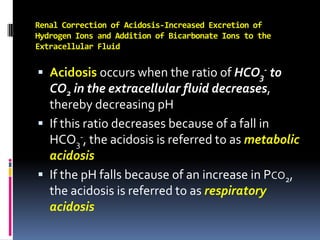
  • 1. Renal Correction of Acidosis-Increased Excretion of Hydrogen Ions and Addition of Bicarbonate Ions to the Extracellular Fluid Acidosis occurs when the ratio of HCO3- to CO2 in the extracellular fluid decreases, thereby decreasing pH If this ratio decreases because of a fall in HCO3-, the acidosis is referred to as metabolic acidosis If the pH falls because of an increase in Pco2, the acidosis is referred to as respiratory acidosis
  • 2. In metabolic acidosis, an excess of H+ over HCO3- occurs in the tubular fluid primarily because of decreased filtration of HCO3- This decreased filtration of HCO3- is caused mainly by a decrease in the extracellular fluid concentration of HCO3- In metabolic acidosis, there is also a decrease in pH and a rise in extracellular fluid H+ concentration
  • 3. The primary compensations include increased ventilation rate, which reduces Pco2, and renal compensation, which, by adding new bicarbonate to the extracellular fluid, helps minimize the initial fall in extracellular HCO3- concentration
  • 4. Metabolic Acidosis Results from Decreased Extracellular Fluid Bicarbonate Concentration Renal Tubular Acidosis This type of acidosis results from a defect in renal secretion of H+ or in reabsorption of HCO3-, or both. These disorders are generally of two types: Inadequate amounts of NH4+ are excreted, so that there is net accumulation of acid in the body fluids Impairment of renal tubular HCO3- reabsorption Some causes of renal tubular acidosis include chronic renal failure, insufficient aldosterone secretion
  • 5. Diarrhea Severe diarrhea is probably the most frequent cause of metabolic acidosis. The cause of this acidosis is the loss of large amounts of sodium bicarbonate into the feces
  • 6. Diabetes Mellitus. Diabetes mellitus is caused by lack of insulin secretion by the pancreas or by insufficient insulin secretion In the absence of sufficient insulin, some of the fats are split into acetoacetic acid, and this is metabolized by the tissues for energy in place of glucose. With severe diabetes mellitus, blood acetoacetic acid levels can rise very high, causing severe metabolic acidosis. In an attempt to compensate for this acidosis, large amounts of acid are excreted in the urine
  • 7. In respiratory acidosis, there is a reduction in pH, an increase in extracellular fluid H+ concentration, and an increase in Pco2, which is the initial cause of the acidosis The compensatory response is an increase in plasma HCO3-, caused by the addition of new bicarbonate to the extracellular fluid by the kidneys. The rise in HCO3- helps offset the increase in Pco2, thereby returning the plasma pH toward normal
  • 8. Respiratory Acidosis is Caused by Decreased Ventilation and Increased PCO2 Damage to the respiratory center in the medulla oblongata can lead to respiratory acidosis Obstruction of the passageways of the respiratory tract, pneumonia, emphysema, as well as any factor that interferes with the exchange of gases between the blood and the alveolar air, can cause respiratory acidosis.
  • 9. Characteristics of primary Acid-Base Disturbances The primary event is indicated by the double arrows (↑↑ or ↓↓). Respiratory acid-base disorder are initiated by the increase in the PCO2, whereas metabolic disorders are initiated by decreases in HCO3-
  • 10. Renal Correction of Alkalosis-Decreased Tubular Secretion of Hydrogen Ions and Increased Excretion of Bicarbonate Ions In alkalosis, the ratio of HCO3- to CO2 in the extracellular fluid increases, causing a rise in pH (a decrease in H+ concentration)
  • 11. In respiratory alkalosis, there is an increase in extracellular fluid pH and a decrease in H+ concentration The cause of the alkalosis is a decrease in plasma Pco2, caused by hyperventilation The reduction in Pco2 then leads to a decrease in the rate of H+ secretion by the renal tubules Consequently, there is not enough H+ to react with all the HCO3- that is filtered Therefore, the compensatory response to a primary reduction in Pco2 in respiratory alkalosis is a reduction in plasma HCO3- concentration, caused by increased renal excretion of HCO3-.
  • 12. Respiratory Alkalosis Results from Increased Ventilation and Decreased Pco2 Respiratory alkalosis is caused by overventilation by the lungs A physiologic type of respiratory alkalosis occurs when a person ascends to high altitude. The low oxygen content of the air stimulates respiration, which causes excess loss of CO2 and development of mild respiratory alkalosis The means for compensation are the chemical buffers of the body fluids and the ability of the kidneys to increase HCO3- excretion
  • 13. In metabolic alkalosis, there is also an increase in plasma pH and a decrease in H+ concentration The cause of metabolic alkalosis, however, is a rise in the extracellular fluid HCO3- concentration In metabolic alkalosis, the primary compensations are decreased ventilation, which raises Pco2, and increased renal HCO3- excretion, which helps compensate for the initial rise in extracellular fluid HCO3- concentration.
  • 14. Metabolic Alkalosis Is Caused by Increased Extracellular Fluid Bicarbonate Concentration Administration of Diuretics. All diuretics cause increased flow of fluid along the tubules, usually causing increased flow in the distal and collecting tubules This leads to increased reabsorption of Na+ from these parts of the nephrons. Because the sodium reabsorption here is coupled with H+ secretion, the enhanced sodium reabsorption also leads to an increase in H+ secretion and an increase in bicarbonate reabsorption.
  • 15. Excess Aldosterone. When large amounts of aldosterone are secreted by the adrenal glands, a mild metabolic alkalosis develops. Aldosterone promotes extensive reabsorption of Na+ from the distal and collecting tubules and at the same time stimulates the secretion of H+ by the intercalated cells of the collecting tubules. This increased secretion of H+ leads to its increased excretion by the kidneys and, therefore, metabolic alkalosis
  • 16. Characteristics of primary Acid-Base Disturbances The primary event is indicated by the double arrows (↑↑ or ↓↓). Respiratory acid-base disorders are initiated by the decrease in the PCO2, whereas metabolic disorders are initiated by an increases in HCO3-
  • 17. Clinical Measurements and Analysis of Acid-Base Disorders The simple acid-base disorders described previously can be diagnosed by analyzing three measurements from an arterial blood sample: pH, plasma bicarbonate concentration, and Pco2
  • 18. A pH less than 7.4 indicates acidosis, whereas a pH greater than 7.4 indicates alkalosis Examine the plasma Pco2 and HCO3- concentration. The normal value for Pco2 is about 40 mm Hg, and for HCO3-, it is 24 mEq/L If the disorder has been characterized as acidosis and the plasma Pco2 is increased, there must be a respiratory component to the acidosis After renal compensation, the plasma HCO3- concentration in respiratory acidosis would tend to increase above normal Therefore, the expected values for a simple respiratory acidosis would be reduced plasma pH, increased Pco2, and increased plasma HCO3- concentration after partial renal compensation
  • 19.
  • 20. Treatment of Acidosis or Alkalosis To neutralize excess acid, large amounts of sodium bicarbonate can be ingested by mouth The sodium bicarbonate is absorbed from the gastrointestinal tract into the blood and increases the bicarbonate portion of the bicarbonate buffer system, thereby increasing pH toward normal For the treatment of alkalosis, ammonium chloride can be administered by mouth. When the ammonium chloride is absorbed into the blood, the ammonia portion is converted by the liver into urea. This reaction liberates HCl, which immediately reacts with the buffers of the body fluids to shift the H+ concentration in the acidic direction. Another substance used occasionally is lysine monohydrochloride.