SlideShare a Scribd company logo
Cerebrum
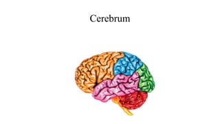
Cerebrum
• Largest part of brain
• Divided into 2 hemispheres
• Controls all conscious thoughts &
intellectual functions
• Processes somatic motor and sensory
information
External features
• Poles
• Surfaces
• Borders
• Sulci & gyri
Poles
• 3 poles
• Frontal
• Occipital
• Temporal
Surfaces
• 3 surfaces
• Superolateral surface
• Medial surface
• Inferior surface
Borders
• 3 borders
• Superomedial
• Inferolaeral
• Inferomedial
• Medial orbital
• Medial occipital
•
Gyrus & sulci
• To increase surface area
• To accommodate more neurons
• Sulcus
• Indentation (groove)
• Gyrus
• Ridge (elevation)
Fissures
• Deep groove
• Longitudinal fissure
• Separates two hemispheres
Grey & white matter
• Outside
• Cortex
• Neurons present
• White
• Axons present
Main Sulci
• On superolateral surface
• Two prominent sulci are present
• Posterior ramus of the lateral sulcus
• Central sulcus
• On medial surface - near occipital pole
• Parietooccipital sulcus
• Upper end of this sulcus reaches
superomedial border
• Small part of it can be seen on superolateral
surface
Lobes
• Frontal lobe
• Anterior to central sulcus and
• Above posterior ramus of lateral sulcus
• Parietal lobe
• Behind central sulcus
• Bounded below by posterior ramus of the
lateral sulcus and by the second
imaginary line and
• Behind by the upper part of the first
imaginary line
Lobes
• Occipital lobe
• Area lying behind the first imaginary
line connecting parieto-occipital sulcus
and pre-occipital notch
• Temporal lobe
• Below posterior ramus of the lateral
sulcus and the second imaginary line
• Separated from the occipital lobe by the
lower part of the first imaginary line
Sulci and gyri of frontal lobe
• Precentral sulcus
• Superior frontal sulcus
• Inferior frontal sulcus
• Precentral gyrus
• Superior frontal gyrus
• Middle frontal gyrus
• Inferior frontal gyrus
Sulci and gyri of parietal lobe
• Postcentral sulcus
• Intraparietal sulcus
• Postcentral gyrus
• Superior parietal lobule
• Inferior parietal lobule
• Supramarginal gyrus
• Angular gyrus
• Arcus temporo-occipitalis
Sulci and gyri of temporal lobe
• Superior temporal
sulcus
• Inferior temporal
sulcus
• Superior temporal gyrus
• Middle temporal gyrus
• Inferior temporal gyrus
• Transverse temporal gyri
Sulci & gyri of occipital lobe
• Lateral occipital sulcus
• Divides the lateral surface into
• Superior occipital gyrus
• Inferior occipital gyrus
• Lunate sulcus
• Transverse occipital sulcus
• Lies posterior to parieto-occipital sulcus
• Gyrus descendens
• Arcus parietooccipitalis
Sulci & gyri on medial surface
• Callosal sulcus
• Cingulate sulcus
• Short sulcus
• Cingulate gyrus
• Paracentral lobule
• Medial frontal gyrus
Sulci & gyri on medial surface
• Suprasplenial sulcus
• Parietooccipital sulcus
• Calcarine sulcus
• Cuneus
• Precuneus
• Isthmus
Structures in inferior surface
• In centre
• Interpeduncular fossa
• Between 2 cerebral
hemispheres
• Inferior surface of
hemisphere can be
divided into
• Orbital & tentorial
surfaces
Interpeduncular fossa
• Lies anterior to midbrain
• Boundaries
• In front by
• Optic chiasma
• Anterolatealrly by
• Right and left optic tracts
• Posterolaterally
• Crus cerebri
• Posteriorly
• Pons
Sulci & gyri on orbital surface
• Olfactory sulcus
• Antero posterior sulcus near medial border
• Olfactory bulb & tract
• Gyrus rectus
• Medial to olfactory sulcus
• Orbital sulcus
• H – shaped
• Anterior, posterior, medial & lateral orbital gyri
Sulci & gyri on tentorial surface
• Collateral sulcus
• Near medial border
• Posteriorly forms lateral boundary for Lingual gyrus
• Anteriorly forms lateral boundary for parahippocampal gyrus
• Rhinal sulcus
• Separates uncus from medial occipito temporal gyrus
• Occipitotemporal sulcus
• Lateral one
• Divides the remaining gyri into
• Medial & lateral occipito temporal gyri
Insula
• Submerged part of cerebral cortex
• Found in depth of lateral sulcus
• Surrounded by circular sulcus
Lentiform nucleus lies deep to it
Functional Areas of Cerebral Cortex
• In 1909, Brodmann’s areas
were described based on
cytoarchitecture
• Later they were found to be
functionally significant
• Cytoarchitecture is based on
• Density of different cortical
neurons and thickness of layers
•
• 6 layers are present in cortex
• Molecular –outermost
• Outer granular – stellate & granule cells
• Outer pyramidal – small & medium
• Inner granular – stellate cells
• Inner pyramidal – medium & large
• Polymorphous – Marinoti cells & multipolar
Functional areas in frontal lobe
• Primary motor
• Premotor
• Frontal eye field
• Supplementary motor
• Prefrontal
• Broca’s area
Functional areas in frontal lobe
• Precentral motor cortex (area 4)
• Infront of central sulcus including anterior
wall of central sulcus
• Anterior part of paracentral lobule
• Has large pyramidal cells of Betz
• Opposite side body is topographically
represented
• In upside down manner (head is lower and leg
is up
• Lower limb and perineum are on medial side
• Controls highly skilled voluntary
movement of opposite half of body
• Voluntary control of micturition &
defecation are in anterior part of
paracentral lobule
Functional areas in frontal lobe
• Premotor cortex (area 6)
• Lies infront of motor cortex
• In posterior parts of superior middle and
inferior frontal gyri
• Inferiorly upto lateral sucus
• Superiorly extends into medial surface
• Anterior to paracentral lobule
• Upper part – writing centre
• Lower part & around ascending ramus
of lateral sulcus
• Motor speech area (44 & 45) (Broca’s area)
Functional areas in frontal lobe
• Frontal eye field (8)
• Ability of the eyes to work together
• To follow an object
• Lies infront of upper part of premotor
area
• In posterior part of middle frontal gyrus
• Controls conjugate movements of eye
• Prefrontal cortex (9, 10 & 11)
• Essential for thought process
• Examples
• Solving mathematical problems
• Having foresight
• Giving judgments
Functional areas in frontal lobe
• Supplementary motor area (24)
• Primary motor area (6) continues on
medial surface
• Function
• Programming sequential motor
movements
• Lesion
• Results loss in coordination in
movements of two limbs
Functional areas in parietal lobe
• Primary somatosensory
• Secondary somatosensory
• Gustatory
• Association
Functional areas in parietal lobe
• Primary somatosensory area (3, 1 & 2)
• Lies in
• Posterior wall (3) and lip of central sulcus (1)
• Post central gyrus (2)
• Extends into paracentral lobule
• Receives sense of bladder & rectum fullness
• Functions
• Tactile localization between the 2 points discrimination & tactile
discrimination between 2 objects
Functional areas in parietal lobe
• Secondary somatosensory
• Lies along
• Upper lip of Posterior ramus of
lateral sulcus
• Functions
• Responds to stroking the skin
by brush or tapping
Functional areas in parietal lobe
• Somesthetic association area (5)
• Lies in
• Anterior part of superior parietal lobule
• Function
• Integrates tactile & visual stimuli
• To determine shape & size of an object
Functional areas in temporal lobe
• Primary auditory (41 & 42))
• Auditory association (22)
• Visual association
• Limbic
Functional areas in temporal lobe
• Primary auditory area (41 & 42)
• Located in
• Transverse temporal gyrus
• In floor of posterior ramus of lateral sulcus &
adjoining superior temporal gyrus
• Function
• Detecting the direction of the sound
Functional areas in temporal lobe
• Auditory association area (22)
• Lies around primary auditory area
• In middle of superior temporal gyrus
Functional areas in occipital lobe
• Primary visual cortex (17)
• Called as striate cortex
• Lies in
• Lips & walls of
• Posterior part of calcarine sulcus
Functional areas in occipital lobe
• Second visual area (18)
• Lies close to primary visual area
• Function
• Understand the written language

More Related Content

What's hot

Cerebrum
CerebrumCerebrum
Cerebrum
Dr.B.B. Gosai
 
Cerebrum
CerebrumCerebrum
Cerebrum
Dr. sana yaseen
 
Thalamus
ThalamusThalamus
Thalamus
Sachin Adukia
 
Cervical Plexus
Cervical PlexusCervical Plexus
Cervical Plexus
Rashmi Priyem Saravanan
 
Norma Verticalis
Norma VerticalisNorma Verticalis
Norma Verticalis
Rashmi Priyem Saravanan
 
Thalamus, anatomy of thalamus, Thalamus PPT
Thalamus, anatomy of thalamus, Thalamus PPTThalamus, anatomy of thalamus, Thalamus PPT
Thalamus, anatomy of thalamus, Thalamus PPT
drasarma1947
 
Medulla Oblongata
Medulla OblongataMedulla Oblongata
Medulla Oblongata
Vijay Kevlani
 
BACK OF NECK & SUB OCCIPITAL TRIANGLE
BACK OF NECK & SUB OCCIPITAL TRIANGLEBACK OF NECK & SUB OCCIPITAL TRIANGLE
BACK OF NECK & SUB OCCIPITAL TRIANGLE
Kanav Bhanot
 
Anatomy of diencephalon
Anatomy of diencephalonAnatomy of diencephalon
Anatomy of diencephalon
Dr. Mohammad Mahmoud
 
Anatomy head and neck(norma frontalis)
Anatomy head and neck(norma frontalis)Anatomy head and neck(norma frontalis)
Anatomy head and neck(norma frontalis)
AreebFatimaPT
 
Spinal cord neuroanatomy
Spinal cord neuroanatomySpinal cord neuroanatomy
Spinal cord neuroanatomy
Siva Pesala
 
Anatomy of the Cerebrum
Anatomy of the CerebrumAnatomy of the Cerebrum
Anatomy of the Cerebrum
profgoodnewszion
 
15 dural venous sinuses
15 dural venous sinuses15 dural venous sinuses
15 dural venous sinuses
Med Study
 
Antomy of Thalamus and hypothalamus
Antomy of Thalamus and hypothalamusAntomy of Thalamus and hypothalamus
Antomy of Thalamus and hypothalamus
Dr Laxman Khanal
 
Basal ganglia
Basal gangliaBasal ganglia
Basal ganglia
Chaitra Chinmayee M K
 
Parotid gland
Parotid gland Parotid gland
Parotid gland
Dr Suyashi
 
Midbrain And Cerebellum
Midbrain And CerebellumMidbrain And Cerebellum
Midbrain And Cerebellum
Dr.Faris Muhammed
 
Deep cervical fascia (fascia colli)
Deep cervical fascia (fascia colli)Deep cervical fascia (fascia colli)
Deep cervical fascia (fascia colli)surajitkundu
 

What's hot (20)

Cerebrum
CerebrumCerebrum
Cerebrum
 
Cerebrum
CerebrumCerebrum
Cerebrum
 
Thalamus
ThalamusThalamus
Thalamus
 
Cervical Plexus
Cervical PlexusCervical Plexus
Cervical Plexus
 
Norma Verticalis
Norma VerticalisNorma Verticalis
Norma Verticalis
 
Thalamus, anatomy of thalamus, Thalamus PPT
Thalamus, anatomy of thalamus, Thalamus PPTThalamus, anatomy of thalamus, Thalamus PPT
Thalamus, anatomy of thalamus, Thalamus PPT
 
Medulla Oblongata
Medulla OblongataMedulla Oblongata
Medulla Oblongata
 
BACK OF NECK & SUB OCCIPITAL TRIANGLE
BACK OF NECK & SUB OCCIPITAL TRIANGLEBACK OF NECK & SUB OCCIPITAL TRIANGLE
BACK OF NECK & SUB OCCIPITAL TRIANGLE
 
Anatomy of diencephalon
Anatomy of diencephalonAnatomy of diencephalon
Anatomy of diencephalon
 
Anatomy head and neck(norma frontalis)
Anatomy head and neck(norma frontalis)Anatomy head and neck(norma frontalis)
Anatomy head and neck(norma frontalis)
 
Spinal cord neuroanatomy
Spinal cord neuroanatomySpinal cord neuroanatomy
Spinal cord neuroanatomy
 
Norma lateralis
Norma lateralisNorma lateralis
Norma lateralis
 
Anatomy of the Cerebrum
Anatomy of the CerebrumAnatomy of the Cerebrum
Anatomy of the Cerebrum
 
15 dural venous sinuses
15 dural venous sinuses15 dural venous sinuses
15 dural venous sinuses
 
Antomy of Thalamus and hypothalamus
Antomy of Thalamus and hypothalamusAntomy of Thalamus and hypothalamus
Antomy of Thalamus and hypothalamus
 
Basal ganglia
Basal gangliaBasal ganglia
Basal ganglia
 
Parotid gland
Parotid gland Parotid gland
Parotid gland
 
Midbrain And Cerebellum
Midbrain And CerebellumMidbrain And Cerebellum
Midbrain And Cerebellum
 
Cerebrum
CerebrumCerebrum
Cerebrum
 
Deep cervical fascia (fascia colli)
Deep cervical fascia (fascia colli)Deep cervical fascia (fascia colli)
Deep cervical fascia (fascia colli)
 

Similar to Cerebrum

HOD cerebrum- areas and limbic system.pptx
HOD cerebrum- areas and  limbic system.pptxHOD cerebrum- areas and  limbic system.pptx
HOD cerebrum- areas and limbic system.pptx
ZainabRasheed15
 
bordder syurfaces, poles and the Sulci and gyri of cerebrum
bordder syurfaces, poles and the Sulci and gyri of cerebrumbordder syurfaces, poles and the Sulci and gyri of cerebrum
bordder syurfaces, poles and the Sulci and gyri of cerebrum
Dr. sana yaseen
 
anatomy cerebrum.pptx
anatomy cerebrum.pptxanatomy cerebrum.pptx
anatomy cerebrum.pptx
Agraj Mishra
 
Cerebrum
CerebrumCerebrum
Cerebrum
Shrikalla Nair
 
Cerebrum (motor and sensory cortex) + basal ganglia.pptx
Cerebrum (motor and sensory cortex) + basal ganglia.pptxCerebrum (motor and sensory cortex) + basal ganglia.pptx
Cerebrum (motor and sensory cortex) + basal ganglia.pptx
ssuser7ff432
 
Motor cortex
Motor cortexMotor cortex
Motor cortex
Asir John Samuel
 
cerebrum ,corpus callosum, basal nuclei .pptx
cerebrum ,corpus callosum, basal nuclei .pptxcerebrum ,corpus callosum, basal nuclei .pptx
cerebrum ,corpus callosum, basal nuclei .pptx
sahilsinghrajput769
 
Cerebrum part of brain anatomy for mbbs and dpt.ppt
Cerebrum part of brain anatomy for mbbs and dpt.pptCerebrum part of brain anatomy for mbbs and dpt.ppt
Cerebrum part of brain anatomy for mbbs and dpt.ppt
TaroTari
 
Ch 12 lecture_outline_a
Ch 12 lecture_outline_aCh 12 lecture_outline_a
Ch 12 lecture_outline_aTheSlaps
 
Ch 12 lecture_outline_a
Ch 12 lecture_outline_aCh 12 lecture_outline_a
Ch 12 lecture_outline_aTheSlaps
 
frontal lobe anatomy and clinical relevance
frontal lobe anatomy and clinical relevancefrontal lobe anatomy and clinical relevance
frontal lobe anatomy and clinical relevance
Imran Rizvi
 
Central Nervous System.pdf
Central Nervous System.pdfCentral Nervous System.pdf
Central Nervous System.pdf
RekhaChaudhary20
 
Orbit-&-it’s-content.pdf
Orbit-&-it’s-content.pdfOrbit-&-it’s-content.pdf
Orbit-&-it’s-content.pdf
Qman3
 
Cerebral cortex & its components
Cerebral cortex & its componentsCerebral cortex & its components
Cerebral cortex & its components
Dr Anupam Mittal
 
Medulla
MedullaMedulla
Medulla
anatomysrs
 
Brodmann's areas of the cerebral cortex
Brodmann's areas of the cerebral cortexBrodmann's areas of the cerebral cortex
Brodmann's areas of the cerebral cortex
Dr. Mohammed Sadiq Azam M.D.
 
Neuroanatomy Cerebrum summary slide.pptx
Neuroanatomy Cerebrum summary slide.pptxNeuroanatomy Cerebrum summary slide.pptx
Neuroanatomy Cerebrum summary slide.pptx
TalentAshjay
 
Frontal lobe
Frontal lobeFrontal lobe
Frontal lobe
Sumit Sharma
 
Tracts+oblongata+2021
Tracts+oblongata+2021Tracts+oblongata+2021
Tracts+oblongata+2021
obaje godwin sunday
 

Similar to Cerebrum (20)

HOD cerebrum- areas and limbic system.pptx
HOD cerebrum- areas and  limbic system.pptxHOD cerebrum- areas and  limbic system.pptx
HOD cerebrum- areas and limbic system.pptx
 
bordder syurfaces, poles and the Sulci and gyri of cerebrum
bordder syurfaces, poles and the Sulci and gyri of cerebrumbordder syurfaces, poles and the Sulci and gyri of cerebrum
bordder syurfaces, poles and the Sulci and gyri of cerebrum
 
anatomy cerebrum.pptx
anatomy cerebrum.pptxanatomy cerebrum.pptx
anatomy cerebrum.pptx
 
Cerebrum
CerebrumCerebrum
Cerebrum
 
Cerebrum (motor and sensory cortex) + basal ganglia.pptx
Cerebrum (motor and sensory cortex) + basal ganglia.pptxCerebrum (motor and sensory cortex) + basal ganglia.pptx
Cerebrum (motor and sensory cortex) + basal ganglia.pptx
 
Motor cortex
Motor cortexMotor cortex
Motor cortex
 
cerebrum ,corpus callosum, basal nuclei .pptx
cerebrum ,corpus callosum, basal nuclei .pptxcerebrum ,corpus callosum, basal nuclei .pptx
cerebrum ,corpus callosum, basal nuclei .pptx
 
Cerebrum part of brain anatomy for mbbs and dpt.ppt
Cerebrum part of brain anatomy for mbbs and dpt.pptCerebrum part of brain anatomy for mbbs and dpt.ppt
Cerebrum part of brain anatomy for mbbs and dpt.ppt
 
Ch 12 lecture_outline_a
Ch 12 lecture_outline_aCh 12 lecture_outline_a
Ch 12 lecture_outline_a
 
Ch 12 lecture_outline_a
Ch 12 lecture_outline_aCh 12 lecture_outline_a
Ch 12 lecture_outline_a
 
frontal lobe anatomy and clinical relevance
frontal lobe anatomy and clinical relevancefrontal lobe anatomy and clinical relevance
frontal lobe anatomy and clinical relevance
 
Motor system
Motor systemMotor system
Motor system
 
Central Nervous System.pdf
Central Nervous System.pdfCentral Nervous System.pdf
Central Nervous System.pdf
 
Orbit-&-it’s-content.pdf
Orbit-&-it’s-content.pdfOrbit-&-it’s-content.pdf
Orbit-&-it’s-content.pdf
 
Cerebral cortex & its components
Cerebral cortex & its componentsCerebral cortex & its components
Cerebral cortex & its components
 
Medulla
MedullaMedulla
Medulla
 
Brodmann's areas of the cerebral cortex
Brodmann's areas of the cerebral cortexBrodmann's areas of the cerebral cortex
Brodmann's areas of the cerebral cortex
 
Neuroanatomy Cerebrum summary slide.pptx
Neuroanatomy Cerebrum summary slide.pptxNeuroanatomy Cerebrum summary slide.pptx
Neuroanatomy Cerebrum summary slide.pptx
 
Frontal lobe
Frontal lobeFrontal lobe
Frontal lobe
 
Tracts+oblongata+2021
Tracts+oblongata+2021Tracts+oblongata+2021
Tracts+oblongata+2021
 

More from anatomysrs

arches of foot.pptx
arches of foot.pptxarches of foot.pptx
arches of foot.pptx
anatomysrs
 
pleura 2022.pptx
pleura 2022.pptxpleura 2022.pptx
pleura 2022.pptx
anatomysrs
 
cerebellum.pptx
cerebellum.pptxcerebellum.pptx
cerebellum.pptx
anatomysrs
 
anterior abdominal wall.pptx
anterior abdominal wall.pptxanterior abdominal wall.pptx
anterior abdominal wall.pptx
anatomysrs
 
diaphragm.pptx
diaphragm.pptxdiaphragm.pptx
diaphragm.pptx
anatomysrs
 
Caecum, appendix inferior mesenteric artery.pptx
Caecum, appendix inferior mesenteric artery.pptxCaecum, appendix inferior mesenteric artery.pptx
Caecum, appendix inferior mesenteric artery.pptx
anatomysrs
 
anal canal.pptx
anal canal.pptxanal canal.pptx
anal canal.pptx
anatomysrs
 
ureter & urinary bladder -.pptx
ureter & urinary bladder -.pptxureter & urinary bladder -.pptx
ureter & urinary bladder -.pptx
anatomysrs
 
sacroiliac .pptx
sacroiliac .pptxsacroiliac .pptx
sacroiliac .pptx
anatomysrs
 
skeletal system21-22.pptx
skeletal system21-22.pptxskeletal system21-22.pptx
skeletal system21-22.pptx
anatomysrs
 
Integumentary system -mbbs.pptx
Integumentary  system -mbbs.pptxIntegumentary  system -mbbs.pptx
Integumentary system -mbbs.pptx
anatomysrs
 
Forearm muscles.pptx
Forearm muscles.pptxForearm muscles.pptx
Forearm muscles.pptx
anatomysrs
 
Brachial plexus.pptx
Brachial  plexus.pptxBrachial  plexus.pptx
Brachial plexus.pptx
anatomysrs
 
Wrist & I carpometacarpal.pptx
Wrist & I carpometacarpal.pptxWrist & I carpometacarpal.pptx
Wrist & I carpometacarpal.pptx
anatomysrs
 
Anatomy of nose
Anatomy of noseAnatomy of nose
Anatomy of nose
anatomysrs
 
Maxillary nerve
Maxillary nerveMaxillary nerve
Maxillary nerve
anatomysrs
 
Extra ocular muscles
Extra ocular musclesExtra ocular muscles
Extra ocular muscles
anatomysrs
 
Suboccipital triangle
Suboccipital triangleSuboccipital triangle
Suboccipital triangle
anatomysrs
 
Cervical fascia
Cervical fasciaCervical fascia
Cervical fascia
anatomysrs
 
Face
FaceFace

More from anatomysrs (20)

arches of foot.pptx
arches of foot.pptxarches of foot.pptx
arches of foot.pptx
 
pleura 2022.pptx
pleura 2022.pptxpleura 2022.pptx
pleura 2022.pptx
 
cerebellum.pptx
cerebellum.pptxcerebellum.pptx
cerebellum.pptx
 
anterior abdominal wall.pptx
anterior abdominal wall.pptxanterior abdominal wall.pptx
anterior abdominal wall.pptx
 
diaphragm.pptx
diaphragm.pptxdiaphragm.pptx
diaphragm.pptx
 
Caecum, appendix inferior mesenteric artery.pptx
Caecum, appendix inferior mesenteric artery.pptxCaecum, appendix inferior mesenteric artery.pptx
Caecum, appendix inferior mesenteric artery.pptx
 
anal canal.pptx
anal canal.pptxanal canal.pptx
anal canal.pptx
 
ureter & urinary bladder -.pptx
ureter & urinary bladder -.pptxureter & urinary bladder -.pptx
ureter & urinary bladder -.pptx
 
sacroiliac .pptx
sacroiliac .pptxsacroiliac .pptx
sacroiliac .pptx
 
skeletal system21-22.pptx
skeletal system21-22.pptxskeletal system21-22.pptx
skeletal system21-22.pptx
 
Integumentary system -mbbs.pptx
Integumentary  system -mbbs.pptxIntegumentary  system -mbbs.pptx
Integumentary system -mbbs.pptx
 
Forearm muscles.pptx
Forearm muscles.pptxForearm muscles.pptx
Forearm muscles.pptx
 
Brachial plexus.pptx
Brachial  plexus.pptxBrachial  plexus.pptx
Brachial plexus.pptx
 
Wrist & I carpometacarpal.pptx
Wrist & I carpometacarpal.pptxWrist & I carpometacarpal.pptx
Wrist & I carpometacarpal.pptx
 
Anatomy of nose
Anatomy of noseAnatomy of nose
Anatomy of nose
 
Maxillary nerve
Maxillary nerveMaxillary nerve
Maxillary nerve
 
Extra ocular muscles
Extra ocular musclesExtra ocular muscles
Extra ocular muscles
 
Suboccipital triangle
Suboccipital triangleSuboccipital triangle
Suboccipital triangle
 
Cervical fascia
Cervical fasciaCervical fascia
Cervical fascia
 
Face
FaceFace
Face
 

Recently uploaded

PrudentRx's Function in the Management of Chronic Illnesses
PrudentRx's Function in the Management of Chronic IllnessesPrudentRx's Function in the Management of Chronic Illnesses
PrudentRx's Function in the Management of Chronic Illnesses
PrudentRx Program
 
Letter to MREC - application to conduct study
Letter to MREC - application to conduct studyLetter to MREC - application to conduct study
Letter to MREC - application to conduct study
Azreen Aj
 
ABDOMINAL COMPARTMENT SYSNDROME
ABDOMINAL COMPARTMENT SYSNDROMEABDOMINAL COMPARTMENT SYSNDROME
ABDOMINAL COMPARTMENT SYSNDROME
Rommel Luis III Israel
 
Time line.ppQAWSDRFTGYUIOPÑLKIUYTREWASDFTGY
Time line.ppQAWSDRFTGYUIOPÑLKIUYTREWASDFTGYTime line.ppQAWSDRFTGYUIOPÑLKIUYTREWASDFTGY
Time line.ppQAWSDRFTGYUIOPÑLKIUYTREWASDFTGY
DianaRodriguez639773
 
Rate Controlled Drug Delivery Systems.pdf
Rate Controlled Drug Delivery Systems.pdfRate Controlled Drug Delivery Systems.pdf
Rate Controlled Drug Delivery Systems.pdf
Rajarambapu College of Pharmacy Kasegaon Dist Sangli
 
Empowering ACOs: Leveraging Quality Management Tools for MIPS and Beyond
Empowering ACOs: Leveraging Quality Management Tools for MIPS and BeyondEmpowering ACOs: Leveraging Quality Management Tools for MIPS and Beyond
Empowering ACOs: Leveraging Quality Management Tools for MIPS and Beyond
Health Catalyst
 
Dimensions of Healthcare Quality
Dimensions of Healthcare QualityDimensions of Healthcare Quality
Dimensions of Healthcare Quality
Naeemshahzad51
 
PET CT beginners Guide covers some of the underrepresented topics in PET CT
PET CT  beginners Guide  covers some of the underrepresented topics  in PET CTPET CT  beginners Guide  covers some of the underrepresented topics  in PET CT
PET CT beginners Guide covers some of the underrepresented topics in PET CT
MiadAlsulami
 
NEEDLE STICK INJURY - JOURNAL CLUB PRESENTATION - DR SHAMIN EABENSON
NEEDLE STICK INJURY - JOURNAL CLUB PRESENTATION - DR SHAMIN EABENSONNEEDLE STICK INJURY - JOURNAL CLUB PRESENTATION - DR SHAMIN EABENSON
NEEDLE STICK INJURY - JOURNAL CLUB PRESENTATION - DR SHAMIN EABENSON
SHAMIN EABENSON
 
Gemma Wean- Nutritional solution for Artemia
Gemma Wean- Nutritional solution for ArtemiaGemma Wean- Nutritional solution for Artemia
Gemma Wean- Nutritional solution for Artemia
smuskaan0008
 
Deep Leg Vein Thrombosis (DVT): Meaning, Causes, Symptoms, Treatment, and Mor...
Deep Leg Vein Thrombosis (DVT): Meaning, Causes, Symptoms, Treatment, and Mor...Deep Leg Vein Thrombosis (DVT): Meaning, Causes, Symptoms, Treatment, and Mor...
Deep Leg Vein Thrombosis (DVT): Meaning, Causes, Symptoms, Treatment, and Mor...
The Lifesciences Magazine
 
Bringing AI into a Mid-Sized Company: A structured Approach
Bringing AI into a Mid-Sized Company: A structured ApproachBringing AI into a Mid-Sized Company: A structured Approach
Bringing AI into a Mid-Sized Company: A structured Approach
Brian Frerichs
 
KEY Points of Leicester travel clinic In London doc.docx
KEY Points of Leicester travel clinic In London doc.docxKEY Points of Leicester travel clinic In London doc.docx
KEY Points of Leicester travel clinic In London doc.docx
NX Healthcare
 
CANSA support - Caring for Cancer Patients' Caregivers
CANSA support - Caring for Cancer Patients' CaregiversCANSA support - Caring for Cancer Patients' Caregivers
CANSA support - Caring for Cancer Patients' Caregivers
CANSA The Cancer Association of South Africa
 
Stem Cell Solutions: Dr. David Greene's Path to Non-Surgical Cardiac Care
Stem Cell Solutions: Dr. David Greene's Path to Non-Surgical Cardiac CareStem Cell Solutions: Dr. David Greene's Path to Non-Surgical Cardiac Care
Stem Cell Solutions: Dr. David Greene's Path to Non-Surgical Cardiac Care
Dr. David Greene Arizona
 
TEST BANK For Accounting Information Systems, 3rd Edition by Vernon Richardso...
TEST BANK For Accounting Information Systems, 3rd Edition by Vernon Richardso...TEST BANK For Accounting Information Systems, 3rd Edition by Vernon Richardso...
TEST BANK For Accounting Information Systems, 3rd Edition by Vernon Richardso...
rightmanforbloodline
 
Nursing education curriculum development.pptx
Nursing education curriculum development.pptxNursing education curriculum development.pptx
Nursing education curriculum development.pptx
sadhanajagtap3
 
NKTI Annual Report - Annual Report FY 2022
NKTI Annual Report - Annual Report FY 2022NKTI Annual Report - Annual Report FY 2022
NKTI Annual Report - Annual Report FY 2022
nktiacc3
 
HUMAN BRAIN.pptx.PRIYA BHOJWANI@GAMIL.COM
HUMAN BRAIN.pptx.PRIYA BHOJWANI@GAMIL.COMHUMAN BRAIN.pptx.PRIYA BHOJWANI@GAMIL.COM
HUMAN BRAIN.pptx.PRIYA BHOJWANI@GAMIL.COM
priyabhojwani1200
 
Trauma Outpatient Center .
Trauma Outpatient Center                       .Trauma Outpatient Center                       .
Trauma Outpatient Center .
TraumaOutpatientCent
 

Recently uploaded (20)

PrudentRx's Function in the Management of Chronic Illnesses
PrudentRx's Function in the Management of Chronic IllnessesPrudentRx's Function in the Management of Chronic Illnesses
PrudentRx's Function in the Management of Chronic Illnesses
 
Letter to MREC - application to conduct study
Letter to MREC - application to conduct studyLetter to MREC - application to conduct study
Letter to MREC - application to conduct study
 
ABDOMINAL COMPARTMENT SYSNDROME
ABDOMINAL COMPARTMENT SYSNDROMEABDOMINAL COMPARTMENT SYSNDROME
ABDOMINAL COMPARTMENT SYSNDROME
 
Time line.ppQAWSDRFTGYUIOPÑLKIUYTREWASDFTGY
Time line.ppQAWSDRFTGYUIOPÑLKIUYTREWASDFTGYTime line.ppQAWSDRFTGYUIOPÑLKIUYTREWASDFTGY
Time line.ppQAWSDRFTGYUIOPÑLKIUYTREWASDFTGY
 
Rate Controlled Drug Delivery Systems.pdf
Rate Controlled Drug Delivery Systems.pdfRate Controlled Drug Delivery Systems.pdf
Rate Controlled Drug Delivery Systems.pdf
 
Empowering ACOs: Leveraging Quality Management Tools for MIPS and Beyond
Empowering ACOs: Leveraging Quality Management Tools for MIPS and BeyondEmpowering ACOs: Leveraging Quality Management Tools for MIPS and Beyond
Empowering ACOs: Leveraging Quality Management Tools for MIPS and Beyond
 
Dimensions of Healthcare Quality
Dimensions of Healthcare QualityDimensions of Healthcare Quality
Dimensions of Healthcare Quality
 
PET CT beginners Guide covers some of the underrepresented topics in PET CT
PET CT  beginners Guide  covers some of the underrepresented topics  in PET CTPET CT  beginners Guide  covers some of the underrepresented topics  in PET CT
PET CT beginners Guide covers some of the underrepresented topics in PET CT
 
NEEDLE STICK INJURY - JOURNAL CLUB PRESENTATION - DR SHAMIN EABENSON
NEEDLE STICK INJURY - JOURNAL CLUB PRESENTATION - DR SHAMIN EABENSONNEEDLE STICK INJURY - JOURNAL CLUB PRESENTATION - DR SHAMIN EABENSON
NEEDLE STICK INJURY - JOURNAL CLUB PRESENTATION - DR SHAMIN EABENSON
 
Gemma Wean- Nutritional solution for Artemia
Gemma Wean- Nutritional solution for ArtemiaGemma Wean- Nutritional solution for Artemia
Gemma Wean- Nutritional solution for Artemia
 
Deep Leg Vein Thrombosis (DVT): Meaning, Causes, Symptoms, Treatment, and Mor...
Deep Leg Vein Thrombosis (DVT): Meaning, Causes, Symptoms, Treatment, and Mor...Deep Leg Vein Thrombosis (DVT): Meaning, Causes, Symptoms, Treatment, and Mor...
Deep Leg Vein Thrombosis (DVT): Meaning, Causes, Symptoms, Treatment, and Mor...
 
Bringing AI into a Mid-Sized Company: A structured Approach
Bringing AI into a Mid-Sized Company: A structured ApproachBringing AI into a Mid-Sized Company: A structured Approach
Bringing AI into a Mid-Sized Company: A structured Approach
 
KEY Points of Leicester travel clinic In London doc.docx
KEY Points of Leicester travel clinic In London doc.docxKEY Points of Leicester travel clinic In London doc.docx
KEY Points of Leicester travel clinic In London doc.docx
 
CANSA support - Caring for Cancer Patients' Caregivers
CANSA support - Caring for Cancer Patients' CaregiversCANSA support - Caring for Cancer Patients' Caregivers
CANSA support - Caring for Cancer Patients' Caregivers
 
Stem Cell Solutions: Dr. David Greene's Path to Non-Surgical Cardiac Care
Stem Cell Solutions: Dr. David Greene's Path to Non-Surgical Cardiac CareStem Cell Solutions: Dr. David Greene's Path to Non-Surgical Cardiac Care
Stem Cell Solutions: Dr. David Greene's Path to Non-Surgical Cardiac Care
 
TEST BANK For Accounting Information Systems, 3rd Edition by Vernon Richardso...
TEST BANK For Accounting Information Systems, 3rd Edition by Vernon Richardso...TEST BANK For Accounting Information Systems, 3rd Edition by Vernon Richardso...
TEST BANK For Accounting Information Systems, 3rd Edition by Vernon Richardso...
 
Nursing education curriculum development.pptx
Nursing education curriculum development.pptxNursing education curriculum development.pptx
Nursing education curriculum development.pptx
 
NKTI Annual Report - Annual Report FY 2022
NKTI Annual Report - Annual Report FY 2022NKTI Annual Report - Annual Report FY 2022
NKTI Annual Report - Annual Report FY 2022
 
HUMAN BRAIN.pptx.PRIYA BHOJWANI@GAMIL.COM
HUMAN BRAIN.pptx.PRIYA BHOJWANI@GAMIL.COMHUMAN BRAIN.pptx.PRIYA BHOJWANI@GAMIL.COM
HUMAN BRAIN.pptx.PRIYA BHOJWANI@GAMIL.COM
 
Trauma Outpatient Center .
Trauma Outpatient Center                       .Trauma Outpatient Center                       .
Trauma Outpatient Center .
 

Cerebrum

  • 2. Cerebrum • Largest part of brain • Divided into 2 hemispheres • Controls all conscious thoughts & intellectual functions • Processes somatic motor and sensory information
  • 3. External features • Poles • Surfaces • Borders • Sulci & gyri
  • 4. Poles • 3 poles • Frontal • Occipital • Temporal
  • 5. Surfaces • 3 surfaces • Superolateral surface • Medial surface • Inferior surface
  • 6. Borders • 3 borders • Superomedial • Inferolaeral • Inferomedial • Medial orbital • Medial occipital •
  • 7. Gyrus & sulci • To increase surface area • To accommodate more neurons • Sulcus • Indentation (groove) • Gyrus • Ridge (elevation)
  • 8. Fissures • Deep groove • Longitudinal fissure • Separates two hemispheres
  • 9. Grey & white matter • Outside • Cortex • Neurons present • White • Axons present
  • 10. Main Sulci • On superolateral surface • Two prominent sulci are present • Posterior ramus of the lateral sulcus • Central sulcus • On medial surface - near occipital pole • Parietooccipital sulcus • Upper end of this sulcus reaches superomedial border • Small part of it can be seen on superolateral surface
  • 11. Lobes • Frontal lobe • Anterior to central sulcus and • Above posterior ramus of lateral sulcus • Parietal lobe • Behind central sulcus • Bounded below by posterior ramus of the lateral sulcus and by the second imaginary line and • Behind by the upper part of the first imaginary line
  • 12. Lobes • Occipital lobe • Area lying behind the first imaginary line connecting parieto-occipital sulcus and pre-occipital notch • Temporal lobe • Below posterior ramus of the lateral sulcus and the second imaginary line • Separated from the occipital lobe by the lower part of the first imaginary line
  • 13. Sulci and gyri of frontal lobe • Precentral sulcus • Superior frontal sulcus • Inferior frontal sulcus • Precentral gyrus • Superior frontal gyrus • Middle frontal gyrus • Inferior frontal gyrus
  • 14. Sulci and gyri of parietal lobe • Postcentral sulcus • Intraparietal sulcus • Postcentral gyrus • Superior parietal lobule • Inferior parietal lobule • Supramarginal gyrus • Angular gyrus • Arcus temporo-occipitalis
  • 15. Sulci and gyri of temporal lobe • Superior temporal sulcus • Inferior temporal sulcus • Superior temporal gyrus • Middle temporal gyrus • Inferior temporal gyrus • Transverse temporal gyri
  • 16. Sulci & gyri of occipital lobe • Lateral occipital sulcus • Divides the lateral surface into • Superior occipital gyrus • Inferior occipital gyrus • Lunate sulcus • Transverse occipital sulcus • Lies posterior to parieto-occipital sulcus • Gyrus descendens • Arcus parietooccipitalis
  • 17. Sulci & gyri on medial surface • Callosal sulcus • Cingulate sulcus • Short sulcus • Cingulate gyrus • Paracentral lobule • Medial frontal gyrus
  • 18. Sulci & gyri on medial surface • Suprasplenial sulcus • Parietooccipital sulcus • Calcarine sulcus • Cuneus • Precuneus • Isthmus
  • 19. Structures in inferior surface • In centre • Interpeduncular fossa • Between 2 cerebral hemispheres • Inferior surface of hemisphere can be divided into • Orbital & tentorial surfaces
  • 20. Interpeduncular fossa • Lies anterior to midbrain • Boundaries • In front by • Optic chiasma • Anterolatealrly by • Right and left optic tracts • Posterolaterally • Crus cerebri • Posteriorly • Pons
  • 21. Sulci & gyri on orbital surface • Olfactory sulcus • Antero posterior sulcus near medial border • Olfactory bulb & tract • Gyrus rectus • Medial to olfactory sulcus • Orbital sulcus • H – shaped • Anterior, posterior, medial & lateral orbital gyri
  • 22. Sulci & gyri on tentorial surface • Collateral sulcus • Near medial border • Posteriorly forms lateral boundary for Lingual gyrus • Anteriorly forms lateral boundary for parahippocampal gyrus • Rhinal sulcus • Separates uncus from medial occipito temporal gyrus • Occipitotemporal sulcus • Lateral one • Divides the remaining gyri into • Medial & lateral occipito temporal gyri
  • 23. Insula • Submerged part of cerebral cortex • Found in depth of lateral sulcus • Surrounded by circular sulcus Lentiform nucleus lies deep to it
  • 24. Functional Areas of Cerebral Cortex • In 1909, Brodmann’s areas were described based on cytoarchitecture • Later they were found to be functionally significant • Cytoarchitecture is based on • Density of different cortical neurons and thickness of layers • • 6 layers are present in cortex • Molecular –outermost • Outer granular – stellate & granule cells • Outer pyramidal – small & medium • Inner granular – stellate cells • Inner pyramidal – medium & large • Polymorphous – Marinoti cells & multipolar
  • 25. Functional areas in frontal lobe • Primary motor • Premotor • Frontal eye field • Supplementary motor • Prefrontal • Broca’s area
  • 26. Functional areas in frontal lobe • Precentral motor cortex (area 4) • Infront of central sulcus including anterior wall of central sulcus • Anterior part of paracentral lobule • Has large pyramidal cells of Betz • Opposite side body is topographically represented • In upside down manner (head is lower and leg is up • Lower limb and perineum are on medial side • Controls highly skilled voluntary movement of opposite half of body • Voluntary control of micturition & defecation are in anterior part of paracentral lobule
  • 27. Functional areas in frontal lobe • Premotor cortex (area 6) • Lies infront of motor cortex • In posterior parts of superior middle and inferior frontal gyri • Inferiorly upto lateral sucus • Superiorly extends into medial surface • Anterior to paracentral lobule • Upper part – writing centre • Lower part & around ascending ramus of lateral sulcus • Motor speech area (44 & 45) (Broca’s area)
  • 28. Functional areas in frontal lobe • Frontal eye field (8) • Ability of the eyes to work together • To follow an object • Lies infront of upper part of premotor area • In posterior part of middle frontal gyrus • Controls conjugate movements of eye • Prefrontal cortex (9, 10 & 11) • Essential for thought process • Examples • Solving mathematical problems • Having foresight • Giving judgments
  • 29. Functional areas in frontal lobe • Supplementary motor area (24) • Primary motor area (6) continues on medial surface • Function • Programming sequential motor movements • Lesion • Results loss in coordination in movements of two limbs
  • 30. Functional areas in parietal lobe • Primary somatosensory • Secondary somatosensory • Gustatory • Association
  • 31. Functional areas in parietal lobe • Primary somatosensory area (3, 1 & 2) • Lies in • Posterior wall (3) and lip of central sulcus (1) • Post central gyrus (2) • Extends into paracentral lobule • Receives sense of bladder & rectum fullness • Functions • Tactile localization between the 2 points discrimination & tactile discrimination between 2 objects
  • 32. Functional areas in parietal lobe • Secondary somatosensory • Lies along • Upper lip of Posterior ramus of lateral sulcus • Functions • Responds to stroking the skin by brush or tapping
  • 33. Functional areas in parietal lobe • Somesthetic association area (5) • Lies in • Anterior part of superior parietal lobule • Function • Integrates tactile & visual stimuli • To determine shape & size of an object
  • 34. Functional areas in temporal lobe • Primary auditory (41 & 42)) • Auditory association (22) • Visual association • Limbic
  • 35. Functional areas in temporal lobe • Primary auditory area (41 & 42) • Located in • Transverse temporal gyrus • In floor of posterior ramus of lateral sulcus & adjoining superior temporal gyrus • Function • Detecting the direction of the sound
  • 36. Functional areas in temporal lobe • Auditory association area (22) • Lies around primary auditory area • In middle of superior temporal gyrus
  • 37. Functional areas in occipital lobe • Primary visual cortex (17) • Called as striate cortex • Lies in • Lips & walls of • Posterior part of calcarine sulcus
  • 38. Functional areas in occipital lobe • Second visual area (18) • Lies close to primary visual area • Function • Understand the written language