
Recommandé
Contenu connexe
Tendances
Tendances (20)
Similaire à Liposomes ppt By Ankit S Parulkar
Similaire à Liposomes ppt By Ankit S Parulkar (20)
Dernier
Dernier (20)
Liposomes ppt By Ankit S Parulkar
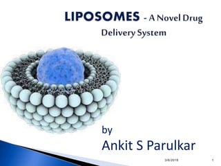
- 1. 3/6/2018 1 by Ankit S Parulkar
- 2. Liposomes are the simple microscopic vesicles in which aqueous layer is enclosed by phospho lipid bilayers that are used to transfer vaccines ,drugs ,enzymes and other substances to targetcells or organs Structually ,liposomes are concentric bilayerd vesicles in which an aqueous volume is entirely enclosed by a membraneous lipid bilayer mainly composed of natural or synthetic phospholipids. Can be produced from cholesterols, non toxic surfactants, sphingolipids, glycolipids, long chain fatty acids and even membrane proteins. 3/6/2018 2
- 3. 3/6/2018 3
- 4. COMPOSITION Phospholipids: Dilauryl phosphotidyl choline (DLPC), Dimyristoyl phosphotidyl choline (DMPC), Dipalmitoy phosphotidyl choline (DPPC), Distearoyl phosphotidyl choline (DSPC), Dioleolyl phosphotidyl choline (DOPC), Dilauryl phosphotidyl glycerol (DLPG), Distearoyl phosphotidyl serine (DSPS). Cholesterol: Act as intercalator with phospholipids molecules. Restrict the confirmational changes of lipids. Membrane stabilizer. 3/6/2018 4
- 5. ADVANTAGES Non-toxic. Biodegradable. Non-immunogenic. Lowers systemic toxicity. Targeted delivery. Protection of sensitive drug molecules. Enhance drug solubilisation ( Amphoterecin, Cyclosporins). Improved pharmacokinetic effects. 3/6/2018 5
- 6. • Leakage of encapsulated drug during storage. • Short half-life. • Batch to batch variation. • Difficult in large scale manufacturing and sterilization. • Production cost is high. • Once administered, liposomes can not be removed. • Some times phospholipids undergoes hydrolysis and oxidation reactions 3/6/2018 6
- 8. 3/6/2018 8
- 9. 3/6/2018 9
- 10. METHODS OF LIPOSOME PREPARATION PASSIVE LOADING Involves loading of the entrapped agents before or during the manufacturing procedure ACTIVE OR REMOTELOADING Certain types of compounds with ionizable groups and those with both lipid and solubility , can be introduced into the liposomes after the formation of the intact vesicles 3/6/2018 10
- 11. 3/6/2018 11
- 12. METHODS OF PREPARATION OF LIPOSOMES All the methods of preparing liposomes involve three or four basic stages • Drying down lipids from organic solvent • Dispersion of lipids in aqueous media • Purification of resultant liposomes • Analysis of final product 3/6/2018 12
- 13. Mechanical Dispersion Methods Hand shaken MLV’s Lipids + solvent ( chloroform: Methanol) ( In 250 ml RBF) Evaporate for 15 min above phase transition temperature(Flush with nitrogen) Till residues dry Add 5 ml buffer containing material to be entrapped Rotate flask at room temp, at 60 RPM for 30 min until lipid removes from wall of RBF Milky white dispersion (stand for 2 hours to get MLV Rotary Evaporator 3/6/2018 13
- 14. Pro liposome Sorbitol / Nacl ( increase surface area of lipid film) + 5ml lipid solution ( fitted to evaporator ) (Evaporation) Again add lipid solution Dry the content using Lyophilizer ( freeze dryer) (Stand over night at room temp) Flushed with nitrogen for drying properly MLVs 3/6/2018 15
- 15. Freeze Drying Lipid + Solvent ( Tertiary butanol) Freeze drying Add Aqueous phase / Saline containing drug Rapid mixing above phase transition temperature MLVs 3/6/2018 16
- 16. Micro emulsification liposome (MEL) •MEL is prepared by the “Micro fluidizer”, which pumps fluid at very high pressure (10,000 psi) through a 5 um orifice. •Then, it is forced along defined micro channels, which direct two streams of fluid to colloid together at right angle at very high velocity. •After a single pass, size reduced to a size 0.1& 0.2 um in diameter. 3/6/2018 17
- 18. Sonicated unilamellar vesicles MLV in test tube Sonicate for 5-10 min above phase transition temp Filter & centrifuge at 100000 rpm for 30 min at 20º c Decant top layer to get Sonicated unilamellar vesicles BATH SONICATOR PROBE SONICATOR 3/6/2018 20
- 19. French Pressure Cell • French pressure cell is invented by ‘Charles Stacy French. • In this technique the large vesicles are converted to small vesicles under very high pressure. •This technique yields uni or oligo lamellar liposomes of intermediate size (30-80 nm in diameter depending on applied pressure). •This liposomes are more stable as compared to sonicated liposomes. 3/6/2018 21
- 20. 3/6/2018 22
- 21. Membrane extrusion liposomes • Mixture of surfactant, cholesterol and dicetyl phosphate in chloroform is made into thin film by evaporation. The film is hydrated with aqueous drug solution and the resultant suspension extruded through polycarbonate membranes, which are placed in series for upto 8 passages. It is a good method for controlling liposome size. • Less pressure is required here (> 100 psi) as compare to French pressure cell. 3/6/2018 23
- 22. Freeze thaw sonication process 3/6/2018 24 The method is based on freezing of a unilamellar dispersion & then thawing at room temp for 15 min. Thus the process ruptures & refuses SUVs during which the solute equilibrates between inside & outside & liposomes themselves fuse & increase in size. Entrapment volume can be up to 30% of the total vol. of dispersion.
- 23. Dried reconstituted vesicle SUV in aqueous phase + Solute Freeze drying DRV method: Rehydration, film stacks dispersed in aqueous phase Solute in uni or oligo lamellar vesicles. 3/6/2018 25
- 24. Ph induced vesiculation MLVs or LUVs ( Ph 2.5-3) Add 1 M NaOH Ph rises to 11 Now add 0.1 M HCl Ph moves down to 7.5 SUV Change in Ph brings about an increase in surface charge density of lipid bilayer, which induces spontaneous vesiculation 3/6/2018 26
- 25. Solvent dispersion method: Ethanol injection Lipid + ethanol solution in the syringe Inject rapidly In the aqueous phase Small unilamellar vesicles 3/6/2018 27
- 26. Ether injection Lipid + ether solution in the syringe Inject slowly In the aqueous phase ( On heated water bath, 60ºc) Large unilamellar vesicles 3/6/2018 28
- 27. Water organic phase: Double emulsion Organic solution + Lipid + Aqueous phase Emulsion (W/O) Hot aqueous solution of buffer Multi compartment vesicle W/O/W (double emulsion) LUVs 3/6/2018 29
- 28. Reverse phase evaporation: (MLV, LUV) Emulsion Evaporation under reduced pressure, rotary evaporator Semi solid gel Shake to get LUVs “Lipid monolayer which enclosed the collapsed vesicle, is contributed to adjacent intact vesicle to form the outer leaflet of bilayer of LUV”. 3/6/2018 30
- 29. Stable plurilamellar vesicle (SPLVs) •It involves preparation of water in organic phase dispersion with an excess of lipid followed by drying under continued bath sonication with stream of nitrogen. •The internal SPLV is different from that of MLV, in that they lack a large aqueous core. •The internal environment of both the vesicle is different from each other. 3/6/2018 31
- 30. DETERGENTDEPLETION(REMOVAL)METHODS: • Detergents associate with the phospholipid molecules and serve to screen the hydrophobic portions of molecule from water. • The structures formed as a result of this association is known as micelles. • A three stage model of interaction for detergents with lipid bilayers: •Stage1: At low concentration detergents equilibrates between vesicular lipid and water phase. •Stage2: After reaching a critical detergent concentration, membrane structure tends to unstable and transforms gradually in to micelles. •Stage3: All lipid exists in mixed micelle form. • Three methods are applied for removal of detergent and transition of mixed micelles to concentric bilayered form. 3/6/2018 32
- 31. DIALYSIS: The molecules of detergent are removed from mixed micelle by dialysis by lowering the concentration of detergent in bulk aqueous phase. eg: sodium cholate. octylglucoside COLUMN CHROMATOGRAPHY: Removal of detergent is achieved by by passing the dispersion over a sephadexg-25 column pre-saturated with constitutive lipids and pre-equilibrated with hydrating buffer. eg: deoxycholate. 3/6/2018 33
- 32. Evaluation of liposomes • The liposomes prepared by various techniques are to be evaluated for their physical, chemical as well as biological properties, has these influence the behavior of liposomes in vivo. • Physical properties 1. Particle size • Both particle size and particle size distribution of liposomes influence their physical stability. These can be determined by the following method. • Laser light scattering • Transmission electron microscopy 2. Surface charge The passive, negative or natural charge on the surface of the liposomes is due to the composition of the head groups. 3/6/2018 34
- 33. •The surface charge of liposomes governs on the extent of distribution in-vivo, as well as interaction with the target cells. •The method involved in the measurement of surface charge is based on free-flow electrophoresis of MLVs. •It utilizes a cellulose acetate plate dipped in sodium borate buffer of Ph 8.8. •About 5n moles of lipid samples are applied on to the plate, which is then subjected to electrophoresis at 4 ͦc for 30 mins. •The liposomes get bifurcated depending on their surface charge. •This technique can be used for determining the heterogeneity of charges in the liposome suspension as well as to detect any impurities such as fatty acids 3/6/2018 35
- 34. 3. Percent drug encapsulated. • Quantity of drug entrapped in the liposomes helps to estimate the behavior of the drugin biological system • Liposomes are misture of encapsulated and unencapsulated drug fractions • The %of drug encapsulation is done by first separating the free drug fraction from encapsulated drug fraction • Themethods usedto separate the free drugfrom the sample are: a. Mini columncentrifugation method b. Protamine aggregated method 4. Phasebehavior •At transition temperature liposomes undergo reversible phase transition • Done by DSC 3/6/2018 36
- 35. 5. Drug Release Rate The rate of drug release from the liposomes can be determined by in-vivo assays which helps to predict the pharmacokinetics and bioavailability of the drug. However in- vivo studies are found to be more complete. 3/6/2018 37
- 36. Chemicalproperties 1. Determination of phospholipids The phospholipid content of liposomes can be determined directly by two assays, Bartlett assay and Steward assay. (a)Bartlett assay • This method of determining the phospholipid is very sensitive and may produce erroneous results in the presence of even trace amounts of inorganic phosphate. Therefore, borosilicate glass tubes and double-distilled water is used. Initially the phosphorous present in the lipid bilayer of the sample is hydrolyzed to inorganic phosphate. 3/6/2018 38
- 37. Then ammonium molybdate is added to convert inorganic phosphate to phosphomolybdic acid(PMA). The sample is then treated with aminonaphthylsulphonic acid to quantitatively reduce the PMA to a blue-coloured compound. The intensity of the blue colour produced can be measured by spectrophotometric means and the value is plotted on the standard curve to obtain the content of phospholipids. (b)Steward assay This assay overcomes the drawbacks of Bratlett assay, but cannot be used to mixture of unknown phospholipids. 3/6/2018 39
- 38. A standard curve is prepared by treating known concentration of phospholipids in chloroform with 0.1 M solution of ammonium ferrothiocyanate 9 reagent. The sample are also treated with the same reagent and the optical density is determined at 485 nm. The absorbance of the sample can be plotted on the standard curve to obtain the concentration of phospholipids. 2. Cholesterol analysis Qualitative analysis Performed using a capillary column filled with fused silica. Quantitative analysis The sample is reacted with a reagent (containing ferric perchlorate, ethyl acetate and H₂SO₄) and the absorbance of purple coloured complex is measured at 610 nm. 3/6/2018 40
- 39. MODES OF LIPOSOME AND CELL INTERACTION: Adsorption Endocytosis Fusion Lipid transfer 3/6/2018 41
- 41. Other applications 1). Liposomes in parasitic diseaseases and infections •Liposomses get digested by phagocytic cells in body making them as ideal vehicles for tageting drug to these macrophages. •Example- leishmaniasis and fungal infections 2). Liposomes inbasic sciences •Liposomes can be used to understand the topology, shape fluctuations, phase behaviour, permeability, fission and fusion of biological mambranes. •They can serve as a model to study vesiculation, vesicle shedding and endo and exo- cytosis of living cells. 3). Application in cosmetics •Liposomes being well hydrated help in reducing dryness of the skin and ageing •Can act as a supply which replenish lipids importantly, linolenic acid. 4).Liposomes in bioengineering •Used in modern genetic engineering and gene recombinant technology i.e. Fragments of DNA and Nucleic acids 5).Liposomes in Agro-food industry • Due to demanding solubility properties of liposomes it can be used in food processing. 3/6/2018 43
- 43. 3/6/2018 45
- 44. Lack long term stability (short shelf life) Physical and chemical instability Freeze dry and pH adjustment Low “Pay Load” - Poor Encapsulation Drugs and drugs without opposite charge Modifications 3/6/2018 46
- 45. COMMERCIAL PRODUCTS • Doxorubicin(DOXIL). • Daunorubicin(DAUNOXOME). • Amphotericin B(APHOTEC, AMBISOME, ABELCET). • Cytarabine(DEPOCYTE). 3/6/2018 47
- 46. CONCLUSION • Liposome over the years have been investigated as major drug delivery system. • The use of liposomes in delivery of drugs and genes to tumour site are promising and may serve as a handle for focus of future research. 3/6/2018 48
- 47. Y. Sultana., Liposomal Drug Delivery Systems: An Update Review. Current Drug Delivery 2007; 4: 297-305. Sharma Shailesh, Sharma Neelam, Kumar Sandeep, Gupta GD., Liposomes: Areview., Journal of Pharmacy Research 2009; 2(7):1163-67. Mohammad Riaz., Liposomes Preparation Methods., Pakistan Journal of Pharmaceutical Sciences January 1996; Vol.19(1): 65- 77. MU Uhumwangho and RS Okor., Current trends in the production and biomedical applications of liposomes: a review .JMBR . June 2005; Vol. 4(1): 9-21. http://www.biopharminternational.com/biopharm/data/article standard/biopharm/032002/7278/article.pdf., web, 28.01.2012 http://www.ias.ac.in/jarch/currsci/68/00000742.pdf.,web, 28.01.2012 D.D. Lasic., Applications of Liposomes. Handbook of Biological Physics., Elsevier Science B.V.., 1995; Vol.1: 1-29 3/6/2018 49
- 48. 3/6/2018 50