2. 1218 CORRELL ET AL.
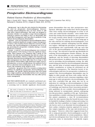
patients in terms of predicting risk of cardiovascular death.8 Table 1. Coded Electrocardiogram (ECG) Abnormalities
Also, abnormal electrocardiograms in patients with docu- Abnormalities n (% of Total ECGs)
mented coronary artery disease or at high risk for coronary
artery disease and undergoing major noncardiac surgery Q waves
Minor 33 (2.9)
were shown to predict long-term outcome.9
Major* 15 (1.3)
The existing literature gives no guidance on age or risk ST junction/segment depression
stratification for minimizing unnecessary preoperative Minor 104 (9.1)
electrocardiogram screening or maximizing its yield and Major* 19 (1.7)
T wave changes
utility. Furthermore, previous studies on the utility of Minor 186 (16.2)
preoperative electrocardiograms have not evaluated the Major* 57 (5.0)
impact on preoperative management as an endpoint. Here, ST segment elevation* 8 (0.7)
the prevalence of electrocardiograms abnormalities in Left axis deviation 65 (5.7)
Right axis deviation 15 (1.3)
1,149 preoperative patients and the correlation between Left ventricular hypertrophy 102 (8.9)
significant abnormalities and a variety of patient risk factors First-degree atrioventricular block 48 (4.2)
is reported. This study was designed to test the hypothesis Mobitz type II or higher blockade* 0 (0)
that significant abnormalities on preoperative electrocar- Short PR interval 6 (0.5)
Pacemaker 13 (1.1)
diograms, i.e., those that would affect preoperative man- Left bundle branch block* 20 (1.7)
agement, do not exist in the absence of specific risk factors. Right bundle branch block 50 (4.4)
In addition, age in the absence of other risk factors was Interventricular condunction delay 65 (5.7)
evaluated as an independent predictor of significant elec- Frequent premature atrial complexes 10 (0.9)
Frequent premature ventricular complexes 22 (1.9)
trocardiograms abnormalities. Atrial fibrillation* 30 (2.6)
Sinus tachycardia 18 (1.6)
Sinus bradycardia 38 (3.3)
Materials and Methods * Significant abnormality requiring further evaluation.
With approval of the Partners Human Research Com-
mittee (Boston, MA), all preoperative electrocardiograms electrocardiograms would result in further assessment or
for patients presenting to the Weiner Center for Preop- evaluation by the preoperative clinician before the pa-
erative Evaluation at Brigham and Women’s Hospital tient could proceed to surgery: major Q waves, major ST
(Boston, MA) during the period of October and Novem- junction/segment depression, major T wave changes, ST
ber 2003 were reviewed. The Weiner Center evaluates segment elevation, Mobitz type II or higher blockade,
more than 85% of all elective surgical patients. All pa- left bundle branch block, and atrial fibrillation. The as-
tients over the age of 50 yrs had an electrocardiograms sessment and evaluation could include the retrieval of a
performed per institutional guidelines. All electrocardio- previous electrocardiograms or cardiac testing for com-
grams at Brigham and Women’s Hospital are officially parison, retrieval of information from the patient’s pri-
interpreted by a staff cardiologist. All electrocardiograms mary care physician or cardiologist, the performance of
used for the study were downloaded from the hospital’s further testing or a change to a patient’s medical therapy
electronic database and coded by two of four possible (e.g., addition of or alteration of a -blocker dose) as
study investigators using the Minnesota Code classifica- previously described by our group.11
tion system10 (table 1). If any coding discrepancies were Patients with significantly abnormal electrocardio-
noted, all four investigators evaluated the electrocardio- grams were then compared to a control group randomly
gram and a majority decision was used to assign a code. selected (using an online true random sequence gener-
Q waves and ST or T wave changes were considered ator) from the remaining patients who had normal or
minor if the electrocardiograms interpretation graded insignificantly abnormal electrocardiograms. The num-
the abnormality as being nonspecific, and they were ber of patients in this group was chosen to be approxi-
considered major if the electrocardiograms interpreta- mately twice the number of patients who had abnormal
tion was suggestive of ischemia or infarct per the official electrocardiograms to have increased power given the
cardiology reading. Frequent premature atrial or ventric- relative scarcity of cases. The control group was deter-
ular complexes were defined as more than one complex mined to be a representative sampling of the entire
in ten beats. Sinus tachycardia was defined as a rate more possible not significantly abnormal and normal electro-
than 100 beats per minute, and sinus bradycardia was cardiograms group because comparisons of age (63.1
defined as a rate less than 50 beats per minute. 9.8 yr for the population) and gender (429 men and 631
The following electrocardiograms abnormalities, deter- women for the population) of the two groups revealed
mined ahead of time, were considered to be “significant” nonsignificant differences of P 0.22 and 0.44, respec-
in that it was the consensus of our anesthesiology and tively. Patient data collected for these two groups in-
cardiology group that their presence on a preoperative cluded age, gender, surgical type, and risk, specific items
Anesthesiology, V 110, No 6, Jun 2009
3. PATIENT FACTORS PREDICTIVE OF ELECTROCARDIOGRAM ABNORMALITIES 1219
from the past medical history, and any postoperative Results
complications. The items recorded included a history of
myocardial infarction (by patient report), anginal symp- A total of 1,149 electrocardiograms were evaluated
toms (by patient report), congestive heart failure (by during the 2-month period. Table 1 lists the incidence
patient report), severe valvular disease (defined as hav- of coded abnormalities. A total of 864 separate abnor-
ing at least moderate regurgitation or stenosis of any malities were identified in a total of 540 patients
valve by a documented echocardiogram or having a (47.0%). Eighty-nine patients (7.7%) had at least one
history of a valve repair), diabetes – insulin-dependant or abnormality that was considered significant. The most
noninsulin-dependant (by patient report), renal insuffi- common abnormality was minor T wave changes seen
ciency (defined as creatinine above the upper limit of in 186 patients (16.2% of the total electrocardio-
normal for age and gender), low functional capacity grams). The most common significant abnormality was
(metabolic equivalents less than four by patient report), major T wave changes seen in 57 patients (5.0% of the
stroke (by patient report), hypertension (by patient re- total electrocardiograms).
port), smoking (current or history by patient report), Table 2 shows the patient demographics for the pa-
high cholesterol (by patient report of being on therapy), tients who had significant electrocardiograms abnormal-
coronary artery disease (by patient report of bypass ities and for the control patients. There were significant
surgery or any percutaneous cardiac intervention in the differences between the groups in terms of age and
absence of a documented myocardial infarction), and pe- gender. Examination of various age thresholds revealed
ripheral vascular disease (by patient report or history of that age of 65 yr or older was the most predictive of
vascular surgery). All risk factors for each patient were having an abnormal electrocardiograms. Table 3 lists the
listed. Postoperative cardiac complications were recorded patient risk factors for the two groups. The most com-
after a retrospective chart review and included evidence of mon risk factor in the significantly abnormal electrocar-
perioperative ischemia/infarction by cardiac enzymes or diograms group was age above 65 yr (69.7%). The most
new rhythm disturbances on electrocardiograms. common risk factor in the control group was hyperten-
sion (42.6%).
Statistical Analysis Table 4 lists the odds ratios for the risk factors corre-
All analyses were performed in SAS 9.1.3 (SAS Insti- lated with having a significantly abnormal electrocardio-
tute, Carey, NC). A two-sample t test was used to com- grams. The patient parameters, listed in order of increas-
pare the age differences among groups. A chi-square test ing influence on the predicted probability of having a
was used to test the gender, patient risk factors, and significantly abnormal electrocardiograms, are as fol-
postoperative cardiac complication differences among lows: high cholesterol, age over 65 yr, severe valvular
groups. A univariate sensitivity analysis was done to disease, myocardial infarction, angina, and congestive
determine the optimal effect of age, specifically, mini- heart failure. Each of these factors was independently
mizing the –2 log likelihood. This age cutpoint was then and significantly associated with an increased proba-
used as an independent risk factor. All categorical data bility of the patient having a significantly abnormal
for surgical type, surgical risk, demographics, and items electrocardiograms.
from the medical history were coded as 0 absent and Table 5 lists the interventions prompted by finding a
1 present. A univariate analysis was done to determine significantly abnormal electrocardiograms at the preop-
which variables were related to having an abnormal erative visit. The 13 patients who were presenting for
electrocardiograms. The variables that were significant open heart surgery (coronary artery bypass grafting or
to P 0.1 by the univariate analysis were then entered valve surgery) are not included because they all would
into a regression analysis. A priori decisions were made have had cardiac testing at our institution preceding
to remove cardiac and vascular surgery from the regres- their operation regardless of electrocardiograms find-
sion analysis because these are already represented ings. In the remaining 76 patients with abnormal elec-
within the patient factors (e.g., myocardial infarction, trocardiograms, there were 19 (25%) who required some
coronary artery disease, valve disease, peripheral vascu- new intervention before proceeding to the operating
lar disease) and thus would have been redundant. In
Table 2. Patient Demographics
addition, high-risk surgery was removed a priori be-
cause most of these surgeries (19 of 25) were within the Significantly Control
cardiac and vascular groups. The multivariate logistic Abnormal ECG
ECG (n 89) (n 195) P Value
regression analysis was carried out by using a manual
backwards selection, with a P value (stay criteria) of less Age, mean SD 69.2 9.1 62.5 10.0 0.0001
Gender, n (%) 0.02
than 0.05 being considered significant in the final model.
Male 54 (60.7) 88 (45.1)
A receiver-operating characteristic curve was con- Female 35 (39.3) 107 (54.9)
structed by plotting sensitivity against the false-positive
rate (1–specificity) over a range of cutpoint values. ECG electrocardiogram; SD standard deviation.
Anesthesiology, V 110, No 6, Jun 2009
4. 1220 CORRELL ET AL.
Table 3. Patient Risk Factors Table 5. Preoperative Management Interventions Performed
for the Patients with a Significantly Abnormal
Significantly Electrocardiogram (ECG)*
Abnormal Control
ECG ECG
Intervention n
(n 89) (n 195) P Value
Retrieval of old electrocardiograms 25
Age 65 yr, n (%) 62 (69.7) 68 (34.9) 0.0001
Retrieval of old cardiac test 32
Angina, n (%) 14 (15.7) 3 (1.5) 0.0001
New cardiac test ordered 14
Congestive heart failure, n (%) 25 (28.1) 6 (3.1) 0.0001
Cardiology consult obtained 3
Severe valve disease, n (%) 16 (18.0) 4 (2.1) 0.0001
-blocker started 2
Myocardial infarction, n (%) 24 (27.0) 9 (4.6) 0.0001
Diabetes, n (%) 27 (30.3) 21 (10.8) 0.0001
Renal insufficiency, n (%) 14 (15.7) 8 (4.1) 0.0007 * Excluding 13 patients having cardiac surgery.
Low functional capacity, n (%) 32 (36.0) 32 (16.4) 0.0003
Stroke, n (%) 8 (9.0) 3 (1.5) 0.0025 and ischemia (table 6). The overall number of cardiac
Hypertension, n (%) 56 (63.0) 83 (42.6) 0.0015
Current smoker, n (%) 13 (14.6) 21 (10.8) 0.3554 complications was extremely small, and the study
Former smoker, n (%) 28 (31.5) 64 (32.8) 0.8203 was not expected to make any conclusions from this
High cholesterol, n (%) 37 (41.6) 35 (17.9) 0.0001 endpoint.
Coronary artery disease, n (%) 14 (15.7) 9 (4.6) 0.0014
The Hosmer and Lemeshow test demonstrates that our
Peripheral vascular disease, n (%) 13 (14.6) 7 (3.6) 0.0008
Cardiac surgery, n (%) 15 (16.9) 3 (1.5) 0.0001 model has adequate goodness-of-fit (P 0.28) as indi-
General surgery, n (%) 22 (24.7) 48 (24.6) 0.9849 cated by a statistically nonsignificant P value. The dis-
Gynecologic surgery, n (%) 6 (6.7) 25 (12.8) 0.1275 criminative capacity of the model to assign true-positives
Neurologic surgery, n (%) 2 (2.2) 9 (4.6) 0.53
Orthopedic surgery, n (%) 12 (13.5) 41 (21.0) 0.1301
is also adequate, with a c statistic or area under the
Other surgery,* n (%) 0 (0) 4 (2.1) 0.4087 receiver-operating characteristic curve of 0.84. The sen-
Otorhinolaryngeal surgery, n (%) 3 (3.4) 17 (8.7) 0.1023 sitivity of the model, defined as the percentage of pa-
Plastic surgery, n (%) 0 (0) 6 (3.1) 0.2195 tients predicted to have a significantly abnormal electro-
Thoracic surgery, n (%) 7 (7.9) 22 (11.3) 0.3777
Urologic surgery, n (%) 13 (14.6) 17 (8.7) 0.1342
cardiograms who really have one (true-positive), is
Vascular surgery, n (%) 9 (10.1) 3 (1.5) 0.0008 87.6%. The specificity of the model, defined as the per-
High risk surgery, n (%) 25 (28.1) 6 (3.1) 0.0001 centage of patients predicted to not have a significantly
abnormal electrocardiograms who do not have it (true-
* Ophthalmology, gastroenterology, radiology, and anesthesiology.
negative), is 59.5%.
ECG electrocardiogram.
room. Two patients had -blockers started; three pa-
tients were seen by a cardiologist who felt no further Discussion
evaluation was needed, and the remaining 14 patients This study was designed to better refine the criterion
had cardiac testing ordered. The tests were nonimaging for preoperative electrocardiograms ordering. Patient
or imaging stress tests in 11 patients, and cardiac cathe- risk factors of age over 65 yr, history of angina, conges-
terization in three patients. Three of the patients could tive heart failure, high cholesterol, myocardial infarction,
not have the test performed before the original surgery and severe valvular disease were found to be predictive
date, leading to postponement of the case. Two of the for having a significantly abnormal electrocardiograms,
patients had their case cancelled, and the results of the defined as major Q waves, major ST junction/segment
workup are not known. The number of cases postponed or depression, major T wave changes, ST segment eleva-
canceled represents 0.4% of the total number of patients tion, Mobitz type II or higher blockade, left bundle
who had electrocardiograms over the study period. branch block, or atrial fibrillation.
There were no statistical differences between the This report is unique in defining significant preopera-
groups in terms of major postoperative cardiac com- tive electrocardiograms abnormalities as those that
plications, including postoperative atrial fibrillation should prompt further action by the preoperative clini-
Table 4. Predictors of Having a Significantly Abnormal cian. Previous studies in this area have defined the im-
Electrocardiogram (ECG) in the Preoperative Period pact of preoperative electrocardiograms as the effect on
Risk Factor P Value Odds Ratio 95% CI Table 6. Postoperative Cardiac Complications
Age 65 yr 0.0001 4.08 2.13–7.79
Significantly Control
Angina 0.0101 7.49 1.62–34.69 Abnormal ECG
Congestive heart failure 0.0001 12.18 3.44–43.11 ECG (n 89) (n 195) P Value
High cholesterol 0.0195 2.26 1.14–4.48
Myocardial infarction 0.0002 6.16 2.34–16.20 Atrial fibrillation, n (%) 2 (2.2) 2 (1) NS
Severe valve disease 0.0259 4.80 1.21–19.10 Ischemia, n (%) 2 (2.2) 0 (0) NS
CI confidence interval. ECG electrocardiogram; NS not significant.
Anesthesiology, V 110, No 6, Jun 2009
5. PATIENT FACTORS PREDICTIVE OF ELECTROCARDIOGRAM ABNORMALITIES 1221
significant postoperative complications or on the delay segment depression, major T wave changes, ST segment
or cancellation of surgical procedures.12 However, in elevation, Mobitz type II or higher blockade, left bundle
actual clinical practice, the point-of-care decision regard- branch block, or atrial fibrillation. These specific abnor-
ing abnormal electrocardiograms by the preoperative malities are based on the group’s evaluation of the ex-
clinician is whether further information or testing is isting literature and clinical experience developed over
needed before allowing the patient to undergo the several years. The management can include requesting
planned procedure. Because collection of this informa- information from the patient’s primary care physician or
tion does not necessarily result in a delay or cancellation cardiologist and previous testing results (electrocardio-
of surgery, delay or cancellation of a procedure are thus grams, noninvasive and invasive cardiac examinations)
insensitive endpoints on which to measure clinician be- or initiating new consultations, cardiac testing, or ther-
havior and resource utilization. This fact is supported by apies (e.g., perioperative -blockade).
this study in that only five patients had surgery post- Several limitations exist for our study. The first is that
poned or cancelled. Therefore, we used the decision for the study was performed in a retrospective manner. It is
further evaluation, which is in actuality the triage point unlikely that this was of significance, however, because
in actual clinical practice, as a metric. a prospective design would not have the ability to
Many surgical institutions use age as the sole criterion change an electrocardiograms or alter the patients’ his-
for performing preoperative electrocardiograms. The im- tories. The patient’s histories were not known at the
pact of these electrocardiograms, however, is limited by time the electrocardiograms were read by the cardiolo-
the arbitrary nature of the age selected and the subse- gist, and agreement between investigators regarding the
quent number of normal or minor abnormalities discov- coding was required.
ered. Moreover, arbitrary age-based thresholds are asso- Another limitation is that it is possible that some risk
ciated with the costs and resources used in providing factors could have been further subdivided or sharp-
electrocardiograms testing, the additional testing pro- ened; however the choice of which categories to subdi-
voked by abnormalities, and the possible delay of surgi- vide was not apparent at the outset of the study. Now
cal procedures. Our hope was that age in the absence of that we know the general categories that are significant,
risk factors was not an independent predictor of signif- it is possible that further research could be done to see
if further sharpening would actually lead to a different or
icant electrocardiograms abnormalities; this would help
more specific list of criteria.
us reduce the number of preoperative electrocardio-
A further limitation is the absence of an analysis of the
grams performed. However, our results indicate that in a
subsequent impact of the clinician’s response to the
population older than 50 yr, an increased odds ratio for
electrocardiograms on postoperative outcomes. Our
independently predicting significant preoperative elec-
study was not intended to evaluate postoperative com-
trocardiograms abnormalities did occur at age greater
plications, which were extremely small in incidence
than 65 yr (table 4). On the basis of our results, age
(table 5). Many studies that have attempted to correlate
cannot be eliminated as a screening factor, which
preoperative electrocardiograms findings with cardiac
sharply differs from the guidelines put forth by the Cen-
events are inconclusive. One study found that a rhythm
ter for Medicare and Medicaid Services, which has
other than sinus or frequent premature ventricular con-
ceased paying for preoperative electrocardiograms
tractions were the only electrocardiograms findings cor-
based on age.# related with postoperative cardiac events.19 electrocar-
The electrocardiograms abnormalities that should diograms findings predictive of sudden cardiac death in
prompt the preoperative clinician to request further the population include abnormalities suggestive of myo-
information, consultation, or testing are controversial. cardial infarction (i.e., Q waves) or an intraventricular
No consensus currently exists in the literature regarding conduction defect in people with overt coronary heart
what is considered a significantly abnormal electrocar- disease, left ventricular hypertrophy and tachycardia in
diograms.13–18 The abnormalities determined to be sig- people without coronary heart disease, and nonspecific
nificant for the purposes of this study were based on a ST-T abnormalities in men without coronary heart dis-
consensus opinion among our perioperative medicine ease.20 In vascular surgery patients, left ventricular hy-
specialists, a group including anesthesiologists and car- pertrophy or ST depression have been shown to be
diologists, at the Brigham and Women’s Hospital (table predictive of postoperative cardiac events.21
1). Our practice is to require further information, evalu- There are circumstances in which a preoperative elec-
ation, or management if the preoperative electrocardio- trocardiograms in patients with none of the risk factors
grams exhibits significant Q waves, major ST junction/ defined in our model may be of value. Some clinicians
desire baseline electrocardiograms before specific types
# Centers for Medicare and Medicaid Services: Medicare National Coverage of surgery, such as cardiac or thoracic, where postoper-
Determinations Manual, Chapter 1, Part 1 (Sections 10 – 80.12). Available at:
http://www.cms.hhs.gov/manuals/downloads/ncd103c1_Part1.pdf. Accessed May
ative electrocardiograms changes frequently occur. Base-
10, 2008. line electrocardiograms may also be of value in patients
Anesthesiology, V 110, No 6, Jun 2009
6. 1222 CORRELL ET AL.
who are on pharmacologic agents known to produce 4. U.S. Preventive Services Task Force: Screening for coronary heart disease:
Recommendations statement. Ann Intern Med 2004; 140:569–72
adverse effects detected by electrocardiograms changes 5. Goldberger AL, O’Konski M: Utility of the routine electrocardiogram before
or correlate with therapeutic responses or disease pro- surgery and on general hospital admission. Ann Intern Med 1986; 105:552–7
6. Pasternak LR, Arens JF, Caplan RA, Connis RT, Fleisher LA, Flowerdew R,
gression.22 Gold BS, Mayhew JF, Nickinovich DG, Rice LJ, Roizen MF, Twersky RS: Practice
It is possible that some clinicians would seek further advisory for preanesthesia evaluation. A report by the American Society of
Anesthesiologists task force on preanesthesia evaluation. ANESTHESIOLOGY 2002;
cardiac information on patients who relate a history of 96:485–96
angina, congestive heart failure, myocardial infarction, 7. Fleisher LA, Beckman J, Brown K, Calkins H, Chaikof E, Fleischmann KE,
Freeman WK, Froehlich JB, Kasper EK, Kersten JR, Riegel B, Robb JF: ACC/AHA
or severe valvular disease even in the absence of an 2007 guidelines on perioperative cardiovascular evalution and cardiac care for
abnormal electrocardiograms. Thus the findings of this noncardiac surgery: A report of the American College of Cardiology/American
Heart Association Task Force on Practice Guidelines. (Writing Committee to
study that history of high cholesterol or age over 65 yr is Revise the 2002 Guidelines on Perioperative Cardiovascular Evaluation for Non-
predictive of abnormal electrocardiograms may be the cardiac Surgery). J Am Coll Cardiol 2007; 50:e159–241
8. Noordzij PG, Boersma E, Bax JJ, Feringa HH, Schreiner F, Schouten O, Kertai
most valuable addition to our understanding of preoper- MD, Klein J, van Urk H, Elhendy A, Poldermans D: Prognostic value of routine
ative assessment. preoperative electrocardiography in patients undergoing noncardiac surgery.
Am J Cardiol 2006; 97:1103–6
Although our list of risk factors is capable of identify- 9. Jeger RV, Probst C, Arsenic R, Lippuner T, Pfisterer ME, Seeberger MD,
ing patients who are at risk of having significant preop- Filipovic M: Long-term prognostic value of the preoperative 12-lead electrocar-
diogram before major noncardiac surgery in coronary artery disease. Am Heart J
erative electrocardiograms abnormalities, it cannot cap- 2006; 151:508–13
ture all patients who have abnormal electrocardiograms. 10. Blackburn H, Keys A, Simonson E, Rautaharju P, Punsar S: The electrocar-
diogram in population studies: A classification system. Circulation 1960; 21:
Five patients (0.44%) in the significantly abnormal group 1160–75
would not have been identified due to their age being 11. Correll DJ, Bader AM, Hull MW, Tsen LC, Hepner DL: The value of
preoperative clinic visits in identifying issues with potential impact on operating
less than 65 yr and the absence of other risk factors room efficiency. ANESTHESIOLOGY 2006; 105:1254–9
defined by the model. Three of these patients were 12. Rabkin SW, Horne JM: Preoperative electrocardiography: Effect of new
abnormalities on clinical decisions. Can Med Assoc J 1983; 128:146–7
presenting for a general surgical procedure, one for a 13. Knutsen R, Knutsen SF, Curb JD, Reed DM, Kautz JA, Yano K: The
thoracic surgery and one for an orthopedic surgery; the predictive value of resting electrocardiograms for 12-year incidence of coronary
heart disease in the Honolulu Heart Program. J Clin Epidemiol 1988; 41:293–302
latter two surgeries were categorized as high-risk. None 14. Cedres BL, Liu K, Stamler J, Dyer AR, Stamler R, Berkson DM, Paul O,
of these 5 patients had a postoperative cardiac compli- Lepper M, Lindberg HA, Marquardt J, Stevens E, Schoenberger JA, Shekelle RB,
Collette P, Garside D: Independent contribution of electrocardiographic abnor-
cation. It will need to be determined if it is acceptable to malities to risk of death from coronary heart disease, cardiovascular diseases, and
limit electrocardiograms to this high-risk population all causes. Findings of three Chicago epidemiologic studies. Circulation 1982;
65:146–53
with the potential to cancel very few cases on the day of 15. Sutherland SE, Gazes PC, Keil JE, Gilbert GE, Knapp RG: Electrocardio-
surgery if a patient is noted to have an abnormality on graphic abnormalities and 30-year mortality among white and black men of the
Charleston heart study. Circulation 1993; 88:2685–92
the preinduction electrocardiograms. 16. Vitelli LL, Crow RS, Shahar E, Hutchinson RG, Rautaharju PM, Folsom AR:
In conclusion, patient risk factors of age above 65 yr, Electrocardiographic findings in a healthy biracial population. Am J Cardiol 1998;
81:453–9
history of angina, congestive heart failure, high cholesterol, 17. Scheidt-Nave C, Barrett-Connor E, Wingard DL: Resting electrocardio-
myocardial infarction, or severe valvular disease are predic- graphic abnormalities suggestive of asymptomatic ischemic heart disease associ-
ated with non-insulin-dependent diabetes mellitus in a defined population. Cir-
tive for having a significantly abnormal electrocardio- culation 1990; 81:899–906
grams defined as major Q waves, major ST junction/ 18. Fleisher LA, Beckman JA, Brown KA, Calkins H, Chaikof E, Fleischmann
KE, Freeman WK, Froehlich JB, Kasper EK, Kersten JR, Riegel B, Robb JF:
segment depression, major T wave changes, ST segment ACC/AHA 2006 guideline update on perioperative cardiovascular evaluation for
elevation, Mobitz type II or higher blockade, left bundle noncardiac surgery: focused update on perioperative beta-blocker therapy. A
Report of the American College of Cardiology/American Heart Association Task
branch block, or atrial fibrillation. Age greater than 65 yr Force on Practice Guidelines (Writing Committee to Update the 2002 Guidelines
in the absence of other risk factors remains an indepen- on Perioperative Cardiovascular Evaluation for Noncardiac Surgery). J Am Coll
Cardiol 2006; 47:2343–55
dent predictor of significant preoperative electrocardio- 19. Goldman L, Caldera DL, Nussbaum SR, Southwick FS, Krogstad D, Murray
grams abnormalities. B, Burke DS, O’Malley TA, Goroll AH, Caplan CH, Nolan J, Carabello B, Slater EE:
Multifactorial index of cardiac risk in noncardiac surgical procedures. N Engl
J Med 1977; 297:845–50
20. Kreger BE, Cupples A, Kannel WB: The electrocardiogram in prediction of
References sudden death: Framingham Study experience. Am Heart J 1987; 113:377–82
21. Landesberg G, Einav S, Cristopherson R, Beattie C, Berlatzky Y, Rosenfeld
1. De Bacquer D, De Backer G, Kornitzer M, Blackburn H: Prognostic value of B, Eidelman LA, Norris E, Anner H, Mosseri M, Cotev S, Luria MH: Perioperative
ECG findings for total, cardiovascular disease, and coronary heart disease death ischemia and cardiac complications in major vascular surgery: Importance of the
in men and women. Heart 1998; 80:570–7 preoperative twelve-lead electrocardiogram. J Vasc Surg 1997; 26:570–8
2. Sox HC, Garber AM, Littenberg B: The resting electrocardiogram as a 22. Schlant RC, Adolph RJ, DiMarco JP, Dreifus LS, Dunn MI, Fisch C, Garson
screening test. Ann Intern Med 1989; 111:489–502 A Jr, Haywood LJ, Levine HJ, Murray JA: Guidelines for electrocardiography: A
3. Liu LL, Dzankic S, Leung JM: Preoperative electrocardiogram abnormalities report of the American College of Cardiology/American Heart Association Task
do not predict postoperative cardiac complications in geriatric surgical patients. Force on Assessment of Diagnostic and Therapeutic Cardiovascular Procedures
J Am Geriatr Soc 2002; 50:1186–91 (Committee on Electrocardiography). J Am Coll Cardiol 1992; 19:473–81
Anesthesiology, V 110, No 6, Jun 2009