SlideShare a Scribd company logo
1 of 12
Download to read offline
Ojvensha E learning Resources-Prepared by Dr.B.B.Gosai
CEREBRUM
Introduction: Cerebrum is the largest part of central nervous system and highest in the
functional hierarchy. It is formed by right and left cerebral hemispheres partly connected in the
midline by band of corpus callosum.
Location: Located in the cranial cavity.
(Above diagram is for explanation only)
External Features:
Cerebrum has two cerebral hemispheres partly connected by corpus callosum and partly
separated by Medial longitudinal fissure. Longitudinal fissure contains falx cerebri (Fold of dura
mater).
On the surface of cerebrum large size folds are seen.
Depressions on the surface of cerebrum are known as Sulci.
Elevations between sulci is known as Gyri.
Ojvensha E learning Resources-Prepared by Dr.B.B.Gosai
Surfaces of Cerebrum:
Three surfaces:
Superolateral surface: Between Superomedial border and inferolateral border.
Medial Surface: Between Superomedial and inferomedial border.
Inferior Surface: Between Inferolateral and inferomedial border.
Three borders:
Superomedial border: between superolateral and medial surface.
Inferolateral border: between superolateral and inferior surface.
Inferomedial border: between inferior and medial surface.
Three poles:
Frontal pole: Anterior most part of cerebrum (frontal lobe)
Occipital pole: Posterior most part of cerebrum (Occipital lobe)
Temporal pole: Anterior most part of temporal lobe.
Sulci, Gyri and Important Functional areas of Cerebrum:
Cerebrum on superolateral surface is divided into three lobes by certain sulci and imaginary
lines.
Main sulci for demarcation of lobes are:
1. Central sulcus: Starts from 1 cm behind the midpoint on superomedial border and runs
obliquely on superolateral surface without interruptions and ends just above the lateral sulcus.
2. Lateral sulcus: It is also seen on superolateral surface.
It has a stem and three rami. Stem is near the anterior end of temporal lobe.
Three rami:
Ojvensha E learning Resources-Prepared by Dr.B.B.Gosai
Anterior ramus runs forwards.
Ascending ramus runs upwards
Posterior ramus is longest and runs backwards.
3. Parieto-occipital sulcus: It is predominantly seen on Medial surface. Its small part is seen on
superolateral surface.
SUPEROLATERAL SURFACE:
It is divided into four lobes:
1. Frontal lobe
2. Parietal lobe
3. Temporal lobe
4. Occipital lobe
1. Frontal lobe: Part of cerebrum in front of central sulcus.
2. Parietal lobe: Part of cerebrum behind the central sulcus.
3. Occipital lobe: Behind the imaginary line joining parito-occipital sulcus and preoccipital
notch.
4. Temporal lobe: Below the posterior ramus of lateral sulcus.
Ojvensha E learning Resources-Prepared by Dr.B.B.Gosai
Frontal pole (Black arrow)
Temporal pole (Red arrow)
Occipital pole (Green arrow)
Ojvensha E learning Resources-Prepared by Dr.B.B.Gosai
Important Figure for Sulci and gyri on superolateral surface
(All Labels are to be mentioned)
Ojvensha E learning Resources-Prepared by Dr.B.B.Gosai
Important Figure for Functional areas in superolateral surface
(All labels are to be mentioned)
Sulci, Gyri and functional areas in Frontal lobe:
Sulci:
Pre-central sulcus: in front of central sulcus and runs parallel to it.
Superior and inferior frontal sulci: In front of pre-central sulcus and runs horizontally.
Anterior and ascending rami of lateral sulcus: in the lower aspect of anterior part of frontal
lobe.
Gyri:
Ojvensha E learning Resources-Prepared by Dr.B.B.Gosai
Pre-central gyrus: Located between the central and pre-central sulcus. It contains Motor areas
of cerebrum.
Superior, Middle and Inferior frontal gyri: The part of frontal lobe in front of precentral gyrus is
divided into superior, middle and inferior frontal gyri by superior and inferior sulci.
Functional areas:
Motor Areas of Cerebrum: (*****Short note)
1. Motor Area of cerebrum:
Location: It is located in the pre-central gyrus between the central sulcus and pre-central
sulcus.
Important characteristics:
a. Body is represented upside down in the area. i.e. head and neck is represented in the lower
part of area and lower limbs are represented in the upper part of the area.
b. In the area movements are represented and not the individual muscles. i.e. for example
flexion movement is represented and not the individual muscle causing flexion.
c. The parts having fine movements are represented larger compared to parts having coarse
movements. i.e. Thumb, fingers, lips tongue and eyes are represented larger compared to
trunk and limbs.
d. Upper 1.5 cm area is supplied by Anterior cerebral artery and rest of the area is supplied by
middle cerebral artery.
e. It controls opposite half of body (Contralateral control). i.e. Right side of area control left
side of the body and left side area control right side of body.
Function: It is responsible for the execution of the movements of body.
Applied Anatomy:
a. Damage to this area in compression by collection of hematoma in fracture of skull leads to
paralysis of the muscles of opposite half of body.
b. Blockage of anterior cerebral artery leads to paralysis of lower limb muscles and loss of
control on sphincters while rest of the body is not affected. (Anterior cerebral supply upper
part of area).
c. Blockage of Middle cerebral artery leads to paralysis of the all the muscles of body except
the lower limb and sphincters.
Ojvensha E learning Resources-Prepared by Dr.B.B.Gosai
2. Pre-motor area: In front of motor area.
Function: Planning of the movements.
3. Broca’s motor area of speech:
Located in triangular area between anterior and ascending rami of lateral sulcus (Pars
Traingularis).
Function: Motor speech of person i. e. articulation of words.
Applied: Damage to this area leads to Motor Aphasia. ( Loss of speech).
Silent Area of Brain (Prefrontal cortex): It is anterior part of frontal lone.
Earlier it was believed to be silent but now it is know that it is part of limbic system and
concerned with social and emotional behaviour of person.
Damage to this area leads to Anti-social behaviour.
Sulci, Gyri and Functional areas in Parietal lobe:
Sulci:
Post-central sulcus: behind the central sulcus and runs parallel to it.
Intraparietal sulcus: Curved sulcus in the middle of parietal lobe.
Gyri:
Post-central gyrus: Located between central and post central sulcus. It contains primary
sensory area.
Superior parietal lobule: Part above the intraparietal sulcus.
Inferior parietal lobule: Part below the intraparietal sulcus.
Functional areas:
1. Primary Sensory area ( Primary somesthetic area) : Located in post central gyrus between
central and post central sulcus.
Function: Identification of all general sensations and taste sensation.
Ojvensha E learning Resources-Prepared by Dr.B.B.Gosai
2. Secondary sensory area (Secondary somesthetic area) : Located behind the primary sensory
area.
Function: It keeps the memory of all sensations experienced by person and coordinated with
incoming new sensations.
3. Wernick’s sensory area of speech: It is cap like area on the posterior end of posterior ramus
of lateral sulcus.
Function: Identification of text from the images and sound and send it to Broca’s area for
articulation.
Applied aspect: Damage to Wernick’s area leads to inability to identify text from images and
sound i.e. Sensory Aphasia. Person can speak but unable to read aloud from book and repeat
spoken words.
Sulci, Gyri and Functional areas in Temporal lobe:
Sulci:
Superior and Inferior Temporal sulci.
Gyri:
Superior, middle and inferior temporal gyri: Temporal lobe ins divided in to superior, middle
and inferior gyri by superior and inferior temporal sulci.
Functional Areas:
1. Primary Auditory area: Located in superior temporal gyrus.
Function: Identification of sound. (Hearing)
2. Auditory association area: Located outer to primary auditory area.
Function: Keeps memory of all sound heard by person and coordinated with new incoming
sound.
Applied anatomy:
Damage to auditory area leads to partial hearing loss.
Abnormal stimulation of Auditory association area leads to Auditory Hallucinations i.e. person
complains of hearing non-existent sound.
Sulci, Gyri and Functional areas in Occipital lobe:
Sulci:
Ojvensha E learning Resources-Prepared by Dr.B.B.Gosai
Small part of calcarine sulcus: Most of the sulcus is seen on medial surface.
Lunate sulcus: Curved sulcus in from of end of calcarine sulcus.
Functional area:
Primary visual area: Mostly seen on medial surface above and below the calcarine sulcus>
Function: Identification of image. ( Vision)
Damage to this area leads to Contralateral homonymous hemianopia.
Visual Association area: located outer to visual area.
Function: Keeps memory of all images seen by person and coordinated with new incoming
images.
Abnormal stimulation of visual association area leads to visual Hallucinations i.e. person
complains of seeing non-existent images.
MEDIAL SURFACE:
Ojvensha E learning Resources-Prepared by Dr.B.B.Gosai
Sulci:
Callosal sulcus: Closely related to corpus callosum.
Cingulate sulcus: Runs parallel to corpus callosum.
Parieto-occipital sulcus and calcarine sulcus forms “Y” shape on posterior aspect of medial
surface.
Upper limb of “Y” is Parieto-occipital sulcus. (Red Arrow)
Lower limb of “Y” is Calcarine sulcus. (Black arrow)
Gyri:
Cingulate gyrus: Between callosal sulcus and cingulated sulcus. It is part of limbic system
concerned with social, emotional behaviour and memory.
Cuneus: part between calcarine and parieto-occipital sulcus.
Pre-cuneus: part in front of parieto-occipital sulcus.
INFERIOR SURFACE:
Ojvensha E learning Resources-Prepared by Dr.B.B.Gosai
Divided in to two parts:
1. Orbital part: Related to roof of orbit. (Anterior smallerpart)
Sulci:
Olfactory sulcus: related to olfactory tract.
Orbital sulci: “H” shaped.
Gyri:
Gyrus rectus: Medial to olfactory sulcus.
Orbital gyri: between orbital sulci.
2. Tentorial part: related to tentorium cerebella (fold of dura mater) (Posterior larger part)
Sulci:
Collateral sulcus: It is medial sulcus bounding the parahippocampal gyrus.
Occipito-temporal sulcus: It is lateral to collateral sulcus.
Rhinal sulcus: small sulcus in anterior part of parahippocampal gyrus.
Gyri:
Parahippocampal gyrus: Medial to collateral sulcus. Part of limbic system. Cocerned with social,
emotional behaviour and memory.
Uncus: hook shaped anterior part of parahippocampal gyrus.
NOTE:
Describe sulci and gyri of cerebrum (only mention sulci and gyri and draw the diagram for the same)
Describe functional cortical areas of cerebrum ( mention the functuional areas in the superolateral
surface with functions and applied aspect)
Short note:
Motor areas of cerebrum (Mention motor area, premotor area and Broca’s motor area of speech
with diagram showing only these areas)
Motor area of cerebrum (only Motor area to be mentioned with diagram)
====================X================

More Related Content

What's hot

Anatomy of brainstem
Anatomy of brainstemAnatomy of brainstem
Anatomy of brainstem
MBBS IMS MSU
 

What's hot (20)

Fourth ventricle
Fourth ventricleFourth ventricle
Fourth ventricle
 
4th ventricle
4th ventricle4th ventricle
4th ventricle
 
Diencephalon 1
Diencephalon 1Diencephalon 1
Diencephalon 1
 
Meninges of brain & spinal cord
Meninges of brain & spinal cordMeninges of brain & spinal cord
Meninges of brain & spinal cord
 
Anatomy of cerebellum
Anatomy of cerebellumAnatomy of cerebellum
Anatomy of cerebellum
 
Cerebral cortex ppt
Cerebral cortex pptCerebral cortex ppt
Cerebral cortex ppt
 
Brain stem dr.gosai
Brain stem dr.gosaiBrain stem dr.gosai
Brain stem dr.gosai
 
Anatomy of midbrain
Anatomy of midbrainAnatomy of midbrain
Anatomy of midbrain
 
The cerebellum
The cerebellumThe cerebellum
The cerebellum
 
Anatomy of brainstem
Anatomy of brainstemAnatomy of brainstem
Anatomy of brainstem
 
Medulla Oblongata
Medulla OblongataMedulla Oblongata
Medulla Oblongata
 
Cerebrum ppt
Cerebrum pptCerebrum ppt
Cerebrum ppt
 
Brain ventricles
Brain ventriclesBrain ventricles
Brain ventricles
 
Cerebellum
CerebellumCerebellum
Cerebellum
 
Spinal cord (2)
Spinal cord (2)Spinal cord (2)
Spinal cord (2)
 
Cerebrum i
Cerebrum iCerebrum i
Cerebrum i
 
Spinal cord
Spinal cordSpinal cord
Spinal cord
 
Cerebrum
CerebrumCerebrum
Cerebrum
 
CEREBRAL CORTEX
CEREBRAL CORTEXCEREBRAL CORTEX
CEREBRAL CORTEX
 
Lateral ventricle of Brain. By Dr.N.Mugunthan.M.S
Lateral ventricle of Brain. By Dr.N.Mugunthan.M.SLateral ventricle of Brain. By Dr.N.Mugunthan.M.S
Lateral ventricle of Brain. By Dr.N.Mugunthan.M.S
 

Viewers also liked

Anatomy of brain sulcus and gyrus - Dr.Sajith MD RD
Anatomy of brain sulcus and gyrus - Dr.Sajith MD RDAnatomy of brain sulcus and gyrus - Dr.Sajith MD RD
Anatomy of brain sulcus and gyrus - Dr.Sajith MD RD
Sajith Selvaganesan
 
Brain cerebrum
Brain cerebrumBrain cerebrum
Brain cerebrum
stewart_j
 
white fibres of cns modified
white fibres of cns modifiedwhite fibres of cns modified
white fibres of cns modified
Med Study
 
19. cerebrum
19. cerebrum19. cerebrum
19. cerebrum
Kim Chen
 
Cerebrum internal features by DR.ARSHAD
Cerebrum internal features by DR.ARSHADCerebrum internal features by DR.ARSHAD
Cerebrum internal features by DR.ARSHAD
SMS_2015
 
Corpus callosum
Corpus callosumCorpus callosum
Corpus callosum
PS Deb
 
Brain ppt
Brain pptBrain ppt
Brain ppt
cnps
 
External Morphology Of The Cerebrum
External Morphology Of The CerebrumExternal Morphology Of The Cerebrum
External Morphology Of The Cerebrum
guest098d461
 
BiologyExchange.co.uk Shared Resource
BiologyExchange.co.uk Shared ResourceBiologyExchange.co.uk Shared Resource
BiologyExchange.co.uk Shared Resource
biologyexchange
 

Viewers also liked (20)

Cerebrum
CerebrumCerebrum
Cerebrum
 
The cerebral hemispheres - Gross Anatomy & Connections
The cerebral hemispheres - Gross Anatomy & ConnectionsThe cerebral hemispheres - Gross Anatomy & Connections
The cerebral hemispheres - Gross Anatomy & Connections
 
Anatomy of brain sulcus and gyrus - Dr.Sajith MD RD
Anatomy of brain sulcus and gyrus - Dr.Sajith MD RDAnatomy of brain sulcus and gyrus - Dr.Sajith MD RD
Anatomy of brain sulcus and gyrus - Dr.Sajith MD RD
 
Brain cerebrum
Brain cerebrumBrain cerebrum
Brain cerebrum
 
Sulci gyri
Sulci gyriSulci gyri
Sulci gyri
 
Cerebrum
CerebrumCerebrum
Cerebrum
 
Sulcal anatomy supratentorial brain, excluding the temporal lobe.
Sulcal anatomy supratentorial brain, excluding the temporal lobe.Sulcal anatomy supratentorial brain, excluding the temporal lobe.
Sulcal anatomy supratentorial brain, excluding the temporal lobe.
 
Histology of cerebrum and cerebellum
Histology of cerebrum and cerebellumHistology of cerebrum and cerebellum
Histology of cerebrum and cerebellum
 
Gross anatomy of human brain
Gross anatomy of human brainGross anatomy of human brain
Gross anatomy of human brain
 
white fibres of cns modified
white fibres of cns modifiedwhite fibres of cns modified
white fibres of cns modified
 
White matter of cerebrum dr.gosai
White matter of cerebrum dr.gosaiWhite matter of cerebrum dr.gosai
White matter of cerebrum dr.gosai
 
19. cerebrum
19. cerebrum19. cerebrum
19. cerebrum
 
Cerebrum internal features by DR.ARSHAD
Cerebrum internal features by DR.ARSHADCerebrum internal features by DR.ARSHAD
Cerebrum internal features by DR.ARSHAD
 
Corpus callosum
Corpus callosumCorpus callosum
Corpus callosum
 
Brain ppt
Brain pptBrain ppt
Brain ppt
 
The human brain presentation
The human brain presentationThe human brain presentation
The human brain presentation
 
Nervous system notes
Nervous system notesNervous system notes
Nervous system notes
 
External Morphology Of The Cerebrum
External Morphology Of The CerebrumExternal Morphology Of The Cerebrum
External Morphology Of The Cerebrum
 
The Nervous System
The Nervous SystemThe Nervous System
The Nervous System
 
BiologyExchange.co.uk Shared Resource
BiologyExchange.co.uk Shared ResourceBiologyExchange.co.uk Shared Resource
BiologyExchange.co.uk Shared Resource
 

Similar to Cerebrum

Functional neuroanatomy of brain
Functional neuroanatomy    of brainFunctional neuroanatomy    of brain
Functional neuroanatomy of brain
Karrar Husain
 

Similar to Cerebrum (20)

Structure and Functions of cerebrum
Structure and Functions of cerebrumStructure and Functions of cerebrum
Structure and Functions of cerebrum
 
NEUROANATOMY 012 Cerebrum Cerebrum brain anatomy .pdf
NEUROANATOMY 012 Cerebrum Cerebrum brain anatomy .pdfNEUROANATOMY 012 Cerebrum Cerebrum brain anatomy .pdf
NEUROANATOMY 012 Cerebrum Cerebrum brain anatomy .pdf
 
Cerebrum external features
Cerebrum external featuresCerebrum external features
Cerebrum external features
 
19-Functions of Cerebral Hemisphere.pdf
19-Functions of Cerebral Hemisphere.pdf19-Functions of Cerebral Hemisphere.pdf
19-Functions of Cerebral Hemisphere.pdf
 
White matter of cerebral hemisphere corpus callosum
White matter of cerebral hemisphere corpus callosum White matter of cerebral hemisphere corpus callosum
White matter of cerebral hemisphere corpus callosum
 
Human Brain
Human BrainHuman Brain
Human Brain
 
brain.pptx
brain.pptxbrain.pptx
brain.pptx
 
Cerebellum
CerebellumCerebellum
Cerebellum
 
Cerebrum
CerebrumCerebrum
Cerebrum
 
introduction to neurology (nervous system, areas, motor and sensory systems)
introduction to neurology (nervous system, areas, motor and sensory systems)introduction to neurology (nervous system, areas, motor and sensory systems)
introduction to neurology (nervous system, areas, motor and sensory systems)
 
Language and brain
Language and brainLanguage and brain
Language and brain
 
Basic Anatomy CNS
Basic Anatomy CNSBasic Anatomy CNS
Basic Anatomy CNS
 
Anatomy lec 2
Anatomy lec   2Anatomy lec   2
Anatomy lec 2
 
Anatomy of the cerebrum; Anatomy - January 2015
Anatomy of the cerebrum; Anatomy - January 2015Anatomy of the cerebrum; Anatomy - January 2015
Anatomy of the cerebrum; Anatomy - January 2015
 
Functional neuroanatomy of brain
Functional neuroanatomy    of brainFunctional neuroanatomy    of brain
Functional neuroanatomy of brain
 
orofacial muscles.pptx
orofacial muscles.pptxorofacial muscles.pptx
orofacial muscles.pptx
 
Cerebellum ibms
Cerebellum ibmsCerebellum ibms
Cerebellum ibms
 
NEUROANATOMY 04 BRAIN STEM neuro anatomy midbrain pons medulla.pdf
NEUROANATOMY 04 BRAIN STEM neuro anatomy midbrain pons medulla.pdfNEUROANATOMY 04 BRAIN STEM neuro anatomy midbrain pons medulla.pdf
NEUROANATOMY 04 BRAIN STEM neuro anatomy midbrain pons medulla.pdf
 
BRAIN ANATOMY SHOWING THE PARTS OF THE BRAIN
BRAIN ANATOMY SHOWING THE PARTS OF THE BRAINBRAIN ANATOMY SHOWING THE PARTS OF THE BRAIN
BRAIN ANATOMY SHOWING THE PARTS OF THE BRAIN
 
Overview of the anatomy of the brain and its physiology
Overview of the anatomy of the brain and its physiologyOverview of the anatomy of the brain and its physiology
Overview of the anatomy of the brain and its physiology
 

More from Dr.B.B. Gosai

Esophagus stomach-dr.gosai
Esophagus stomach-dr.gosaiEsophagus stomach-dr.gosai
Esophagus stomach-dr.gosai
Dr.B.B. Gosai
 

More from Dr.B.B. Gosai (20)

AN 1.1 introduction anatomical terminology-Dr.Gosai
AN 1.1 introduction anatomical terminology-Dr.GosaiAN 1.1 introduction anatomical terminology-Dr.Gosai
AN 1.1 introduction anatomical terminology-Dr.Gosai
 
Sexual differentiation-Dr.B.B.Gosai
Sexual differentiation-Dr.B.B.GosaiSexual differentiation-Dr.B.B.Gosai
Sexual differentiation-Dr.B.B.Gosai
 
Vestibular and auditory apparatus-Dr.B.B.Gosai
Vestibular and auditory apparatus-Dr.B.B.GosaiVestibular and auditory apparatus-Dr.B.B.Gosai
Vestibular and auditory apparatus-Dr.B.B.Gosai
 
Brachial and lumbosacral plexus-Dr.B.B.Gosai
Brachial and lumbosacral plexus-Dr.B.B.GosaiBrachial and lumbosacral plexus-Dr.B.B.Gosai
Brachial and lumbosacral plexus-Dr.B.B.Gosai
 
Lower limb bones-joints-muscles-Dr.B.B.Gosai
Lower limb bones-joints-muscles-Dr.B.B.GosaiLower limb bones-joints-muscles-Dr.B.B.Gosai
Lower limb bones-joints-muscles-Dr.B.B.Gosai
 
Upper limb bones-joints-muscles-Dr.B.B.Gosai
Upper limb bones-joints-muscles-Dr.B.B.GosaiUpper limb bones-joints-muscles-Dr.B.B.Gosai
Upper limb bones-joints-muscles-Dr.B.B.Gosai
 
Introduction human movement apparatus-Dr.B.B.Gosai
Introduction human movement apparatus-Dr.B.B.GosaiIntroduction human movement apparatus-Dr.B.B.Gosai
Introduction human movement apparatus-Dr.B.B.Gosai
 
Spinal cord dr.gosai
Spinal cord dr.gosaiSpinal cord dr.gosai
Spinal cord dr.gosai
 
Ventricles of brain dr.gosai
Ventricles of brain dr.gosaiVentricles of brain dr.gosai
Ventricles of brain dr.gosai
 
Classification of joints dr.gosai
Classification of joints dr.gosaiClassification of joints dr.gosai
Classification of joints dr.gosai
 
Anatomy of Breast in clinical perspective-Dr.Gosai
Anatomy of Breast in clinical perspective-Dr.GosaiAnatomy of Breast in clinical perspective-Dr.Gosai
Anatomy of Breast in clinical perspective-Dr.Gosai
 
Pericardium and heart
Pericardium and heartPericardium and heart
Pericardium and heart
 
Larynx
LarynxLarynx
Larynx
 
Pharynx dr.gosai
Pharynx dr.gosaiPharynx dr.gosai
Pharynx dr.gosai
 
Extraocular muscles dr.gosai
Extraocular muscles dr.gosaiExtraocular muscles dr.gosai
Extraocular muscles dr.gosai
 
Blood supply of brain dr.gosai
Blood supply of brain dr.gosaiBlood supply of brain dr.gosai
Blood supply of brain dr.gosai
 
Cerebellum dr.gosai
Cerebellum dr.gosaiCerebellum dr.gosai
Cerebellum dr.gosai
 
Basal ganglia dr.gosai
Basal ganglia dr.gosaiBasal ganglia dr.gosai
Basal ganglia dr.gosai
 
Introduction abdomen-dr.gosai
Introduction abdomen-dr.gosaiIntroduction abdomen-dr.gosai
Introduction abdomen-dr.gosai
 
Esophagus stomach-dr.gosai
Esophagus stomach-dr.gosaiEsophagus stomach-dr.gosai
Esophagus stomach-dr.gosai
 

Recently uploaded

Beyond the EU: DORA and NIS 2 Directive's Global Impact
Beyond the EU: DORA and NIS 2 Directive's Global ImpactBeyond the EU: DORA and NIS 2 Directive's Global Impact
Beyond the EU: DORA and NIS 2 Directive's Global Impact
PECB
 
The basics of sentences session 3pptx.pptx
The basics of sentences session 3pptx.pptxThe basics of sentences session 3pptx.pptx
The basics of sentences session 3pptx.pptx
heathfieldcps1
 
Seal of Good Local Governance (SGLG) 2024Final.pptx
Seal of Good Local Governance (SGLG) 2024Final.pptxSeal of Good Local Governance (SGLG) 2024Final.pptx
Seal of Good Local Governance (SGLG) 2024Final.pptx
negromaestrong
 
Activity 01 - Artificial Culture (1).pdf
Activity 01 - Artificial Culture (1).pdfActivity 01 - Artificial Culture (1).pdf
Activity 01 - Artificial Culture (1).pdf
ciinovamais
 

Recently uploaded (20)

Application orientated numerical on hev.ppt
Application orientated numerical on hev.pptApplication orientated numerical on hev.ppt
Application orientated numerical on hev.ppt
 
General Principles of Intellectual Property: Concepts of Intellectual Proper...
General Principles of Intellectual Property: Concepts of Intellectual  Proper...General Principles of Intellectual Property: Concepts of Intellectual  Proper...
General Principles of Intellectual Property: Concepts of Intellectual Proper...
 
How to Give a Domain for a Field in Odoo 17
How to Give a Domain for a Field in Odoo 17How to Give a Domain for a Field in Odoo 17
How to Give a Domain for a Field in Odoo 17
 
Holdier Curriculum Vitae (April 2024).pdf
Holdier Curriculum Vitae (April 2024).pdfHoldier Curriculum Vitae (April 2024).pdf
Holdier Curriculum Vitae (April 2024).pdf
 
Energy Resources. ( B. Pharmacy, 1st Year, Sem-II) Natural Resources
Energy Resources. ( B. Pharmacy, 1st Year, Sem-II) Natural ResourcesEnergy Resources. ( B. Pharmacy, 1st Year, Sem-II) Natural Resources
Energy Resources. ( B. Pharmacy, 1st Year, Sem-II) Natural Resources
 
Beyond the EU: DORA and NIS 2 Directive's Global Impact
Beyond the EU: DORA and NIS 2 Directive's Global ImpactBeyond the EU: DORA and NIS 2 Directive's Global Impact
Beyond the EU: DORA and NIS 2 Directive's Global Impact
 
Micro-Scholarship, What it is, How can it help me.pdf
Micro-Scholarship, What it is, How can it help me.pdfMicro-Scholarship, What it is, How can it help me.pdf
Micro-Scholarship, What it is, How can it help me.pdf
 
2024-NATIONAL-LEARNING-CAMP-AND-OTHER.pptx
2024-NATIONAL-LEARNING-CAMP-AND-OTHER.pptx2024-NATIONAL-LEARNING-CAMP-AND-OTHER.pptx
2024-NATIONAL-LEARNING-CAMP-AND-OTHER.pptx
 
The basics of sentences session 3pptx.pptx
The basics of sentences session 3pptx.pptxThe basics of sentences session 3pptx.pptx
The basics of sentences session 3pptx.pptx
 
Z Score,T Score, Percential Rank and Box Plot Graph
Z Score,T Score, Percential Rank and Box Plot GraphZ Score,T Score, Percential Rank and Box Plot Graph
Z Score,T Score, Percential Rank and Box Plot Graph
 
Seal of Good Local Governance (SGLG) 2024Final.pptx
Seal of Good Local Governance (SGLG) 2024Final.pptxSeal of Good Local Governance (SGLG) 2024Final.pptx
Seal of Good Local Governance (SGLG) 2024Final.pptx
 
Unit-IV- Pharma. Marketing Channels.pptx
Unit-IV- Pharma. Marketing Channels.pptxUnit-IV- Pharma. Marketing Channels.pptx
Unit-IV- Pharma. Marketing Channels.pptx
 
Unit-IV; Professional Sales Representative (PSR).pptx
Unit-IV; Professional Sales Representative (PSR).pptxUnit-IV; Professional Sales Representative (PSR).pptx
Unit-IV; Professional Sales Representative (PSR).pptx
 
Presentation by Andreas Schleicher Tackling the School Absenteeism Crisis 30 ...
Presentation by Andreas Schleicher Tackling the School Absenteeism Crisis 30 ...Presentation by Andreas Schleicher Tackling the School Absenteeism Crisis 30 ...
Presentation by Andreas Schleicher Tackling the School Absenteeism Crisis 30 ...
 
Mixin Classes in Odoo 17 How to Extend Models Using Mixin Classes
Mixin Classes in Odoo 17  How to Extend Models Using Mixin ClassesMixin Classes in Odoo 17  How to Extend Models Using Mixin Classes
Mixin Classes in Odoo 17 How to Extend Models Using Mixin Classes
 
On National Teacher Day, meet the 2024-25 Kenan Fellows
On National Teacher Day, meet the 2024-25 Kenan FellowsOn National Teacher Day, meet the 2024-25 Kenan Fellows
On National Teacher Day, meet the 2024-25 Kenan Fellows
 
Activity 01 - Artificial Culture (1).pdf
Activity 01 - Artificial Culture (1).pdfActivity 01 - Artificial Culture (1).pdf
Activity 01 - Artificial Culture (1).pdf
 
TỔNG ÔN TẬP THI VÀO LỚP 10 MÔN TIẾNG ANH NĂM HỌC 2023 - 2024 CÓ ĐÁP ÁN (NGỮ Â...
TỔNG ÔN TẬP THI VÀO LỚP 10 MÔN TIẾNG ANH NĂM HỌC 2023 - 2024 CÓ ĐÁP ÁN (NGỮ Â...TỔNG ÔN TẬP THI VÀO LỚP 10 MÔN TIẾNG ANH NĂM HỌC 2023 - 2024 CÓ ĐÁP ÁN (NGỮ Â...
TỔNG ÔN TẬP THI VÀO LỚP 10 MÔN TIẾNG ANH NĂM HỌC 2023 - 2024 CÓ ĐÁP ÁN (NGỮ Â...
 
Sociology 101 Demonstration of Learning Exhibit
Sociology 101 Demonstration of Learning ExhibitSociology 101 Demonstration of Learning Exhibit
Sociology 101 Demonstration of Learning Exhibit
 
Grant Readiness 101 TechSoup and Remy Consulting
Grant Readiness 101 TechSoup and Remy ConsultingGrant Readiness 101 TechSoup and Remy Consulting
Grant Readiness 101 TechSoup and Remy Consulting
 

Cerebrum

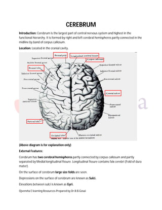
  • 1. Ojvensha E learning Resources-Prepared by Dr.B.B.Gosai CEREBRUM Introduction: Cerebrum is the largest part of central nervous system and highest in the functional hierarchy. It is formed by right and left cerebral hemispheres partly connected in the midline by band of corpus callosum. Location: Located in the cranial cavity. (Above diagram is for explanation only) External Features: Cerebrum has two cerebral hemispheres partly connected by corpus callosum and partly separated by Medial longitudinal fissure. Longitudinal fissure contains falx cerebri (Fold of dura mater). On the surface of cerebrum large size folds are seen. Depressions on the surface of cerebrum are known as Sulci. Elevations between sulci is known as Gyri.
  • 2. Ojvensha E learning Resources-Prepared by Dr.B.B.Gosai Surfaces of Cerebrum: Three surfaces: Superolateral surface: Between Superomedial border and inferolateral border. Medial Surface: Between Superomedial and inferomedial border. Inferior Surface: Between Inferolateral and inferomedial border. Three borders: Superomedial border: between superolateral and medial surface. Inferolateral border: between superolateral and inferior surface. Inferomedial border: between inferior and medial surface. Three poles: Frontal pole: Anterior most part of cerebrum (frontal lobe) Occipital pole: Posterior most part of cerebrum (Occipital lobe) Temporal pole: Anterior most part of temporal lobe. Sulci, Gyri and Important Functional areas of Cerebrum: Cerebrum on superolateral surface is divided into three lobes by certain sulci and imaginary lines. Main sulci for demarcation of lobes are: 1. Central sulcus: Starts from 1 cm behind the midpoint on superomedial border and runs obliquely on superolateral surface without interruptions and ends just above the lateral sulcus. 2. Lateral sulcus: It is also seen on superolateral surface. It has a stem and three rami. Stem is near the anterior end of temporal lobe. Three rami:
  • 3. Ojvensha E learning Resources-Prepared by Dr.B.B.Gosai Anterior ramus runs forwards. Ascending ramus runs upwards Posterior ramus is longest and runs backwards. 3. Parieto-occipital sulcus: It is predominantly seen on Medial surface. Its small part is seen on superolateral surface. SUPEROLATERAL SURFACE: It is divided into four lobes: 1. Frontal lobe 2. Parietal lobe 3. Temporal lobe 4. Occipital lobe 1. Frontal lobe: Part of cerebrum in front of central sulcus. 2. Parietal lobe: Part of cerebrum behind the central sulcus. 3. Occipital lobe: Behind the imaginary line joining parito-occipital sulcus and preoccipital notch. 4. Temporal lobe: Below the posterior ramus of lateral sulcus.
  • 4. Ojvensha E learning Resources-Prepared by Dr.B.B.Gosai Frontal pole (Black arrow) Temporal pole (Red arrow) Occipital pole (Green arrow)
  • 5. Ojvensha E learning Resources-Prepared by Dr.B.B.Gosai Important Figure for Sulci and gyri on superolateral surface (All Labels are to be mentioned)
  • 6. Ojvensha E learning Resources-Prepared by Dr.B.B.Gosai Important Figure for Functional areas in superolateral surface (All labels are to be mentioned) Sulci, Gyri and functional areas in Frontal lobe: Sulci: Pre-central sulcus: in front of central sulcus and runs parallel to it. Superior and inferior frontal sulci: In front of pre-central sulcus and runs horizontally. Anterior and ascending rami of lateral sulcus: in the lower aspect of anterior part of frontal lobe. Gyri:
  • 7. Ojvensha E learning Resources-Prepared by Dr.B.B.Gosai Pre-central gyrus: Located between the central and pre-central sulcus. It contains Motor areas of cerebrum. Superior, Middle and Inferior frontal gyri: The part of frontal lobe in front of precentral gyrus is divided into superior, middle and inferior frontal gyri by superior and inferior sulci. Functional areas: Motor Areas of Cerebrum: (*****Short note) 1. Motor Area of cerebrum: Location: It is located in the pre-central gyrus between the central sulcus and pre-central sulcus. Important characteristics: a. Body is represented upside down in the area. i.e. head and neck is represented in the lower part of area and lower limbs are represented in the upper part of the area. b. In the area movements are represented and not the individual muscles. i.e. for example flexion movement is represented and not the individual muscle causing flexion. c. The parts having fine movements are represented larger compared to parts having coarse movements. i.e. Thumb, fingers, lips tongue and eyes are represented larger compared to trunk and limbs. d. Upper 1.5 cm area is supplied by Anterior cerebral artery and rest of the area is supplied by middle cerebral artery. e. It controls opposite half of body (Contralateral control). i.e. Right side of area control left side of the body and left side area control right side of body. Function: It is responsible for the execution of the movements of body. Applied Anatomy: a. Damage to this area in compression by collection of hematoma in fracture of skull leads to paralysis of the muscles of opposite half of body. b. Blockage of anterior cerebral artery leads to paralysis of lower limb muscles and loss of control on sphincters while rest of the body is not affected. (Anterior cerebral supply upper part of area). c. Blockage of Middle cerebral artery leads to paralysis of the all the muscles of body except the lower limb and sphincters.
  • 8. Ojvensha E learning Resources-Prepared by Dr.B.B.Gosai 2. Pre-motor area: In front of motor area. Function: Planning of the movements. 3. Broca’s motor area of speech: Located in triangular area between anterior and ascending rami of lateral sulcus (Pars Traingularis). Function: Motor speech of person i. e. articulation of words. Applied: Damage to this area leads to Motor Aphasia. ( Loss of speech). Silent Area of Brain (Prefrontal cortex): It is anterior part of frontal lone. Earlier it was believed to be silent but now it is know that it is part of limbic system and concerned with social and emotional behaviour of person. Damage to this area leads to Anti-social behaviour. Sulci, Gyri and Functional areas in Parietal lobe: Sulci: Post-central sulcus: behind the central sulcus and runs parallel to it. Intraparietal sulcus: Curved sulcus in the middle of parietal lobe. Gyri: Post-central gyrus: Located between central and post central sulcus. It contains primary sensory area. Superior parietal lobule: Part above the intraparietal sulcus. Inferior parietal lobule: Part below the intraparietal sulcus. Functional areas: 1. Primary Sensory area ( Primary somesthetic area) : Located in post central gyrus between central and post central sulcus. Function: Identification of all general sensations and taste sensation.
  • 9. Ojvensha E learning Resources-Prepared by Dr.B.B.Gosai 2. Secondary sensory area (Secondary somesthetic area) : Located behind the primary sensory area. Function: It keeps the memory of all sensations experienced by person and coordinated with incoming new sensations. 3. Wernick’s sensory area of speech: It is cap like area on the posterior end of posterior ramus of lateral sulcus. Function: Identification of text from the images and sound and send it to Broca’s area for articulation. Applied aspect: Damage to Wernick’s area leads to inability to identify text from images and sound i.e. Sensory Aphasia. Person can speak but unable to read aloud from book and repeat spoken words. Sulci, Gyri and Functional areas in Temporal lobe: Sulci: Superior and Inferior Temporal sulci. Gyri: Superior, middle and inferior temporal gyri: Temporal lobe ins divided in to superior, middle and inferior gyri by superior and inferior temporal sulci. Functional Areas: 1. Primary Auditory area: Located in superior temporal gyrus. Function: Identification of sound. (Hearing) 2. Auditory association area: Located outer to primary auditory area. Function: Keeps memory of all sound heard by person and coordinated with new incoming sound. Applied anatomy: Damage to auditory area leads to partial hearing loss. Abnormal stimulation of Auditory association area leads to Auditory Hallucinations i.e. person complains of hearing non-existent sound. Sulci, Gyri and Functional areas in Occipital lobe: Sulci:
  • 10. Ojvensha E learning Resources-Prepared by Dr.B.B.Gosai Small part of calcarine sulcus: Most of the sulcus is seen on medial surface. Lunate sulcus: Curved sulcus in from of end of calcarine sulcus. Functional area: Primary visual area: Mostly seen on medial surface above and below the calcarine sulcus> Function: Identification of image. ( Vision) Damage to this area leads to Contralateral homonymous hemianopia. Visual Association area: located outer to visual area. Function: Keeps memory of all images seen by person and coordinated with new incoming images. Abnormal stimulation of visual association area leads to visual Hallucinations i.e. person complains of seeing non-existent images. MEDIAL SURFACE:
  • 11. Ojvensha E learning Resources-Prepared by Dr.B.B.Gosai Sulci: Callosal sulcus: Closely related to corpus callosum. Cingulate sulcus: Runs parallel to corpus callosum. Parieto-occipital sulcus and calcarine sulcus forms “Y” shape on posterior aspect of medial surface. Upper limb of “Y” is Parieto-occipital sulcus. (Red Arrow) Lower limb of “Y” is Calcarine sulcus. (Black arrow) Gyri: Cingulate gyrus: Between callosal sulcus and cingulated sulcus. It is part of limbic system concerned with social, emotional behaviour and memory. Cuneus: part between calcarine and parieto-occipital sulcus. Pre-cuneus: part in front of parieto-occipital sulcus. INFERIOR SURFACE:
  • 12. Ojvensha E learning Resources-Prepared by Dr.B.B.Gosai Divided in to two parts: 1. Orbital part: Related to roof of orbit. (Anterior smallerpart) Sulci: Olfactory sulcus: related to olfactory tract. Orbital sulci: “H” shaped. Gyri: Gyrus rectus: Medial to olfactory sulcus. Orbital gyri: between orbital sulci. 2. Tentorial part: related to tentorium cerebella (fold of dura mater) (Posterior larger part) Sulci: Collateral sulcus: It is medial sulcus bounding the parahippocampal gyrus. Occipito-temporal sulcus: It is lateral to collateral sulcus. Rhinal sulcus: small sulcus in anterior part of parahippocampal gyrus. Gyri: Parahippocampal gyrus: Medial to collateral sulcus. Part of limbic system. Cocerned with social, emotional behaviour and memory. Uncus: hook shaped anterior part of parahippocampal gyrus. NOTE: Describe sulci and gyri of cerebrum (only mention sulci and gyri and draw the diagram for the same) Describe functional cortical areas of cerebrum ( mention the functuional areas in the superolateral surface with functions and applied aspect) Short note: Motor areas of cerebrum (Mention motor area, premotor area and Broca’s motor area of speech with diagram showing only these areas) Motor area of cerebrum (only Motor area to be mentioned with diagram) ====================X================