1. Periodontology 2000, Vol. 41, 2006, 177–187 Copyright Ó Blackwell Munksgaard 2006
Printed in Singapore. All rights reserved PERIODONTOLOGY 2000
Prospects for tooth regeneration
S I L V I O E. D U A I L I B I , M O N I C A T. D U A I L I B I , J O S E P H P. V A C A N T I &
P A M E L A C. Y E L I C K
Regenerative dental medicine uses an integrated bud, cap, bell, crown, and root (70, 71) (Fig. 2). The
sciences approach, involving developmental and coordinated development of tooth supporting struc-
molecular/cellular biology, molecular genetics and tures, including periodontal ligament and alveolar
chemical engineering (5, 25, 28, 30, 41, 62, 65, 75). bone, begins around the bell stage (68).
Recent advances in the fields of dental tissue The tooth germ is first identifiable as a localized
engineering, materials science and stem cell biology, thickening and proliferation of the oral epithelium
suggest that tooth regeneration will be possible (Fig. 2A). The dental epithelium forms a bud that
in the foreseeable future. Published reports have extends into the underlying dental mesenchyme,
demonstrated that dental tissue progenitor cells marking the first stage of tooth development. The
present in the pulp tissue of deciduous and adult dental epithelium subsequently undergoes signifi-
teeth can be used to generate dentin and alveolar cant proliferative activity (Fig. 2B), extending around
bone (59, 84), while those present in immature tooth the periphery to form a cap-like structure (Fig. 2C).
buds can be used to bioengineer small, anatomically During this process, the nonproliferating enamel
correct, whole tooth crowns consisting of enamel, knot signaling center (72) becomes identifiable, as
dentin and pulp tissue (13, 81–83). These promising epithelial cells organize themselves into three
results, along with many other studies reporting distinct regions, namely the outer epithelium, the
dental tissue regeneration, suggest a means for the inner epithelium, and central cell layers called
eventual regeneration of replacement teeth in the stratum intermedium and stellate reticulum
humans. However, before this can occur, certain (Fig. 2C,D). The ectomesenchymal cells of the dental
obstacles must first be overcome. Below, we will papilla condense beneath the invaginating dental
review recent progress in tooth-regeneration efforts epithelium, eventually giving rise to dentin and pulp
to date, and identify challenges that must be met in tissues. The dental follicle forms around the enamel
order for this approach to reach clinical relevance in organ and dental papilla, eventually forming the
humans. periodontal tissues (Fig. 2C,D).
The bell stage is characterized by continued
proliferation and histodifferentiation of the dental
epithelium (Fig. 2D). Inner dental epithelial cells
Tooth development as a model for assume a cuboidal shape and produce high levels
tooth regeneration of glycogen, adjacent stratum intermedium pro-
duces high levels of alkaline phosphatase, and the
The interactions of the dental epithelium and stellate reticulum assumes a distinctive star shape,
mesenchyme during natural tooth development surrounded by the outer epithelial cell layer
provide insight into how bioengineered tooth for- (Fig. 2D).
mation may be facilitated. Although human incisor, As tooth development proceeds through differen-
canine, premolar and molar teeth exhibit distinct tiation stages, dental mesenchyme-derived odonto-
morphologies, (Fig. 1), they develop in basically the blasts differentiate and elaborate the dentin matrix,
same manner. Tooth development, the result of and epithelial cell-derived ameloblasts cells secrete
reciprocal interactions between the dental epithe- the enamel matrix for enamel production (Fig. 2E).
lium and the neural crest cell-derived ectomesen- After the tooth crown has formed, tooth root struc-
chyme, is initiated by the dental epithelium and tures develop from the rudimentary Hertwig’s epi-
proceeds through five distinct morphological stages: thelial root sheath, forming dentin, cementum,
177
2. Duailibi et al.
Deciduous Teeth Permanent Teeth
11 years
7 years
12 years
8 years
15 years
9 years
21 years
10 years
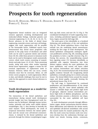
Fig. 1. Human deciduous and adult tooth from 7 to 21 years of age. Deciduous and permanent human teeth form and
´ ˜
erupt as shown. (Adapted from: Picosse M. Anatomia dentaria, 2nd edition. Sao Paulo: Sarvier, 1977: 66–67.)
periodontal ligament and alveolar bone (Fig. 2F). Human replacement teeth form as a localized
The co-ordinated processes of tooth root maturation proliferation of the dental lamina of a pre-existing
and tooth eruption proceed in an interdependent deciduous tooth (68). Humans form only one set of
manner that is not well understood at the present replacement teeth, which do not exhibit subsequent
time (68). regenerative capabilities. The molecular signaling
178
3. Prospects for tooth regeneration
Thus, natural primary and replacement tooth
formation provides a foundation upon which tooth-
regeneration strategies can be based. It is anticipa-
ted that tooth-regeneration strategies need not
necessarily be as complex as natural tooth devel-
opment, but rather can mimic certain aspects of
natural tooth formation to facilitate tooth-regener-
ation therapies.
Regenerative capabilities of naturally
formed dental tissues
Human deciduous and adult tooth tissues exhibit a
limited degree of regenerative capacity. Each type of
mineralized dental tissue – enamel, dentin, cemen-
tum and alveolar bone – exhibits distinct properties.
Enamel is formed by a process called amelogenesis,
dentin formation is termed dentinogenesis, cemen-
tum formation is termed cementogenesis, and alve-
olar bone forms by osteogenesis (26).
Enamel, the most highly mineralized dental tissue,
is composed of crystalline calcium phosphate, and is
approximately 96% mineral with the remaining 4%
consisting of organic components and water. Its
hardness, when allowed to dry, is comparable with
brittle steel. The basic structural unit of enamel is an
enamel rod, which is tightly packed and mechanically
adherent to other rods, providing high resistance to
stress fractures (68). The interwoven architecture of
Fig. 2. Human tooth developmental stages. (A) Tooth enamel crystals provides both strength and protec-
bud forms as a proliferation of the dental epithelium (de) tion for the tooth (43). The enamel-forming progen-
into the underlying dental mesenchyme (dm). (B) The
itor cells – ameloblasts – undergo apoptosis as they
dental epithelium continues to proliferate, and the
underlying dental mesenchyme undergoes condensation. elaborate the enamel matrix, such that by the time
EK, enamel knot signaling center. (C) The cap stage tooth the enamel is fully formed, no ameloblasts remain
exhibits distinct outer epithelium (oe), and inner epi- (60). Enamel regeneration therefore is not possible in
thelium (ie) layers, surrounding the condensed dental erupted teeth because the progenitor cells are no
mesenchyme (dm). (D) The bell-stage tooth exhibits dif-
longer present.
ferentiated enamel organ, consisting of distinct inner
enamel (ie), stratum intermedium (si) and stellate reti- Dentin, the mineralized tissue underlying enamel,
culum (sr) cell layers, which surround the dental papilla is characterized by distinctive fluid-filled dentin tu-
(dp). (E) Differentiation-stage teeth exhibit polarized bules (7, 68), and is approximately 74% mineral, with
odontoblasts (od) and ameloblasts (am), which elaborate the organic phase consisting mostly of type-1 colla-
dentin and enamel, respectively. (F) Late differentiation-
gen, with small amounts of dentin proteins and water
stage teeth display rudimentary root structures, called
Hertwig’s epithelial root sheath (hers), periodontal liga- (68). Primary dentin consists of two distinct miner-
ment tissues (pdl), polarized odontoblasts (od), and alized types – circumpulpal dentin, which surrounds
mineralized dentin (d) and enamel (e) layers. (Courtesy the pulp chamber, and mantle dentin, which is
of Katchiburian E, Arana VE. Histologia e Embriologia located at the dentin–enamel junction (44). Dentin is
´
Oral, 2nd edition. Guanabara – Koogan: Medica Pan-
composed of millions of tubules (approximately
americana S.A.C.F., 2004. Reproduced with permission
from the editors.) 60 000 tubules/mm2), which extend through a colla-
gen- and calcium-rich zone of intertubular dentin,
cascades regulating the fascinating process of from the pulpal wall to the dentin–enamel junction
replacement tooth formation remain largely unchar- (66). The tubule diameter at the dentin–enamel
acterized as a result of the lack of a suitable animal junction is 0.06 lm, and widens to approximately
model. 3.0 lm at the pulpal wall. Dentin tubules are fluid
179
4. Duailibi et al.
filled and may contain an odontoblast process, col- supply, localized inflammation or unfavorable pros-
lagen and nonmyelinated pulpal nerves (68). The thesis pressure (1, 14).
distinct architecture of the fluid-filled dentin tubules In summary, in naturally formed teeth, enamel –
is thought to provide a means of communication the only mineralized tooth tissue derived from the
from the enamel layer to the tooth pulp. Tertiary dental epithelium – exhibits no regenerative proper-
dentin consists of reactionary dentin (formed by pre- ties, while the remaining mineralized periodontal
existing odontoblasts) or reparative dentin (formed and dental tissues, including dentin, pulp, cemen-
from newly differentiated odontoblasts) (64). Once tum, periodontal ligament and alveolar bone, all of
erupted, teeth maintain a limited capacity to form which are formed from neural crest-derived dental
reparative (or tertiary) dentin, when progenitor cells ectomesenchyme, each exhibit a certain degree of
are recruited from the pulp to elaborate localized regenerative capability. Therefore, while the devel-
dentin matrix at the site of injury (68). opment of clinically relevant regeneration strategies
Cementum, the thin layer of mineralized tissue for all types of dental tissues remains a challenging
that covers the dentin of tooth roots, is also highly task, that of enamel is the greatest, owing to the
mineralized, but softer than dentin, consisting of absence of dental epithelial progenitor cells in
approximately 45% inorganic material, 33% organic erupted teeth.
material and 22% water. Cementum, formed by
cementoblasts, is sandwiched between the inner
Postnatal dental stem cells and dental
dentin surface and the outer periodontal ligament
tissue regeneration
surface, and serves to secure the tooth, via the per-
iodontal ligament, to the alveolar bone. Cementum Progenitor stem cells have been identified in hun-
consists of thin, plate-like hydroxyapatite crystals, dreds of human postnatal tissues (10–12, 52, 54, 55,
approximately 55-nm wide and 8-nm thick, and is 85). Stem cells are defined as quiescent cell popula-
similar in chemical composition and physical prop- tions present in low numbers in normal tissue, which
erties to bone. Three types of cementum are present exhibit the distinct characteristic of asymmetric cell
on the tooth: acellular cementum, which covers one- division, resulting in the formation of two distinct
third to one-half of the tooth root adjacent to the daughter cells – a new progenitor/stem cell, and an-
cemento-enamel junction (the area where cemen- other daughter cell capable of forming differentiated
tum and enamel meet); afibrillar cementum, which tissue (18, 32). In this way, stem cells are able to self-
is present at the cemento-enamel junction; and renew and maintain themselves in an undifferenti-
cellular cementum, which typically covers the apical ated state, while also giving rise to differentiating
one-half to two-thirds of the tooth root. Cementum daughter cells. Progenitor cells differ from stem cells
naturally regenerates, slowly forming throughout in that they exhibit a finite life span rather than
the life of the tooth, allowing for continual re- existing throughout the life of the organism, and
attachment of the periodontal ligament fibers, exhibit limited differentiative potential, with the
although the identification of the cementoblast capacity to form only limited tissue types.
progenitor cells remains somewhat controversial at The characterization of dental progenitor/stem
the present time (6). cells has increased significantly over the past 5–
Alveolar bone, formed from neural crest cell- 10 years. Dental mesenchymal progenitor cells have
derived dental mesenchymal cells, functions as the been characterized using transgenic mouse reporter
primary support for teeth, and is composed of models (3, 38), and mesenchymal stem/progenitor
bundles of bone that are built up in layers in a par- cells have been identified and characterized in dental
allel orientation to the coronal–apical direction of the pulp obtained from both deciduous and adult human
tooth. Alveolar bone exhibits rapid turnover in teeth (16, 42, 59). Evidence for both common and
response to mechanical stimulation (79). This char- distinct progenitor cells for periodontal tissues has
acteristic plasticity of alveolar bone distinguishes it been reported (4, 6, 56, 67), as described in more
from other types of bone and allows for constant detail in other chapters of this volume. Preliminary
minor accommodation of tooth movements during characterization of postnatal epithelial and mesen-
mastication (68). Alveolar bone resorption following chymal dental stem/progenitor cells present in
tooth loss can be significant – estimated to be immature tooth buds demonstrated the ability to
40–60% during the first 3 years, with an additional generate bioengineered, anatomically correct tooth
0.25–0.5% loss every year thereafter (1, 2) – presum- crowns containing enamel, dentin, pulp and alveolar
ably as a result of disuse atrophy, decreased blood bone, as described below.
180
5. Prospects for tooth regeneration
cells isolated from both rat and pig tooth buds, which
Regenerative therapies for dental were cultured in vitro for 6 days (Fig. 3), seeded onto
tissues biodegradable scaffolds, and implanted and grown in
the omenta of adult rat hosts. Cultured rat tooth bud
Tissue-engineering approaches have proven to be cells formed distinctly mineralized tissues in 12 weeks,
useful for dental tissue- and whole-tooth-regener- while pig tooth bud cells formed tooth crowns in
ation strategies (33). Based on preclinical cell- and approximately 20–30 weeks (Fig. 4). Histologic and
gene-therapy strategies used for soft tissue organs, immunohistochemical analyses revealed that bio-
reports of the emerging use of tissue-engineering engineered rat and pig dental tissues exhibited many
strategies for dentin, pulp and cementum, as an characteristics of naturally formed dental tissues (13,
alternative to commonly used root canal and crown 81–83). The fact that rat tooth bud cells were cultured
therapies, are becoming more numerous. Advances for up 6 days before being used to generate bioengi-
in vital pulp therapies to regenerate the dentin–pulp neered dental tissues suggests that progenitor dental
complex without the removal of the whole pulp, in- stem cells can be maintained in culture for at least
clude application of exogenous growth factors and/or this long.
stem/progenitor cells (46, 68). The formation of multiple small tooth crowns in
We have previously shown that tooth bud cells can our bioengineered tooth constructs, rather than one
be used to bioengineer anatomically correct tooth large tooth, reveals a number of challenges that
crowns (13, 19, 81–83). Our approach used tooth bud need to be addressed before this approach can
Fig. 3. Cultured tooth bud cells. (A) Rat tooth bud cells cells. (B) After 5 days in culture, small epithelial colonies
cultured for 1 day contain fibroblastic dental mesenchy- are evident (de), surrounded by confluent dental mesen-
mal (dm) cells and small, rounded, dental epithelial (de) chymal (dm) cells.
Fig. 4. Bioengineered mammalian tooth crowns. (A) Bio- ameloblasts (am) and stellate reticulum (sr). (B) Bioengi-
engineered pig tooth exhibits distinct cell layers and tis- neered rat tooth crowns exhibits distinct pulp (pu), pre-
sues observed in naturally formed teeth, including: dentin (pd), dentin (d), and enamel (e) layers.
odontoblasts (od), predentin (pd), dentin (d), enamel (e),
181
6. Duailibi et al.
reliably be used for tooth-regeneration applications. are all highly significant properties. A variety of
First, as bioengineered tooth crown formation re- hydrophilic polymers have been synthesized that
quires the interactions of both dental epithelial cell provide cell support and guidance. Importantly,
progenitors and mesenchymal cell progenitors (as scaffold materials provide a three-dimensional
in natural tooth formation), the ability to bioengi- macromolecular structure to guide the final shape of
neer a tooth of specified size and shape will depend bioengineered tissues. Poly-L-lactic acid and poly
on the ability to first identify, and then guide, the lactic co-glycolic acid co-polymers have been used
interactions of both types of cells. As the cultured to generate composite scaffolds that degrade within
tooth bud cell populations used in our studies were a period of a few weeks up to 1 year (37). Poly-L-
quite heterogeneous, methods to generate purified lactic acid sponges can support the growth of
dental stem cell populations must be developed. chondrocytes in a uniform cellular distribution, their
Next, methods to guide the interactions of epithelial utility has been demonstrated in cartilage tissue
and mesenchymal postnatal dental stem cells to regeneration (36), and polyglycolic acid and poly-
form dentin and enamel layers characteristic of lactic acid have been shown to support the growth of
natural teeth, using modified scaffold materials and biopsied neonatal intestine cells into functional,
designs, for example, must be developed. Finally, as small intestinal tissue (9).
our bioengineered teeth consisted of fairly well- Optimized polymer fabrication techniques have
developed tooth crowns, while the tooth roots were been used to generate three-dimensional structures
relatively undeveloped, we also need to devise composed of an intercommunicating network of
methods to improve bioengineered tooth root for- pores, where the resulting morphology and mech-
mation. Progress in each of these areas is discussed anical properties of the scaffold walls were found to
below. influence tissue engineering applications (29, 34).
Others authors (20) investigated the use of macro-
molecular materials of natural origin (i.e. collagen,
Generation of enriched epithelial alginate, and agarose), derived from hyaluronic acid
and mesenchymal dental stem cell and fibrin glue, to identify polymers that provide the
populations best guidance for cellular differentiation and prolif-
eration (with the objective of replacing tissues by
We are currently using two methods to generate en- cellular transplantation). Natural silk proteins have
riched postnatal dental stem cell populations. The been successfully used to generate scaffolds suitable
first method uses the stem cell antibody, STRO-1 for bone tissue engineering (35). Each type of scaffold
(61), to generate enriched, STRO-1-positive stem cell has unique features that provide flexibility for a
populations from cultured tooth bud cell prepara- variety of tissue-engineering applications. We are
tions. The second method uses side population currently testing a variety of scaffold materials and
profiling to generate enriched dental stem cell designs for guided dentin–enamel junction forma-
populations, based on the demonstrated ability for tion, and optimized whole tooth tissue-engineering
stem cells to efflux Hoechst dye, while nonstem cell applications, based on morphologic cell movements
populations cannot (15). Fluorescent-activated cell of natural tooth development.
sorting allows the separation of Hoechst-negative
stem cells from dye retaining nonstem cell popula-
tions. Clonal cell lines are being established from Functional bioengineered tooth
cells sorted by both methods for future testing in root formation
dental tissue engineering applications.
Our current bioengineered tooth model exhibits
crowns that are much more developed than the tooth
Scaffold materials and design for roots. To improve bioengineered tooth root forma-
whole tooth tissue regeneration tion, we designed hybrid tooth/bone constructs to
test whether the co-ordinated alveolar bone forma-
The importance of scaffold materials and design for tion could improve bioengineered tooth root devel-
tissue engineering has long been recognized. Scaf- opment (82). These studies demonstrated that
fold porosity, biocompatibility and biodegradability, bioengineered tooth roots generated from the hybrid
the ability to support cell growth, and use as a tooth/bone constructs were, in fact, more developed
controlled gene- and protein-delivery vehicle (45) than those formed by tooth constructs alone, indi-
182
7. Prospects for tooth regeneration
cating that this approach is promising. Further in zebrafish is morphologically very similar to adult
modifications of our whole tooth tissue-engineering tooth formation in humans, where adult teeth form
methods are likely to reveal distinct properties of as a dental laminar proliferation from a deciduous
epithelial and mesenchymal dental progenitor cells, tooth (Fig. 5). Although humans generate only two
which can be manipulated to improve current tooth- sets of teeth, zebrafish continuously regenerate teeth
regeneration efforts. throughout their lives. Replacement teeth form
approximately every 7–14 days in juvenile zebrafish,
and more slowly in adults (77), providing the means
Animal models for tooth to study molecular signaling pathways that are per-
regeneration missive for continuous tooth regeneration. The
significant nucleotide and amino acid sequence
Analyses of tooth development in a variety of animal identity, and genomic synteny shared between ze-
models have provided significant insight into tooth- brafish and humans, make the zebrafish a pertinent
regeneration strategies. Characterizations of epithe- model for elucidation of molecular strategies for
lial dental stem cell populations have largely been human replacement tooth formation. These charac-
limited to rodent models exhibiting continuous teristics, together with the molecular/genetic mani-
incisor and molar eruption, including the rat, mouse, pulations possible in zebrafish, make them a superb
rabbit and vole (17, 27, 73). These recent reports model for in vivo analyses of replacement tooth
demonstrate that the epithelial dental stem cell niche formation.
is a specialized epithelial structure located at the
apical end of the tooth, termed the apical bud, and
provide models for identification of genes that Autologous tissues for dental
maintain the epithelial dental stem cell niche (17). tissue-regeneration applications
The vole, which exhibits continuously erupting mo-
lars as well as incisors, has proven useful for com- A major challenge for autologous dental tissue-
paring molecular signaling cascades regulating tooth engineering applications in humans is the identifi-
crown versus tooth root cell fates (73). These studies cation of suitable cell populations to use for these
revealed that the notch signaling pathway, in partic- applications. Reports documenting the successful
ular, is important for maintaining dental stem cell bioengineering of dental tissues include the following
populations, and that the absence of notch signaling provocative findings. Human dental pulp tissues,
results in the terminal differentiation of dental stem isolated from both adult and juvenile teeth, exhibit
cells to tooth root tissue fates. the capacity to differentiate, in vitro and in subcu-
The zebrafish has also proven useful as a model for taneous implants, into dental mesenchyme-derived
tooth regeneration (21, 22, 31, 50, 51, 76, 78, 80). Al- tissues, including dentin, cementum and bone, as
though the zebrafish teeth are pharyngeal, located in well as nerve and vascular endothelium (8, 16, 39, 57,
the pharynx rather than the jaw, their development is 58). Mesenchymal stem cells have been identified in
remarkably similar to that of mammals (51). In a variety of adult tissues as a population of pluripo-
addition, zebrafish continuously regenerate teeth tential self-renewing cells isolated from bone mar-
throughout their lives. Replacement tooth formation row, which exhibit the capacity to differentiate into
Successional Tooth Germ
Dental Lamina Fig. 5. Human and zebrafish
replacement tooth formation. (A)
dl Human successional tooth germs
Dental Organ
form as a localized proliferation of
the dental lamina (arrow). (B) Simi-
pu larly, zebrafish replacement teeth
Dental Papilla
form as a localized proliferation of
d the dental lamina (dl, arrow) of an
Dental Follicle existing functional tooth. Functional
zebrafish teeth exhibit distinct pulp
A B (pu) and dentin (d).
183
8. Duailibi et al.
bone, cartilage, muscle, tendon and adipose tissue. the highly mineralized tooth organ, the product of
Recent studies have identified and characterized reciprocal signaling events between the dental epi-
stem cell populations for cementum, dentine and thelium and dental mesenchyme (23, 69). The need
periodontal ligament (53). As a result of the suc- for alternative dental tissue-replacement therapies
cessful generation of bioengineered reparative den- is evident in recent reports (48, 74), which reveal
tin, it is anticipated that this technique may become startling statistics regarding the high incidence of
clinically relevant in the near future (47). Reports tooth decay and tooth loss in the USA. Recent
demonstrating the use of embryonic stem cells for advances in the identification and characterization of
dental tissue formation reveal the potential utility of dental stem cells, and in dental tissue-engineering
embryonic stem cells for tooth tissue-engineering strategies, suggest that within the next decade, bio-
applications (40, 49). Whole tooth tissue-engineering engineering approaches may successfully be used to
studies revealed that dissociated mammalian tooth regenerate dental tissues (25) and whole teeth (13,
bud cells retain a cell-autonomous developmental 81–83).
program, even when dissociated into single-cell Interest in dental tissue-regeneration applications
suspensions and grown in culture (13, 19, 81, 83). continues to increase as clinically relevant methods
These studies suggest the potential for human tooth for the generation of bioengineered dental tissues,
buds for dental tissue-engineering applications. The and whole teeth, continue to improve. Although
recent identification and characterization of dental obvious practical obstacles remain to be overcome
stem cells suggests the potential use of dental stem before routine clinical treatments become com-
cells for tooth tissue-replacement therapies. Gene- monly available, dental tissue regeneration research
delivery techniques have demonstrated the potential efforts provide an example of how advances in basic
for periodontal ligament regeneration, as well as research can be translated into clinically relevant
for dentin and alveolar bone (24, 47). Methods to dental therapies (63). Tissue engineering offers
generate enriched dental stem cell populations are exciting opportunities for innovative collaborative
suggested by studies of transgenic mice expressing research efforts, integrating the fields of medicine,
green fluorescent protein under the direction of the developmental biology, and physical sciences.
collagen type I gene promoter, which exhibit a Clearly, the future application of regenerative and
population of green fluorescent protein-expressing tissue-engineering techniques to dentistry is one of
dental pulp cells that exhibit stem cell-like charac- immense potential, capable of meeting a variety of
teristics (8, 38). patient needs. High-quality basic dental research is
In contrast to dental mesenchymal dental stem paramount to ensuring that the development of
cells, the identification and characterization of epi- novel clinical treatments is supported by robust
thelial dental stem cells has proven more elusive, mechanistic data and that such approaches are
partly owing to the fact that human enamel does not effective. These efforts reveal how successful inno-
exhibit regenerative capabilities in vivo, reflecting the vations in the field of dentistry can be guided by
fact that epithelial dental stem cells are no longer advances in basic research, highlighting the need for
present in functional human teeth. The recent suc- close partnerships between basic research and clin-
cessful bioengineering of whole tooth crowns con- ical scientists.
taining pulp, dentin and enamel, from immature pig
and rat tooth buds, provides the first evidence that
postnatal epithelial and mesenchymal dental stem Acknowledgments
cells may exhibit utility for whole tooth tissue
engineering (13, 82, 83). This work was supported by the Center for the In-
tegration of Medicine and Innovative Technology
(CIMIT), NIH grants R41DE015445, R21DE16370, and
The future of regenerative dentistry R01DE016132-01A1, CAPES grant SAUX-PE 1295/
2005, and FAPESP grant 04/08924-08.
Recent advances in the fields of tissue engineering
and surgery have merged to create a promising new
era in which it is possible that bioengineered tissues References
and organs may routinely be used to replace con-
genitally missing tissues and organs, and/or those 1. Ashman A, Lopinto J. Placement of implants into ridges
lost to injury or disease. These advances also apply to grafted with bioplant HTR synthetic bone: histological
184
9. Prospects for tooth regeneration
long-term case history reports. J Oral Implantol 2000: 26: 21. Huysseune A, Van der heyden C, Sire JY. Early develop-
276–290. ment of the zebrafish (Danio rerio) pharyngeal dentition
2. Ashman A, LoPinto J, Rosenlicht J. Ridge augmentation for (Teleostei, Cyprinidae). Anat Embryol (Berl) 1998: 198:
immediate postextraction implants: eight year retrospec- 289–305.
tive study. Pract Periodontics Aesthet Dent 1995: 7: 85–94. 22. Jackman WR, Draper BW, Stock DW. Fgf signaling is re-
3. Balic A, Mina M. Analysis of developmental potentials of quired for zebrafish tooth development. Dev Biol 2004: 274:
dental pulp in vitro using GFP transgenes. Orthod 139–157.
Craniofac Res 2005: 8: 252–258. 23. Jernvall J, Thesleff I. Reiterative signaling and patterning
4. Berry JE, Zhao M, Jin Q, Foster BL, Viswanathan H, during mammalian tooth morphogenesis. Mech Dev 2000:
Somerman MJ. Exploring the origins of cementoblasts and 92: 19–29.
their trigger factors. Connect Tissue Res 2003: 44 (Suppl. 1): 24. Jin Q, Anusaksathien O, Webb SA, Printz MA, Giannobile
97–102. WV. Engineering of tooth-supporting structures by deliv-
5. Bohl KS, Shon J, Rutherford B, Mooney DJ. Role of syn- ery of PDGF gene therapy vectors. Mol Ther 2004: 9: 519–
thetic extracellular matrix in development of engineered 526.
dental pulp. Biomater Sci Polymer Ed 1998: 9: 749–764. 25. Kaigler D, Mooney DJ. Tissue engineering impact on den-
6. Bosshardt DD. Are cementoblasts a subpopulation of tistry. J Dent Educ 2001: 65: 456–462.
osteoblasts or a unique phenotype? J Dent Res 2005: 84: 26. Katchburian E, Arana V. Histologia e Embriologia Oral, 2nd
390–406. edn. Brazil: Editorial Medica Panamericana. Guanabara
7. Brannstrom M, Linden LA, Astrom A. The hydrodynamics Koogan, 2005: 151–179.
of the dental tubule and of pulp fluid. A discussion of its 27. Kawano S, Saito M, Handa K, Morotomi T, Toyono T, Seta
significance in relation to dentinal sensitivity. Caries Res Y, Nakamura N, Uchida T, Toyoshima K, Ohishi M, Harada
1967: 1: 310–317. H. Characterization of dental epithelial progenitor cells
8. Braut A, Kollar EJ, Mina M. Analysis of the odontogenic and derived from cervical-loop epithelium in a rat lower inci-
osteogenic potentials of dental pulp in vivo using a col1a1– sor. J Dent Res 2004: 83: 129–133.
2.3-GFP transgene. Int J Dev Biol 2003: 47: 281–292. 28. Kim SS, Vacanti JP. The current status of tissue engin-
9. Choi RS, Riegler M, Pothoulakis C, Kim BS, Mooney D, eering as potential therapy. Semin Pediatr Surg 1999: 8:
Vacanti M, Vacanti JP. Studies of brush border enzymes, 119–123.
basement membrane components and electrophysiology 29. Kim SS, Utsunomiya H, Koski JA, Wu BM, Cima MJ, Sohn J,
of tissue-engineered neo-intestine. J Pediatr Surg 1998: 33: Mukai K, Griffith LG, Vacanti JP. Survival and function of
991–997. hepatocytes on a novel three dimensional synthetic bio-
10. Deans RJ, Moseley AB. Mesenchymal stem cells: biology degradable polymer scaffold with an intrinsic network of
and potential clinical uses. Exp Hematol 2000: 28: 875–884. channels. Ann Surg 1998: 28: 8–13.
11. deWynter EA, Emmerson AJ, Testa NG. Properties of 30. Langer R, Vacanti JP. Tissue engineering. Science 1993: 260:
peripheral blood and cord blood stem cells. Baillieres Best 920–926.
Pract Res Clin Haematol 1999: 12: 1–17. 31. Laurenti P, Thaeron C, Allizard F, Huysseune A, Sire JY.
12. Dua HS, Azuara-Blanco A. Limbal stem cells of the corneal Cellular expression of eve1 suggests its requirement for the
epithelium. Surv Ophthalmol 2000: 44: 415–425. differentiation of the ameloblasts and for the initiation and
13. Duailibi MT, Duailibi SE, Young CS, Bartlett JD, Vacanti JP, morphogenesis of the first tooth in the zebrafish (Danio
Yelick PC. Bioengineered teeth from cultured rat tooth bud rerio). Dev Dyn 2004: 230: 727–733.
cells. J Dent Res 2004: 83: 523–528. 32. Lin H. The self-renewing mechanism of stem cells in the
14. Fenton AH, Zarb GA, MacKay HF. Overdenture oversights. germline. Curr Opin Cell Biol 1998: 10: 687–693.
Dent Clin North Am 1979: 23: 117–130. 33. Lynch SE, Genco RJ, Marx RE., eds. Tissue engineering.
15. Goodell MA, Rosenzweig M, Kim H, Marks DF, DeMaria M, Applications in maxillofacial surgery and periodontics, 1st
Paradis G, Grupp SA, Sieff CA, Mulligan RC, Johnson RP. edn. Carol Stream, IL: Quintessence Publishing, 1999.
Dye efflux studies suggest that hematopoietic stem cells 34. Ma PX, Choi JW. (AQ) Biodegradable polymer scaffolds
expressing low or undetectable levels of CD34 antigen exist with well defined interconnected spherical pore network.
in multiple species. Nat Med 1997: 3: 1337–1345. Tissue Eng 2001: 7: 23–33.
16. Gronthos S, Mankani M, Brahim J, Gehron Robey P, Shi S. 35. Meinel L, Fajardo R, Hofmann S, Langer R, Chen J, Snyder
Postnatal human dental pulp stem cells (DPSCs) in vitro B, Vunjak-Novakovic G, Kaplan D. Silk implants for the
and in vivo. Proc Natl Acad Sci USA 2000: 97: 13625–13630. healing of critical size bone defects. Bone 2005: 37: 688–
17. Harada H, Ohshima H. New perspectives on tooth devel- 698.
opment and the dental stem cell niche. Arch Histol Cytol 36. Mikos AG, Sarakinos G, Leite SM, Vacanti JP, Langer R.
2004: 67: 1–11. Laminated three-dimensional biodegradable foams for
18. Hawkins N, Garriga G. Asymmetric cell division: from a to use in tissue engineering. Biomaterials 1993: 14: 323–
z. Genes Dev 1998: 12: 3625–3638. 330.
19. Honda MJ, Sumita Y, Kagami H, Ueda M. Histological and 37. Mikos AG, Lyman MD, Freed LE, Langer R. Wetting of poly
immunohistochemical studies of tissue engineered (L-lactic acid) and poly (DL-lactic-co-glycolic acid) foams
odontogenesis. Arch Histol Cytol 2005: 68: 89–101. for tissue culture. Biomaterials 1993: 15: 55–58.
20. Hutmacher DW, Goh JCH, Tech SH. An introduction to 38. Mina M, Braut A. New insight into progenitor/stem cells in
biodegradable materials for tissue engineering applica- dental pulp using Col1a1-GFP transgenes. Cells Tissues
tions. Ann Acad Med Singapore 2001: 30: 183–191. Organs 2004: 176: 120–133.
185
10. Duailibi et al.
39. Miura M, Gronthos S, Zhao M, Lu B, Fisher LW, Robey PG, 60. Shibata S, Suzuki S, Tengan T, Yamashita Y. A histo-
Shi S. SHED: stem cells from human exfoliated deciduous chemical study of apoptosis in the reduced ameloblasts
teeth. Proc Natl Acad Sci USA 2003: 100: 5807–5812. of erupting mouse molars. Arch Oral Biol 1995: 40: 677–
40. Modino SA, Sharpe PT. Tissue engineering of teeth using 680.
adult stem cells. Arch Oral Biol 2005: 50: 255–258. 61. Simmons PJ, Torok-Storb B. Identification of stromal cell
41. Mooney DJ, Mikos AG. Growing new organs. Sci Am 1999: precursors in human bone marrow by a novel monoclonal
280: 60–65. antibody, STRO-1. Blood 1991: 78: 55–62.
42. Mooney DJ, Powell C, Piana J, Rutherford B. Engineering 62. Sittinger M, Bujia J, Rotter N, Reitzel D, Minuth WW,
dental pulp-like tissue in vitro. Biomaterials 1996: 12: 865– Burmester GR. Tissue engineering and autologous trans-
868. plant formation: practical approaches with resorbable
43. Moradian-Oldak J. Amelogenins: assembly, processing and biomaterials and new cell culture techniques. Biomaterials
control of crystal morphology. Matrix Biol 2001: 20: 293– 1996: 17: 237–242.
305. 63. Smith AJ. Tooth tissue engineering and regeneration – a
44. Moss ML. Studies on dentin. I. Mantle dentin. Acta Anat translational vision! J Dent Res 2004: 83: 517.
(Basel) 1999: 87: 481–507. 64. Smith AJ, Murray PE, Sloan AJ, Matthews JB, Zhao S. Trans-
45. Murphy WL, Mooney DJ. Controlled delivery of inductive dentinal stimulation of tertiary dentinogenesis. Adv Dent
proteins, plasmid DNA and cells from tissue engineering Res 2001: 15: 51–54.
matrices. J Periodontal Res 1999: 34: 413–419. 65. Stock UA, Vacanti JP. Tissue engineering: current state and
46. Nakashima M, Akamine A. The application of tissue prospects. Annu Rev Med 2001: 52: 143–151.
engineering to regeneration of pulp and dentin in endo- 66. Sumikawa DA, Marshall GW, Gee L, Marshall SJ. Micro-
dontics. J. Endod 2005: 31: 711–718. structure of primary tooth dentin. Pediatr Dent 1999: 21:
47. Nakashima M, Iohara K, Ishikawa M, Ito M, Tomokiyo A, 439–444.
Tanaka T, Akamine A. Stimulation of reparative dentin 67. Taba M Jr, Jin Q, Sugai JV, Giannobile WV. Current con-
formation by ex vivo gene therapy using dental pulp stem cepts in periodontal bioengineering. Orthod Craniofac Res
cells electrotransfected with growth/differentiation factor 2005: 8: 292–302.
11 (Gdf11). Hum Gene Ther 2004: 15: 1045–1053. 68. Ten Cate. Ten Cate’s oral histology: development, structure
48. National Institute for Dental and Craniofacial Research. and function. CV Moseby, 2003.
Strategic Plan-2003–2008. J Am Coll Dent 2003: 70: 43–55. 69. Thesleff I. Epithelial-mesenchymal signaling regulating
49. Ohazama A, Modino SA, Miletich I, Sharpe PT. Stem-cell- tooth morphogenesis. J Cell Sci 2003: 116: 1647–1648.
based tissue engineering of murine teeth. J Dent Res 2004: 70. Thesleff I, Partanen AM, Vainio S. Epithelial-mesenchymal
83: 518–522. interactions in tooth morphogenesis: the roles of extracel-
50. Payne TL, Skobe Z, Yelick PC. Regulation of tooth devel- lular matrix, growth factors, and cell surface receptors.
opment by the novel type I TGFbeta family member J Craniofac Genet Dev Biol 1991: 11: 229–237.
receptor Alk8. J Dent Res 2001: 80: 1968–1973. 71. Thesleff I, Vaahtokari A, Kettunen P, Aberg T. Epithelial-
51. Perrino MA, Yelick PC. Immunolocalization of Alk8 during mesenchymal signaling during tooth development. Con-
replacement tooth development in zebrafish. Cells Tissues nect Tissue Res 1995: 32: 9–15.
Organs 2004: 176: 17–27. 72. Thesleff I, Keranen S, Jernvall J. Enamel knots as signaling
52. Rao MS. Multipotent and restricted precursors in the centers linking tooth morphogenesis and odontoblast dif-
central nervous system. Anat Rec 1999: 257: 137–148. ferentiation. Adv Dent Res 2001: 15: 14–18.
53. Risbud MV, Shapiro IM. Stem cells in craniofacial and 73. Tummers M, Thesleff I. Root or crown: a developmental
dental tissue engineering. Orthod Craniofac Res 2005: 8: choice orchestrated by the differential regulation of the
54–59. epithelial stem cell niche in the tooth of two rodent species.
54. Sainio K, Raatikainen-Ahokas A. Mesonephric kidney – Development 2003: 130: 1049–1057.
stem cell factory? Int J Dev Biol 1999: 43: 435–439. 74. US Department of Health and Human Services. Oral health
55. Seale P, Rudnicki MA. A new look at the origin, function, in America: a report of the surgeon general. Rockville, MD:
and ÔÔstem-cell’’ status of muscle satellite cells. Dev Biol US Department of Health and Human Services, NIDCR,
2000: 218: 115–124. NIH, 2000.
56. Seo BM, Miura M, Gronthos S, Bartold PM, Batouli S, 75. Vacanti MP, Leonard JL, Dore B, Bonassar LJ, Cao Y,
Brahim J, Young M, Robey PG, Wang CY, Shi S. Investiga- Stachelek SJ, Vacanti JP, O’Connell F, Yu CS, Farwell AP,
tion of multipotent postnatal stem cells from human Vacanti CA. Tissue-engineered spinal cord. Transplant Proc
periodontal ligament. Lancet 2004: 364: 149–155. 2001: 33: 592–598.
57. Shi S, Gronthos S. Perivascular niche of postnatal mesen- 76. Van der Heyden C, Huysseune A. Dynamics of tooth for-
chymal stem cells in human bone marrow and dental pulp. mation and replacement in the zebrafish (Danio rerio)
J Bone Miner Res 2003: 18: 696–704. (Teleostei, Cyprinidae). Dev Dyn 2005: 219: 486–496.
58. Shi S, Robey PG, Gronthos S. Comparison of human dental 77. Van der Heyden C, Wautier K, Huysseune A. Tooth suc-
pulp and bone marrow stromal stem cells by cDNA cession in the zebrafish (Danio rerio). Arch Oral Biol 2001:
microarray analysis. Bone 2001: 29: 532–539. 46: 1051–1058.
59. Shi S, Bartold PM, Miura M, Seo BM, Robey PG, Gronthos 78. Van der Heyden C, Allizard F, Sire JY, Huysseune A. Tooth
S. The efficacy of mesenchymal stem cells to regenerate development in vitro in two teleost fish, the cichlid
and repair dental structures. Orthod Craniofac Res 2005: 8: Hemichromis bimaculatus and the cyprinid Danio rerio.
191–199. Cell Tissue Res 2005: 321: 375–389.
186
11. Prospects for tooth regeneration
79. Vavidovitch Z. Bone metabolism associated with tooth 83. Young CS, Kim S-W, Taylor R, Vacanti JP, Bartlett JD, Yelick
eruption and orthodontic tooth movement. J Periodontol PC. Developmental analysis and three-dimensional com-
1979: 50 (4 Spec No): 22–29. puter modeling of tooth crowns grown on biodegradable
80. Yelick PC, Schilling TF. Molecular dissection of craniofacial polymer scaffolds. Arch Oral Biol 2005: 50: 259–265.
development using zebrafish. Crit Rev Oral Biol Med 2002: 84. Zhang W, Walboomers XF, Wolke JG, Bian Z, Fan MW,
13: 308–322. Jansen JA. Differentiation ability of rat postnatal dental
81. Young CS, Terada S, Vacanti JP, Honda M, Bartlett JD, pulp cells in vitro. Tissue Eng 2005: 11: 357–368.
Yelick PC. Tissue engineering of complex tooth structure 85. Zuk PA, Zhu M, Mizuno H, Huang J, Futrell JW, Katz AJ,
on biodegradable polymer scaffolds. J Dent Res 2002: 81: Benhaim P, Lorenz HP, Hedrick MH. Multilineage cells
695–700. from human adipose tissue: implications for cell-based
82. Young CS, Abukawa H, Asrican R, Ravens MS, Troulis MJ, therapies. Tissue Eng 2001: 7: 211–228.
Kaban LB, Vacanti JP, Yelick PC. Tissue engineered hybrid
tooth and bone. Tissue Eng 2005: 11: 1599–1610.
187