2. 644
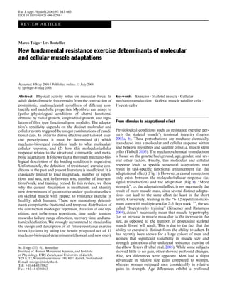
Fig. 1 SimpliWed model for the
transduction of exercise-related a exercise stimulus (e) e = f(x1...xi)
skeletal muscle perturbations x1...xi, mechano-biological exercise
into structural adaptations with stimulus determinants
associated adaptational eVect.
An exercise stimulus with deW-
ned mechano-biological charac- b response matrix (m) m = f(G, Y, S, A, M, T, H, N, I,...)
teristics (described in this paper)
(a) is mechano-chemically trans- Genotype (G )
duced (c) into a quantitative Age (Y )
and/or qualitative adaptation Gender (S )
(d) of the muscle phenotype, Muscular architecture (A )
based on the respective response Muscular subsystem (M )
matrix (b). The adaptation (d) is Muscular anthropometry (T )
associated with the correspond- Hormonal status (H )
ing adaptational eVect (e). A Nutritional status (N )
Immunological status (I )
causal connection exists be-
tween (c) and (d) (red shading)
c signal transduction (s) s = f(e, m, ...)
d adaptation (a) a = f(s)
e adaptational effect (z) z = f(a)
impact on the molecular response to resistance exercise, conditions that have quantitative and/or qualitative
too (Hameed et al. 2003). Following a single bout of high eVects on skeletal muscle phenotype. These mechano-
load knee extensor resistance exercise, mechanogrowth biological determinants are proposed for standardised
factor (MGF) response was attenuated in older subjects. description of resistance exercise stimuli in both aca-
This is indicative of age-related desensitivity to mechani- demic and recreational settings.
cal loading. Also, various human polymorphisms at
genetic loci have a quantitative eVect on muscle pheno-
type (Thompson et al. 2004). Such polymorphisms, also
known as muscle quantitative trait loci, drive the genetic Fundamental mechanical stimuli decoded by skeletal
variation that underlies muscle size and strength. muscle
Therefore, several prerequisites for the identiWcation
of eVective and speciWc exercise perturbations with Basically, muscles can adopt three strategies of quantita-
desired functional enhancement are to be met. First, tive or qualitative eVect on muscular phenotype to adjust
causal relationships between changes at the molecular for altered functional demands (Goldspink 1985): (1)
and cellular level and the resulting adaptation need to be positive or negative longitudinal growth; (2) positive or
identiWed. These relationships need to be established on negative radial growth and (3) contractile [myosin heavy
the basis of several factors such as genetics, age, sex, etc. chain (MyHC)] and metabolic tuning. These adaptational
Further, it must be ascertained whether the adaptation strategies can be adopted concurrently or separately,
leads to functional enhancement. Finally, the various depending on the speciWcity of the (patho-)physiological
causal connections between signal transduction and condition. Generally, exercise-induced physiological con-
adaptation can be interrogated by multiple exercise ditions can be viewed as the perturbations of the muscle
perturbations in order to identify the most eVective and cells’ tensional integrity. Perturbation of tensional integ-
speciWc exercise perturbations. However, for the identi- rity occurs by increasing or decreasing myocellular active
Wcation of eVective and speciWc exercise perturbations it and/or passive tension, as well as energy production or
is imperative to unequivocally deWne and describe the absorption. Additionally, these tensional and energetic
exercise stimulus in mechano-biological relevant terms. alterations can be sustained for diVerent duration. Thus,
These mechano-biological terms should be directly asso- every exercise condition is coded by a speciWc combina-
ciated with the molecular response. Unfortunately, in tion of changes in constant or intermittent active and/or
current investigations into the molecular response to passive tension of diVerent duration. These temporal
exercise, perturbation design and deWnition remain changes in active and/or passive tension, together with
largely undeWned or ignored. This review aims at identi- the inferred structural insults, are then decoded at the
fying mechano-biological determinants of exercise molecular level and transduced into an appropriate
3. 645
adaptational response. In the following sections, we muscle denervation, and eccentric exercise in vivo
provide a detailed description on how the three funda- (CARP/MARP1) (Aihara et al. 2000; Barash et al.
mental adaptations are regulated at the molecular and 2004; Baumeister et al. 1997; Kuo et al. 1999); strain
cellular levels. From this, we derive the relevant mec- in culture, immobilisation at stretched muscle length,
hano-biological determinants for resistance exercise- and eccentric exercise in vivo (Ankrd2/Arpp/
induced muscular conditioning. MARP2) (Barash et al. 2004, 2005; Kemp et al. 2000;
McKoy et al. 2005); or during recovery after meta-
bolic challenge (DARP/MARP3) (Ikeda et al. 2003),
respectively.
Molecular and cellular determinants of longitudinal (3) is likely to be involved in the regulation of ubiquitin-
muscle growth proteasome-mediated myoWbrillar protein degrada-
tion (see “Atrophy signalling”). MuRF1 was found
Mechanical measurements of rabbit muscle strips before to bind the C-terminal immunoglobulin domains of
and after skinning indicate that total passive tension titin (Centner et al. 2001), and in the nucleus MuRF1
increases with increasing sarcomere length (second-order can bind the transcription factor, glucocorticoid
polynomial) (Prado et al. 2005), i.e. with increased strain. modulatory element binding protein-1 (McElhinny
Total passive tension is believed to develop due to the et al. 2002). Furthermore, mechanical tension and the
lengthening of extramyoWbrillar elements (especially the titin catalytic domain have been shown to regulate
collagen content in the extracellular matrix) as well as to the nuclear localisation of MuRF2 and serum
the lengthening of titin. Titin is a giant (» 3–3.7 MDa) response factor (SRF) (Lange et al. 2005).
sarcomeric protein that contains a series of spring ele-
ments within its I-band region, which contribute to the Recently, it has also been demonstrated in an in vivo rat
elastic properties of myoWbrils (Prado et al. 2005). Exter- model that CARP and MLP are sensitive to both muscle
nal or internal forces applied to the myoWbril lengthen or tissue stress and contraction mode, while Ankrd2/Arpp
shorten the myoWbril to above or below the slack length, is sensitive only to contraction mode (Barash et al. 2005).
respectively. The lengthening or shortening of the myoW- This raises the possibility that strain can be sensed inde-
bril creates a titin force, which is directed to restore rest- pendently of stress.
ing length (Miller et al. 2003). MyoWbrils can be Further evidence for the molecular transduction of
lengthened actively or passively. This means that myoW- passive tension comes from the » 1.5- and » 2-fold
brils can lengthen while contracting (“lengthening con- increase in the serine/threonine kinase Akt [protein
traction”, “eccentric contraction”, “active stretch”) or kinase B (PKB)] activity following 5 and 10–20 min of
without concurrent contraction. In contrast, myoWbrils passive stretch, respectively, of the fast-twitch rat exten-
can only actively shorten, i.e. while contracting (“short- sor digitorum longus muscle but not in the slow-twitch
ening contraction”, “concentric contraction”). Conse- soleus muscle (Sakamoto et al. 2003). Once activated,
quently, passive tension in myoWbrils can develop with or Akt phosphorylates an array of substrates, including
without concurrent lengthening contraction or with proteins that mediate protein synthesis, gene transcrip-
shortening contraction. tion, cell proliferation, and survival (Vivanco and Saw-
yers 2002). In mammals, there are three forms of Akt,
Molecular sensing of myoWbrillar strain (Akt1-3), encoded by distinct genes (Vivanco and Saw-
yers 2002). Expression of a constitutively active form of
In line with titin’s structural and elastic properties, Akt1 in skeletal muscle cells, either in vitro (Takahashi
mounting evidence indicates that myoWbrillar strain et al. 2002) or in mice and rats (Bodine et al. 2001b; Lai
mediates signalling pathways that involve titin’s Z-line et al. 2004; Pallafacchina et al. 2002), causes hypertro-
region (Miller et al. 2003). Signalling pathways comprise phy. Conversely, Akt1 inhibits atrophy in vitro and in
(1) the titin-muscle LIM protein (MLP) pathway, (2) the mice (Sandri et al. 2004; Stitt et al. 2004). Based on their
N2A-muscle ankyrin repeat protein (MARP) pathway, Wnding that passive stretch has no eVect on Akt activity
and (3) the titin-muscle RING Wnger protein (MuRF) in rat slow-twitch soleus muscle (in contrast to rat
pathway: extensor digitorum longus muscle), the authors sug-
gested that susceptibility to mechanical stretch is Wbre
(1) is part of a stretch-dependent myocardial signalling type-speciWc (Sakamoto et al. 2003). This notion is also
pathway whose impairment contributes to the patho- supported by the recent Wnding that the relative impor-
genesis of a subset of dilated cardiomypathies in tance of titin and the extracellular matrix for total pas-
humans (Knoll et al. 2002) and is also induced by sive tension can vary between diVerent adult rabbit
skeletal muscle injury due to eccentric exercise skeletal muscles (Prado et al. 2005). Slow-twitch rabbit
(Barash et al. 2004, 2005; Chen et al. 2002; Hentzen muscles exhibit low titin-based passive tension but this
et al. 2006). tension is highly variable in fast-twitch muscles. Fur-
(2) involves 3 homologous MARPs: CARP/MARP1, thermore, titin-based passive tension, but not extramyo-
Ankrd2/Arpp/MARP2, and DARP/MARP3. MARPs Wbrillar passive tension correlates with the muscle type
show cytokine-like induction following cardiac injury, (Prado et al. 2005).
4. 646
Cellular sensing of mechanical stretch sedentary rats had intermediate counts, although closer
to the climbing group. In another series of experiments,
Satellite cells are muscle precursor cells that lie between rat vastus intermedius muscles were tested mechanically
the basal lamina and sarcolemma of skeletal muscle while still in situ, i.e. attached to the bones, but with all
Wbres (Mauro 1961). In normal adult muscle, satellite other muscles about the knee joint removed. As a result,
cells are mitotically and metabolically quiescent (Schultz in descending-trained rats, the knee angle for optimum
et al. 1978). With appropriate environmental signals, sat- torque generation corresponded to longer muscle lengths
ellite cells enter into the cell cycle, i.e. are “activated”, to than in climbing-trained rats. It follows from these
provide the precursors needed for new muscle formation results that eccentric exercise (lengthening contractions)
in growth and repair (Charge and Rudnicki 2004; Hill leads to accretion of serial sarcomeres. Conversely, exer-
et al. 2003; McKinnell et al. 2005). Results from in vitro cise comprising only shortening contractions leads to a
stretch assays demonstrate that mechanical stretch can decrease in the number of serial sarcomeres. Due to the
result in satellite cell activation (Anderson 2000; Ander- Wnding that 38% of the diVerence in sarcomere numbers
son and Pilipowicz 2002; Tatsumi et al. 2001; Wozniak between decline- and incline-trained rats does not appear
et al. 2003). This mechanical stretch induces hepatocyte as a diVerence in optimum angle, the authors suggest
growth factor (HGF) release from its tethering in the that it has been taken up by shortened tendons (Lynn
extracellular matrix in a nitric oxide-dependent manner et al. 1998).
(Tatsumi and Allen 2004; Tatsumi et al. 2002). Once Recently, the contraction type-dependent diVerential
released, HGF binds to the c-met receptor which is serial sarcomere number adaptation has been conWrmed
located on the plasma membrane of the satellite cells. by measuring vastus intermedius and vastus lateralis
This interaction initiates a cascade of signalling events muscle Wbre dynamics of up- and downhill-running rats
that lead to DNA synthesis, and, thus, to satellite cell in vivo (ButterWeld et al. 2005). It was shown in that
proliferation. study that vastus intermedius and vastus lateralis mus-
cles of uphill-walking rats undergo repeated concentric
Structural adaptation to strain perturbation contractions, and therefore they suVer no contraction-
induced injury. Conversely, the vastus intermedius and
It has long been known that muscles adapt to a new vastus lateralis muscles during downhill walking
functional length by adding or removing sarcomeres in undergo repeated eccentric contractions (ButterWeld
series at the ends of the existing myoWbrils (Dix and et al. 2005). Accordingly, short muscle lengths for uphill
Eisenberg 1990; GriYn et al. 1971; Tabary et al. 1972; concentric-biased contractions result in a loss of serial
Williams and Goldspink 1971). Immobilisation at long sarcomeres, while long muscle lengths for downhill
muscle length results in an increase in the number of sar- eccentric-biased contractions result in a gain of serial
comeres in series. Conversely, immobilisation at short sarcomeres (ButterWeld et al. 2005).
muscle length leads to a decrease in the number of sarco- In humans, the optimum angle for torque generation
meres in series. Furthermore, remodelling of the connec- can be measured reliably, e.g. by isokinetic dynamome-
tive tissue following immobilisation has been try. By that means, an angle-torque curve is measured
demonstrated multiple times in mice, rats, rabbits, and during maximum voluntary contraction with constant
cats (Goldspink 1985; Tabary et al. 1976; Tardieu et al. velocity shortening. As determined by this measure, a
1977, 1982; Williams and Goldspink 1984). However, series of eccentric contractions (“hamstring lowers”) of
both the occurrence and the extent of remodelling seem human hamstring muscles produced a signiWcant shift of
to depend on the connective tissue type (series elastic ele- approximately 7° in optimum knee angle for torque gen-
ment and/or parallel elastic element), species, age, muscle eration to longer muscle lengths (Brockett et al. 2001).
length during immobilisation, and time period of immo- The shift in optimum knee angle for torque generation
bilisation. was parallelled by delayed-onset muscle soreness
In exercise physiology, serial sarcomere number mod- (DOMS) in the hamstrings. The shift occurred immedi-
ulation has been a neglected topic so far (Morgan and ately after exercise and persisted 8 days postexercise,
Proske 2004). Only recently has serial sarcomere number consistent with a training eVect. The mechanism by
modulation been investigated in the context of exercise. which eccentric exercise produces muscle damage,
Direct evidence for exercise-induced modulation of serial DOMS, and increased optimum length for torque gener-
sarcomere number has come from treadmill-trained rat ation has been postulated in the “popping sarcomere
vastus intermedius muscles, the postural knee extensors hypothesis” (Morgan 1990). The popping sarcomere
(Lynn and Morgan 1994; Lynn et al. 1998). Rats were hypothesis states that stretch-induced muscle damage
trained by running on a climbing or descending treadmill results from nonuniform lengthening of sarcomeres,
for approximately 10 min day¡1 for 5 days. The latter when active muscle is stretched beyond optimum length.
had previously been shown to cause muscle damage. If sarcomeres are beyond optimum length, then the lon-
Subsequently, serial sarcomere analysis for single Wbres gest sarcomeres will be the weakest and, so, will be
was performed by laser diVraction. As a result, the stretched more rapidly than the others. Thus, they will
descending-trained rats had the largest sarcomere count, become weaker, until rising passive tension compensates
the climbing-trained rats had the smallest count, and for falling active tension. For at least some muscles this
5. 647
corresponds to lengths beyond Wlament overlap. The musculoskeletal and cardiovascular function. Hence, in
term “popping” is used to describe the uncontrolled, vir- order to increase or preserve a functional ROM by serial
tually instantaneous lengthening of a sarcomere from a sarcomere number modulation, eccentric resistance exer-
length commensurate with its passive length to a length cise covering the functional articular range might be the
where passive structures primarily support the tension. method of choice.
Because the weakest sarcomeres are not at the same In conclusion, there is a substantial body of evidence
point along each myoWbril, this nonuniform lengthening that muscle Wbres and satellite cells can sense changes in
leads to a shearing of myoWbrils, exposing membranes, length. Accordingly, active and passive excursions from
especially T-tubules, to large deformations. This is resting length are transduced into a molecular and cellu-
thought to lead to loss of Ca2+ homeostasis, and, hence, lar response with subsequent structural adaptation. How-
damage, either through tearing of membranes or open- ever, with respect to active excursions from resting length,
ing of stretch-activated channels (Allen et al. 2005). Sup- the response at the molecular, cellular, and structural
port for the nonuniform lengthening of sarcomeres level is dependent on the contraction mode. It follows
comes from a recent study of myoWbrils from rabbit directly that muscle length change as well as contraction
psoas muscle and left ventricles of guinea pig during acti- mode are two mechano-biological determinants of exer-
vation and relaxation (Telley et al. 2006a). However, cise-induced skeletal muscle length adaptation. Therefore,
these authors also show that albeit half-sarcomeres of these two mechano-biological determinants, among oth-
contracting single rabbit psoas myoWbrils lengthen to ers (described below), need to be speciWed in reports com-
diVerent extents during a stretch, rapid elongation of ing from investigations into the plasticity of skeletal
individual sarcomeres beyond Wlament overlap (pop- muscle following (resistance) exercise. As a measure of
ping) does not occur. Moreover, in contrast to predic- muscle length change we suggest to specify ROM (x11,
tions of the popping sarcomere hypothesis, they Table 1) during exercise and the number of length excur-
postulate that a stretch rather stabilises the uniformity of sions [i.e. the number of repetitions (x2, Table 1)]. It must
half-sarcomere lengths and sarcomere symmetry (Telley be pointed out, however, that ROM might not always be
et al. 2006b). indicative of fascicle length excursion. The reason for this
With respect to eccentric exercise, it is postulated that is that length changes of the muscle-tendon units do not
the structural adaptation consists of an increase in the necessarily correspond to the length changes in the mus-
number of sarcomeres in series so that a given muscle cle fascicles (Hoyt et al. 2005). This means that the mus-
length corresponds to a shorter sarcomere length (Mor- cle-tendon-unit may lengthen, while the contracting
gan and Talbot 2002). Whether and how serial sarco- muscle is shortening or isometric. As a measure of con-
mere adaptation in humans following eccentric exercise traction mode we propose to report the fractional distri-
is parallelled by changes in tendinous and/or muscle bution of the three contraction types [shortening
belly connective tissue remains to be established. (concentric), isometric, lengthening (eccentric)] per repeti-
A consequence of the popping sarcomere hypothesis tion in terms of occurrence and temporal requirement (x7,
is that the unloaded shortening velocity of muscle Wbres Table 1). Also, the number of contractions should be
should increase with eccentric training. The reason for reported (x2 and x3, Table 1). For example, did the exer-
this is that the unloaded shortening velocity of a Wbre is cise comprise one set of several, only lengthening contrac-
the sum of the velocities of its sarcomeres. Thus, the tions or was one repetition composed of one shortening,
more sarcomeres in series, the faster the unloaded short- one isometric, and one lengthening contraction? How
ening velocity, provided that no alterations in MyHC much time did it take to perform one repetition and how
composition occur. However, this will have to be demon- was this time distributed over the respective contraction
strated in future experiments, especially with respect to modes? The importance of the latter point for inducing
the MyHC isoform gene switching associated with muscle hypertrophy and gains in strength has been dem-
stretch and force production (Goldspink et al. 1991). onstrated in studies where the eVect of fast lengthening
Another consequence of the popping sarcomere hypoth- contractions versus slow lengthening or slow shortening
esis is that signiWcant muscle damage also can occur with contractions has been investigated (Farthing and Chili-
endurance exercise, provided that the duration (mara- beck 2003; Shepstone et al. 2005).
thon running) or mode (downhill running) of exercise is
extreme. Therefore, it is predicted that under certain cir-
cumstances, endurance exercise can lead to serial sarco-
mere accretion with concurrent increase in unloaded Molecular and cellular determinants of radial muscle
Wbre shortening velocity. On the contrary, “conven- hypertrophy
tional” endurance exercise, which is associated with a
bias towards shortening contractions at short muscle Previous work showed that if the tibialis anterior in the
lengths, will lead to a decrease in the serial sarcomere mature rabbit was electrically stimulated while held in
number and, thus, to shorter muscle lengths. Short mus- the stretched position by plaster cast immobilisation, it
cle lengths come with a reduction in functional range of increased in mass by 35% within 7 days (Goldspink et al.
motion (ROM). In general, a reduction in ROM is not 1992). Thus, if the lengthened (stretched) rodent muscle
desirable in health-based settings that aim at increasing is additionally subjected to electrical stimulation, it
6. 648
Table 1 Mechano-biological descriptors of resistance exercise stim- mechano-biologically signiWcant descriptors (x7-x13) are taken into
uli. In order to better discriminate between the signiWcant stimula- account, it is obvious that the two conditions diVer. These two diVer-
tory cues leading to distinct muscular adaptations, we propose to ring conditions will lead to distinct mechano-chemical signal trans-
design and describe resistance exercise-related muscular perturba- ductions in the subjects’ muscles, even if the two subjects had the
tions based on mechano-biological descriptors (a). Example of how same potential to adapt (i.e. the same response matrix characteristics
inaccurate exercise stimulus description might lead to wrong conclu- [see Fig. 1]). Thus, the two diVerring mechano-chemical signal trans-
sions (b). Based on the traditional resistance exercise descriptors (x1- ductions will most likely lead to distinct muscular adaptations and
x6), two hypothetical subjects (denoted A and B) receive exactly the adaptational eVects. Consequently, such results could be misinter-
same resistance exercise stimulus. However, when further missing preted
a
Classical set of descriptors
x1, load magnitude
x2, number of repetitions
Complete set of mechano-biological descriptors
x3, number of sets
x4, rest in-between sets ([s] or [min])
x5, number of exercise interventions (per [d] or week)
x6, duration of the experimental period ([d] or weeks)
x7, fractional and temporal distribution of the contraction
modes per repetition and duration [s] of one repetition
New set of descriptors
x8, rest in-between repetitions ([s] or [min])
x9, time under tension ([s] or [min])
x10, volitional muscular failure
x11, range of motion
x12, recovery time in-between exercise sessions ([h] or [d])
x13, anatomical definition of the exercise (exercise form)
b
x1 x2 x3 x4 x5 x6 x7 x8 x9 x10 x11 x12 x13
2 per week 2 per week
10 weeks
A 75% 1RM 6 1 - 2 s shortening - 24+5 s no 60% 24 h no
0 s isometric
2 s lengthening
10 weeks
B 75% 1RM 6 1 - 10 s shortening - 96+10 s yes 100% 72 h yes
2 s isometric
4 s lengthening
increases in girth as well as length. The distinct role of essence, the maintenance of skeletal muscle mass is the
active tension in generating radial growth is evidenced result of the dynamic balance between muscle protein
by this Wnding. Conversely, when muscle contractile synthesis and muscle protein degradation. Thus, these
activity is reduced by means of immobilisation (e.g. cast- two opposite processes are believed to hold the key to
ing) or unloading (bed rest, space Xight), rapid muscle the understanding of the mechanisms involved in the reg-
loss (atrophy) occurs (Booth and Kelso 1973; Thomason ulation of skeletal muscle mass.
and Booth 1990). Muscle loss is accentuated when immo- Resistance exercise in humans and relevant animal
bilisation occurs at short muscle length and attenuated models such as functional overload via synergist abla-
when immobilisation occurs at long muscle length, i.e. in tion can produce a signiWcant increase in the mass of the
a stretched position (Dupont Salter et al. 2003). There- overloaded muscles. In contrast to endurance exercise,
fore, muscle growth and muscle atrophy are two oppos- resistance exercise is associated with high-intensity-
ing phenomena that are mechanistically linked. Either short-duration workloads. The high-intensity-short-
the activity or inactivity of a common set of molecules duration workloads placed on skeletal muscle during
controlling a few cellular pathways determines whether resistance exercise are at or near maximal capacity, and
the skeletal muscle tissue will respond to deWned stimuli as such produce signiWcant perturbations to the skeletal
with increased protein synthesis and stimulation of cell muscle Wbres and the associated extracellular matrix.
growth or with increased protein breakdown and These perturbations can lead to signiWcant muscle dam-
reduced cell proliferation (Glass 2003a, b, 2005; Nader age, especially if lengthening contractions (eccentric
2005; Rennie et al. 2004; Sartorelli and Fulco 2004). In exercise) with supramaximal loads are performed. How-
7. 649
ever, while both resistance and endurance exercise can Set muscle
result in muscle injury, resistance exercise is more likely girth/length
to be associated with increases in Wbre cross-sectional
area and mass. The reasons for the response’s speciWcity
a Hypertrophy signalling Atrophy signalling
point to diVerences in the integration of hormonal, meta-
bolic, mechanical, neuronal, and immune responses,
which are all likely involved in the distinct transcrip-
Set muscle
tional responses that characterise endurance and resis- girth/length
tance training.
Molecular determinants of skeletal muscle hypertrophy
b Hypertrophy signalling Atrophy signalling
and atrophy
Exercise-induced hypertrophy mediators upstream of Akt
Set muscle
girth/length
Resistance exercise-induced muscle hypertrophy results
when muscle protein synthesis exceeds muscle protein
degradation. In contrast, muscle atrophy is the result c Hypertrophy signalling Atrophy signalling
from increased muscle protein degradation over muscle
protein synthesis. The integration of both anabolic and
catabolic signals that lead to the increase or decrease in Set muscle
skeletal muscle mass (Fig. 2) is believed to occur at the girth/length
molecular nodal point Akt (Nader 2005). Thus, activated
(phosphorylated) Akt is both an eVector of anabolic sig-
nals and a dominant inhibitor of catabolic signals. Acti- d Hypertrophy signalling Atrophy signalling
vation of Akt is mediated by the insulin-like growth
factor 1 (IGF-1)/phosphatidylinositol-3 kinase (PI3K)
pathway. The IGF-1/PI3K pathway is triggered by
increased muscle loading and subsequent expression of Muscle size
the gene encoding IGF-1 in both animal models (DeVol
et al. 1990) and humans (Bamman et al. 2001). On the Fig. 2 SimpliWed model for the relationship between muscle Wbre
size and the balance between anabolic and catabolic stimuli. Muscle
basis of their mRNA transcripts, three human muscle size (girth and/or length) is set by the balance between activity-in-
IGF-1 isoforms have been identiWed so far: IGF-1Ea duced hypertrophy (anabolic) (blue) and counteracting atrophy
(“liver type” isoform), IGF-1Eb, and IGF-1Ec (MGF) (catabolic) (red) signals. In normal muscle, subjected to some
(Goldspink 2005; Hameed et al. 2003). In overloaded amount of tear and wear, hypertrophy and atrophy signals are in
balance (a). Resistance exercise perturbs the balance by inducing
rodent muscle there are two clearly identiWed transcripts, hypertrophy signals over atrophy signals (b), or by inhibiting atro-
IGF-1Ea and IGF-1Eb, of which IGF-1Eb has been phy signals (c), or both (d), thus driving hypertrophy. This model
termed MGF (Goldspink 2005). Rodent MGF diVers does not take into account changes in the contractile and metabolic
slightly from the human MGF sequence as it contains a proWle that may occur following resistance exercise
52 base pair insert in exon 5 (Goldspink 2005). Other
terms such as “mIGF-1”, which corresponds to the IGF-
1Ea isoform, have also been used to describe the diVerent triphosphate and activation of Akt via 3 -phosphoinosi-
isoforms (Musaro et al. 2001). However, only MGF tide-dependent protein kinase 1 (PDK1). However,
appears to be activated by mechanical signals (Yang whether IGF-1 acts as an extracellular cue in muscle
et al. 1996). biology depends on its availability for muscle IGFR.
These muscle-speciWc isoforms of IGF-1 are believed Indeed, the availability of IGF-1 for muscle IGFR is
to be suYcient to induce hypertrophy through either controlled by IGF-1-binding proteins (IGFBPs). Binding
autocrine or paracrine mechanisms (DeVol et al. 1990). of IGF-1 to IGFBPs can lead either to stimulation or
Transgenic mice engineered to overexpress systemic or inhibition of IGF-1 eVects.
liver type IGF-1 under the control of a muscle-speciWc
promoter have skeletal muscles that are twofold greater Hypertrophy mediators downstream of PI3K and Akt
in mass than those seen in normal mice (Coleman et al.
1995; Musaro et al. 2001). Binding of the cytokine IGF-1 Two pathways downstream of PI3K and Akt are
induces a conformational change in the muscle IGF-1 believed to mediate hypertrophy (Glass 2005; Nader
receptor (IGFR) tyrosine kinase, resulting in its trans- 2005): the Akt/mammalian target of rapamycin (mTOR)
phosphorylation and the subsequent phosphorylation of pathway, and the Akt/glycogen synthase kinase 3 beta
insulin receptor substrate 1 (IRS-1). In turn, this results (GSK3 ) pathway. Both pathways lead to marked
in the activation of PI3K. Finally, activation of PI3K hypertrophy through activation of the protein synthetic
results in the production of phosphatidylinositol-3,4,5- machinery. Activation of mTOR by phosphorylated Akt
8. 650
results in an increase in protein translation by two mech- kappa B (NF- B) system. At present, it remains unclear
anisms: Wrst, mTOR activates 70 kDa ribosomal S6 pro- what the relative contribution of these systems to the
tein kinase (S6K1/p70S6k), a positive regulator of protein atrophy process are, and which speciWc roles they may
translation; second, mTOR inhibits the activity of play during each particular disease state or context in
PHAS-1 (also known as 4E-BP1), a negative regulator of which muscle atrophy develops. However, among the
the protein initiation factor eIF-4E. Conversely, phos- various systems involved in muscle proteolysis during
phorylation of Akt results in the inactivation of GSK3 . atrophy, the ubiquitin-proteasome system is thought to
GSK3 blocks protein translation initiated by the eIF2B play a major role (Jagoe et al. 2002). In addition to ubiq-
protein. Therefore, GSK3 inhibition may induce hyper- uitin, three distinct enzymatic components are required,
trophy by stimulating protein synthesis independent of an E1 ubiquitin-activating enzyme, an E2 ubiquitin-con-
the mTOR pathway. jugating enzyme, and an E3 ubiquitin-ligating enzyme
(Glickman and Ciechanover 2002). The E3 ubiquitin
Other growth-signalling pathways in skeletal muscle ligases are the components which confer substrate speci-
Wcity. In multiple models of skeletal muscle atrophy, the
Other signal transduction pathways shown to be acti- expression levels of two genes increased signiWcantly:
vated in response to various forms of muscle contraction Muscle Ring Finger 1 (MuRF1) (Bodine et al. 2001a) and
include those involving the mitogen-activated protein Muscle Atrophy F-box (MAFbx) (Bodine et al. 2001a)
kinase (MAPK) signalling pathways (Long et al. 2004). [also called Atrogin-1 (Gomes et al. 2001)]. Both MuRF1
The MAPK-signalling pathways constitute a network of and MAFbx/Atrogin were shown to encode E3 ubiquitin
phosphorylation cascades that link cellular stress to ligases and to be speciWcally expressed in skeletal muscle
changes in transcriptional activity. Relevant to the pres- (Bodine et al. 2001a). However, the upregulation of
ent review is the observation that exercise leads to the MuRF1 and MAFbx/Atrogin requires the nuclear trans-
activation of at least three MAPK-signalling pathways, location and activity of a family of transcription factors
i.e. extracellular signal-regulated kinases (ERK)1/2, p38 termed Forkhead box O (FOXO). Indeed, in the context
MAPK, c-JUN NH2-terminal kinase (JNK), in skeletal of skeletal muscle atrophy, an increase in FOXO1
muscle (Aronson et al. 1998; Boppart et al. 1999; Wide- mRNA in addition to several other atrophy-related genes
gren et al. 2000). Further, AMP-activated protein kinase was reported (Lecker et al. 2004). Also, FOXO3 activa-
(AMPK) activity has been shown to be increased during tion was demonstrated to be suYcient to induce atrophy
contractions and exercise both in rodents and humans (Sandri et al. 2004). However, recent evidence shows that
(Winder 2001). However, the relevance of the AMPK- FOXO transcription factors are excluded from the
signalling pathway has recently been questioned (Brooks nucleus when phosphorylated by Akt, and translocate to
2005; Wadley et al. 2006). With respect to calcineurin sig- the nucleus upon dephosphorylation. Thus, muscle atro-
nalling in working skeletal muscle, the reader is referred phy is prevented by FOXO inhibition through nuclear
to chapter “Molecular and cellular determinants of con- exclusion by phosphorylation through Akt. This Wnding
tractile and metabolic tuning”. highlights Akt’s role as a molecular checkpoint for the
integration of both anabolic and catabolic signals that
Atrophy signalling lead either to the increase or decrease in skeletal muscle
mass. However, although there is a distinct set of genes
As with protein synthesis, degradation of cellular pro- which are inversely regulated by hypertrophy and atro-
teins is an essential process for the maintenance of myo- phy (Latres et al. 2005), distinct transcriptional pathways
cellular homeostasis. However, in some speciWc are activated in skeletal muscle atrophy. These distinct
situations, when protein degradation exceeds protein syn- transcriptional pathways are not necessarily the converse
thesis, skeletal muscle mass loss occurs. This process of of those seen during hypertrophy. Thus, it seems that
mass loss is termed atrophy. Skeletal muscle atrophy is a atrophy is not simply the converse of hypertrophy.
serious consequence of various conditions such as micro-
gravity, hindlimb suspension, immobilisation, and Muscle mass enhancement by inhibition
numerous diseases, including cancer and AIDS (Baracos of negative regulators
2001; Booth and Kelso 1973; Miro et al. 1997; Thomason
and Booth 1990). Muscle loss is parallelled by profound Myostatin, also known as growth and diVerentiation
transcriptomic (Bey et al. 2003; Cros et al. 2001; St- factor 8 (GDF-8), is a transforming growth factor–
Amand et al. 2001; Stein et al. 2002; Stevenson et al. (TGF- ) family member. It inhibits the progression of
2003; Wittwer et al. 2002) and proteomic (Isfort et al. myoblasts from G1- to S-phase of the cell cycle through
2000, 2002a, b; Toigo et al. 2005) changes. Over the years, upregulation of p21, the only cyclin-dependent kinase 2
several studies have identiWed at least Wve diVerent sys- (Cdk2) inhibitor (McCroskery et al. 2003). Myostatin
tems involved in the degradation of proteins during mus- also inhibits myoblast diVerentiation by downregulation
cle atrophy (Jackman and Kandarian 2004; Kandarian of MyoD/Myogenin expression (Langley et al. 2002).
and Jackman 2006). These are the lysosomal system, the Consequently, myostatin acts as a negative regulator of
calpain system, the caspase or apoptotic protease system, skeletal muscle mass in (1) cattle, (2) mice, and (3)
the ubiquitin proteasome system, and the nuclear factor humans. (1) Naturally occurring mutations in the myo-
9. 651
statin gene are responsible for the “double-muscling” magnitude of the tensional load for exercise. In a scien-
phenotype, which is characterised by a dramatic tiWc setting, we suggest to construct maximal voluntary
increase in muscle mass of certain breeds of cattle torque (MVT)-angle curves, whenever possible. Based on
(McPherron and Lee 1997). (2) Myostatin-null mice these MVT-angle curves, the respective choices with
show an increase in muscle mass due to muscle hyper- respect to tension magnitude can be legitimated. More-
plasia and hypertrophy (McPherron et al. 1997). (3) over, MVT intramuscular imbalances (joint angles of
Recently, a child with muscle hypertrophy was found to disproportionate torque) can be detected and pre-/post-
have a loss-of-function mutation in the myostatin gene MVT-angle curves can be compared with respect to
(Schuelke et al. 2004). This individual showed a quadri- MVT as well as optimum angle for torque generation
ceps cross-sectional area 7.2 standard deviations above (see “Structural adaptation to strain perturbation”). In
the mean for age- and sex-matched controls and the most other settings it might be more practical to report
ability to hold two 3 kg dumbbells in “horizontal sus- the load magnitude in terms of 1RM [e.g. % 1RM (x1,
pension with arms extended” at the age of 4.3 years Table 1)]. Importantly, information about the 1RM
(Schuelke et al. 2004). As suggested, other less dramatic should always be combined with information about the
changes in the myostatin gene (or heterozygosity for the time under tension (TUT) (x9, Table 1) until failure. That
splice site mutation) may confer enhanced athletic is, how many seconds the exercise can maximally be sus-
prowess in a less conspicuous manner (McNally 2004). tained prior to volitional failure (x10, Table 1). This will
However, the child’s mutation has not been found in additionally give important information about the meta-
any other individual, and is therefore not a polymor- bolic changes occurring with training (see “Molecular
phism-driving normal human variation (Gordon et al. and cellular determinants of contractile and metabolic
2005). Furthermore, genetic association studies with tuning”). However, load magnitude per se is not a mea-
myostatin polymorphisms have consistently failed to sure of muscular loading. Only an anatomically perfect
demonstrate any statistically signiWcant relationship technique will allow the eYcient “delivery” of the load to
with any human muscle trait (Ferrell et al. 1999; Ivey the muscle under investigation. It follows that a sound
et al. 2000; Thomis et al. 2004). anatomical deWnition of the exercise in terms of joint
In summary, skeletal muscle mass depends on the positions, movement velocity (movement control), etc.
dynamic balance of protein synthesis versus protein should be an integral part of the exercise stimulus
breakdown. Whether muscle Wbre protein synthesis out- descriptions (x13, Table 1). It is imperative to know if the
weighs protein degradation depends on the activity of muscle was under permanent tension and how much of
intracellular hypertrophy- and atrophy-inducing media- the load eVectively “reached” the target muscle.
tors (Fig. 2). The activity of intracellular hypertropy- and
atrophy-inducing mediators is coordinated at molecular Cellular determinants of muscle hypertrophy
checkpoints within the myoWbre. These molecular check- and atrophy
points integrate anabolic and catabolic signals that are
triggered by (patho-)physiological conditions. Resistance As mentioned above, satellite cells are lineage-commit-
exercise is a physiological condition that aims at induc- ted adult muscle stem cells, located between the basal
ing hypertrophy signalling while repressing atrophy sig- lamina and the sarcolemma of myoWbres. Satellite cells
nalling (Fig. 2), Wnally leading to myoWbre hypertrophy. contribute to postnatal muscle growth and muscle
Resistance exercise is associated with high active tension regeneration after injury (Charge and Rudnicki 2004;
that is imposed on skeletal muscle. As shown, active ten- Dhawan and Rando 2005; McKinnell et al. 2005;
sion through muscular contraction is per se a potent ana- Wagers and Conboy 2005). Upon myotrauma, quiescent
bolic stimulus for myoWbre hypertrophy. However, the satellite cells become activated, proliferate, and ulti-
levels of active tension required to induce graded hyper- mately fuse to existing damaged muscle Wbres or among
trophic eVects or to prevent atrophy are most likely to themselves to form new myoWbres. Satellite cells are
diVer. Therefore, such graded tensional eVects must be activated in response to hypertrophic stimuli, such as
investigated at the molecular and cellular level, if speciWc those occurring during muscle mechanical overload
exercise regimens, e.g. for the prevention or treatment of (Darr and Schultz 1987; Moss and Leblond 1971; Schi-
sarcopenia, are to be developed. Consequently, the level aVino et al. 1976). In several animal models of compen-
of active tension that is imposed on skeletal muscle dur- satory hypertrophy (Hanzlikova et al. 1975; Snow 1990)
ing resistance exercise is a further signiWcant mechano- or after resistance training in humans (Kadi et al. 1999a,
biological determinant of skeletal muscle size adaptation b, 2004; Roth et al. 2001), the total number of activated
(x1, Table 1). As such, it should be quantiWed in resis- satellite cells is substantially increased. The mechanisms
tance exercise reports. However, the quantiWcation of the leading to satellite cell activation during muscle hyper-
load magnitude poses some problems, since usually, load trophy are not entirely understood. It is postulated that
magnitude is reported in terms of the one-repetition- extensive physical activity, such as resistance training or
maximum (1RM), e.g. 75% 1RM. It is beyond the scope muscle overloading (chronic stretch, agonist muscle
of this review to discuss issues related to the 1RM. ablation, tenotomy), inXicts muscle injury (Allen et al.
SuYce it to say that in a scientiWc setting we do not con- 2005; Armstrong et al. 1991; Faulkner et al. 1993;
sider the 1RM an appropriate measure to determine the Gibala et al. 1995). Consequently, muscle injury, similar
10. 652
to more severe muscle damage, may initiate a process of bes that become innervated. Following myoblast fusion,
regeneration. An indirect proof of muscle damage after no further mitotic divisions occur within the myotubes
mechanical stress is given by an increase of serum mark- or muscle Wbres. Thus, under normal biological condi-
ers such as muscle creatine kinase, an enzyme that is tions, adult skeletal muscle is an extremely stable tissue
usually restricted to the myoWbre cytosol. Muscle injury with little turnover of nuclei (Decary et al. 1997; Sch-
initiates an inXammatory response with the attraction of malbruch and Lewis 2000). These Wndings about the
nonmuscle mononucleated cells, such as neutrophils and postmitotic and multinucleated nature of muscle Wbres
macrophages, into the damaged zone (Fielding et al. have led to the concept of a DNA unit or myonuclear
1993). Subsequently, several growth factors are released domain (Allen et al. 1999; Cheek 1985; Hall and Ralston
either by the inWltrating cells or by the damaged myoW- 1989). The myonuclear domain is the theoretical amount
bres themselves. These growth factors may ultimately of cytoplasm supported by a single myonucleus. How-
regulate satellite cell proliferation and diVerentiation. ever, the concept of a myonuclear domain is a theoretical
Indeed, several cytokines have been described that mod- one since regulation of the expression and distribution of
ulate proliferation and diVerentiation of satellite cells in individual proteins within the muscle Wbre is dependent
vitro or during regeneration after (exercise-induced) on a number of diVerent variables related to the nature
muscle injury (Charge and Rudnicki 2004; Vierck et al. of each protein. Nonetheless, since each muscle Wbre is
2000). As mentioned above, HGF is considered to be a made up of many myonuclear domains, muscle Wbre
key regulator of satellite cell activity during muscle radial or longitudinal hypertrophy could conceivably
regeneration (Allen et al. 1995; BischoV 1997). HGF is result from either an increase in the number of domains
secreted by damaged tissue during the early phase of (by increasing myonuclear number) or by an increase in
muscle regeneration in amounts proportional to the the size of existing domains (Edgerton and Roy 1991)
extent of muscle injury (Sheehan and Allen 1999; Tats- (Fig. 3). Research to date has strongly supported the
umi et al. 1998). It seems that HGF directly regulates former concept by showing that satellite cell activation is
satellite cell activation. As described, a large body of evi- required for muscle hypertrophy. The requirement of
dence supports the importance of IGF-1 in the genesis satellite cell activation was Wrst demonstrated by an
of skeletal muscle hypertrophy. IGF-1 can promote approach in which mild -irradiation was employed to
both proliferation and diVerentiation of cultured satel- block satellite cell proliferation. In response to functional
lite cells, and these Wndings have been conWrmed in ani- overload, myonuclear number or muscle size was not
mal models (Charge and Rudnicki 2004). Experiments increased in irradiated rat and mice muscles (Adams
showed that muscle-localised expression of IGF-1Ea et al. 2002; Rosenblatt and Parry 1992). However, recent
(“mIGF-1”) prevented, through an increase of the reports indicate that the size of myoWbres can increase
regenerative potential of satellite cells, the age-related without the addition of new myonuclei (Kadi et al. 2004;
loss of muscle mass (Musaro et al. 2001). Also, satellite Wada et al. 2003; Zhong et al. 2005). It was found that
cells derived from mice overexpressing IGF-1Ea display following 30 and 90 days of resistance exercise, the Wbre
an increased proliferative potential (Chakravarthy et al. area controlled by each myonucleus gradually increased
2000b). Increased proliferative potential seems to be throughout the training period and returned to pretrain-
mediated by activation of the IGF-1/PI3K/Akt path- ing values during detraining (Kadi et al. 2004). No alter-
way, which results in the inactivation (phosphorylation) ations in the number of myonuclei were detected.
of FOXO1 (Machida et al. 2003). Inactivation of Moreover, it has been shown that under normal physio-
FOXO1 downregulates the activation of the p27Kip1 logical conditions myonuclear domain size might vary
promoter (Chakravarthy et al. 2000a). Therefore, the throughout mouse lifespan (Wada et al. 2003) and that
molecular pathways activated by IGF-1 in the muscle myonuclear domain size is not constant during rat soleus
Wbres to promote increased protein translation appear muscle atrophy (Zhong et al. 2005).
also to be activated in satellite cells. However, IGF-1 Whether the increase in mass during hypertrophy
action on satellite cells seems to be IGF-1 isoform-spe- results from an increase in the size of each Wbre (hyper-
ciWc with apparently diVerent expression kinetics trophy) or by an increase in Wbre number (hyperplasia),
(Goldspink 2005). After exercise and/or damage, the has been under debate. Although the evidence has been
IGF-1 gene is Wrst spliced towards MGF but after a day somewhat contradictory, there has been some suggestion
or so becomes completely spliced towards the systemic that an increase in Wbre number may occur in some ani-
IGF-1 isoforms, which in human muscle are IGF-1Ea mals under certain experimental conditions. Indeed, a
and IGF1-Eb (Goldspink 2005; Haddad and Adams review of several investigations into skeletal muscle
2002; Hill and Goldspink 2003; Hill et al. 2003; Yang growth concluded that in several animal species certain
and Goldspink 2002). forms of mechanical overload increases muscle Wbre
number (Kelley 1996). However, it has been suggested
Concepts of myocellular enlargement that some reports have misinterpreted the intricate
arrangements of elongating Wbres as increases in Wbre
Muscle Wbres, i.e. multinucleated muscle cells, develop number (Paul and Rosenthal 2002). Indeed, studies
during embryonic diVerentiation, when mononucleated reporting an increase in the number of muscle Wbres used
myoblasts Wrst proliferate and then fuse to form myotu- avian or cat muscles (Kelley 1996). Both avian and cat
11. 653
Fig. 3 Hypothetical model of
skeletal muscle Wbre cytoplas-
mic enlargement. Schematically
depicted are satellite cells (red/
orange) lying beneath the basal
lamina and sarcolemma of mul-
tinucleated (dark blue) skeletal
muscle Wbres (cross and longitu-
dinal sections). According to the
concept of the myonuclear do-
main (see text for details), mus-
cle Wbre hypertrophy could
conceivably result from either
an increase in the size of existing
domains (dark blue shading)
(a!b and a!b for radial and
longitudinal hypertrophy,
respectively) or by an increase in
the number of domains by addi-
tion of new myonuclei (dark
blue) provided by the satellite
cells (red) (b!c / a!c and
b !c / a !c for radial and
longitudinal hypertrophy,
respectively)
muscles have multiple endplate bands and Wbres that do the incomplete fusion of Wbres regenerating within the
not insert into both tendons but terminate intrafascicu- same basal lamina (Blaivas and Carlson 1991; Bourke
larly (Paul 2001). Thus, it remains to be determined and Ontell 1984).
whether the radial growth of muscles with intrafascicu- Summarising, mechanical stress through high-inten-
larly terminating Wbres in larger mammals arises from sity resistance exercise (especially, but not exclusively
new Wbre formation as assumed previously, or from supramaximal eccentric exercise) inXicts myotrauma.
elongation of existing Wbres as recently proposed (Paul Upon myotrauma, quiescent satellite cells become acti-
and Rosenthal 2002). However, most human muscle fas- vated: (1) through anabolic cytokines that are released
cicles, despite their great length, consist of Wbres that by the perturbed extracellular matrix; (2) by inWltrating
extend continuously from one tendon to the other with a cells involved in the inXammatory response; (3) by the
single nerve endplate band. Therefore, muscle hypertro- damaged myoWbres; (4) in an autocrine manner by the
phy in the adult human apparently can be accounted for satellite cells themselves. Following activation, the satel-
predominantly by hypertrophy of existing Wbres via lite cells proliferate, and ultimately fuse to existing mus-
addition of newly constructed myoWbrils to the contrac- cle Wbres or among themselves for tissue repair/
tile apparatus. Accordingly, it has been suggested that regeneration. Thus, besides myoWbre hypertrophy due to
hyperplasia does not occur in humans following resis- increased protein synthesis/decreased protein degrada-
tance exercise (MacDougall et al. 1984; McCall et al. tion (see “Cellular determinants of muscle hypertrophy
1996). However, hyperplasia still remains a thinkable and atrophy”), satellite cell-based myoplasmic enlarge-
mechanism of muscle enlargement. Thus, more conclu- ment is a further mechanism in adult skeletal muscle
sive evidence might come from future investigations into hypertrophy. It is consistent with the concept of the myo-
the plasticity of human muscle Wbre number. Fibre split- nuclear domain, where the satellite cells provide the
ting or branching is also a characteristic feature of mus- additional DNA for the establishment of additional
cle regeneration (Charge and Rudnicki 2004). Fibre myonuclear domains during myoplasmic enlargement. It
splitting is commonly observed in muscles from patients follows that muscle damage is a further signiWcant mec-
suVering neuromuscular diseases, in hypertrophied mus- hano-biological determinant of skeletal muscle size
cles, and in ageing mouse muscles, all of which are asso- adaptation. Thus, load magnitude (as a measure of extra-
ciated with abnormal regenerative capacity (Bockhold cellular matrix and satellite cell perturbation) as well as
et al. 1998; Charge et al. 2002; SchiaVino et al. 1979). It the number of lengthening contractions (as a measure or
has been hypothesised that Wbre splitting occurs due to eccentric damage) need to be speciWed in resistance exer-
12. 654
cise reports (x1, x2, x7, Table 1). Additionally, successful dent pathways. The key signalling pathways downstream
recovery (repair) from muscle injury depends on the bal- of the elevation in intracellular Ca2+ that translate this
ance between the degenerative and regenerative pro- signal into a transcriptional response include the Ca2+/
cesses. Importantly, degenerative and regenerative calmodulin(CaM)-dependent phosphatase calcineurin
processes are time-dependent. Thus, if resistance exercise (Cn), Ca2+/calmodulin-dependent kinase II (CaMKII),
perturbations are delivered at very short time intervals, Ca2+/calmodulin-dependent kinase IV (CaMKIV), and
the degenerative processes prevail. If the degenerative Ca2+-dependent protein kinase C (PKC) (Chin 2005). In
processes prevail, muscle mass is lost. As a consequence, turn, these Ca2+-dependent key signalling pathways will
studies in which subjects are resistance-trained over a determine the set of genes expressed, thus providing a
determinate period of time (x6, Table 1) should report mechanism for tightly coupling the extent of muscle exci-
recovery times in between the exercise sessions (x12, tation to regulation of transcription (i.e. excitation–tran-
Table 1) and number of exercise interventions per week scription coupling) (Chin 2005). Many Ca2+-sensitive
(x5, Table 1), as a function of exercise-induced muscle target genes have been identiWed in skeletal muscle.
damage (exercise intensity). The importance of these Downstream Ca2+-sensitive target genes of varied
three factors (x6, x12, x5, Table 1) has been demonstrated expression levels between Wbres include the nicotinic ace-
in studies, in which the duration of elevated protein turn- tylcholine receptor (nAChR), glucose transporter 4
over following resistance exercise sessions was investi- (GLUT4), sarcoplasmic reticulum (SR) Ca2+ ATPase
gated (Chesley et al. 1992; MacDougall et al. 1995; (SERCA1), MyHC isoforms, oxidative enzymes, as well
Phillips et al. 1997). as genes that regulate mitochondrial biogenesis. Apart
from the transcriptional regulation, it has been suggested
that muscle Wbre contractile characteristics might also be
regulated by posttranslational modiWcation of contrac-
Molecular and cellular determinants of contractile tile proteins (Canepari et al. 2005). In addition to the role
and metabolic tuning of Cn signalling in the determination of muscle Wbre type
characteristics, this phosphatase is known to play an
Muscle Wbre has multiple, complex functional gene important role in muscle hypertrophy (Dunn et al. 1999;
groupings adapting independently to environmental Michel et al. 2004). Cn dephosphorylates the transcrip-
stimuli (Spangenburg and Booth 2003). The molecular tion factor nuclear factor of activated T cells (NFAT),
regulation of these functional gene groupings is Wbre enabling its nuclear translocation and DNA binding.
type-speciWc and results in the Wbre type’s phenotypical The Cn–NFAT pathway has been linked to Ca2+-
characteristics. Such characteristics comprise, e.g. con- induced skeletal muscle hypertrophy, at least in cultured
tractile protein isoforms, mitochondrial volume, myoglo- skeletal muscle (Semsarian et al. 1999). With regard to
bin levels, capillary density, and oxidative enzyme skeletal muscle hypertrophy in animal models, the role of
capacity. Each of these characteristics could be consid- Cn remains controversial. Current thought suggests that
ered a functional gene domain within the respective Wbre hyperactivation of Cn alone is not suYcient to induce
type (Spangenburg and Booth 2003). Not only can these skeletal muscle hypertrophy but that the activation of
functional modules be regulated within diVerent myoW- accessory parallel signalling pathways for growth is
bres but also MyHC protein expression can be heteroge- required (Michel et al. 2004).
neous within a single Wbre (Pette and Staron 2000;
Talmadge et al. 1996), resulting in “hybrid” (Baldwin The size principle of motor recruitment—consequences
and Haddad 2001) or “polymorphic” (Caiozzo et al. for resistance exercise
2003) Wbres. It is believed that contractile activity follow-
ing neural activation induces changes in common regula- As mentioned above, amplitude and duration of Ca2+-
tory factors within a subpopulation of genes (i.e. gene transients in skeletal muscle as a function of MU Wring
“modules”) to modify the muscle Wbre phenotype frequency are decoded at the molecular level, resulting in
(Spangenburg and Booth 2003). More speciWcally, neural expression changes of Wbre type-speciWc functional gene
activation of skeletal muscle results in the release of ace- modules. For mammals, it has been shown that for many
tylcholine from the neuromuscular junction and depolar- activities there is a graded level of muscle recruitment
isation of the plasma membrane, which activates force that is driven by the diVerent thresholds of the motor
production by a process known as excitation–contrac- neurones: “the size principle of motor recruitment”
tion coupling. The frequency and duration of stimulation (Denny-Brown and Pennybacker 1938; Henneman et al.
determine the amplitude and duration of the Ca2+ tran- 1965, 1974). The size principle predicts that MU recruit-
sients and, as a result, the level of force output by the ment is determined by force requirements. The majority
muscle. Thus, both the amplitude and duration of the of units are, in fact, recruited voluntarily in the order of
Ca2+ transient in skeletal muscle are determined by the increasing size (Monster and Chan 1977; Tanji and Kato
motor unit (MU) Wring frequency. The increases in 1973). Typically, small MUs are type I units, and unit
amplitude, as well as the duration for which these ampli- size increases with progression through the Wbre types:
tudes are achieved, are thought to encode signals that I < IIA < IIX (according to the MyHC isoform classiW-
will be recognised by diVerent downstream Ca2+-depen- cation system in humans). Therefore, when low force is
13. 655
required, only type I MUs will be active. Only when force tion produces the recruitment of additional MUs. The
is high will recruitment demand involvement of the recruited MUs thus become active at a lower torque level
larger MUs. It follows that slow muscle Wbres are acti- than their initial threshold, and the recruitment thresh-
vated for low-force contractions and fast muscle Wbres old continues to decrease in subsequent contractions as
are additionally activated to supply greater force the force production of the active MUs continues to
demands. Several researchers have shown that MU decrease (Adam and De Luca 2003). Indeed, it could be
recruitment is completed by » 50% of maximum volun- shown that the reduction in peak tetanic torque is line-
tary contraction (MVC) in small muscles [adductor polli- arly correlated with the decrease in the mean MU
cis (Kukulka and Clamann 1981), and Wrst dorsal recruitment threshold at corresponding endurance times
interosseus (De Luca et al. 1982a, b, 1996; Milner-Brown (beginning, middle, and end of fatiguing contractions)
et al. 1973)], and 70–80% of MVC in large muscles (Adam and De Luca 2003). However, these data do not
[biceps (Kukulka and Clamann 1981), deltoid (De Luca prove a causal relationship between changes in muscle
et al. 1982a, b), and tibialis anterior (De Luca et al. 1996; force output and changes in MU recruitment. Nonethe-
Erim et al. 1996)]. The size principle applies when either less, it can reasonably be assumed that the drop in peak
slow-ramp force is exerted or constant low forces are tetanic torque that comes with muscular fatique corre-
compared with constant higher forces. The size principle sponds to an increase in the relative force requirement to
of orderly recruitment is also preserved during exercise sustain the target torque level.
(Gollnick et al. 1974a, b; Vollestad and Blom 1985; Vol- Based on these Wndings, we propose a theoretical
lestad et al. 1984, 1992), whether the comparison is of model of muscle fatigue and MU recruitment during
sustained contractions at diVerent intensities, or at diVer- resistance exercise progression (Fig. 4). Provided that
ent times during exercise at the same submaximal force exercise is performed to volitional muscular failure, the
sustained until exhaustion (Adam and De Luca 2003). three diVerent load magnitudes in Fig. 4a–c will lead to
However, during some forms of voluntary or reXex similar, i.e. “complete” MU recruitment (Fig. 4d), and,
contraction, motor neurone size may not be the sole fac- thus, to a similar stimulation of protein synthesis.
tor determining excitation threshold: (1) Specialisation Indeed, preliminary studies aimed at delineating the
of the synaptic input among the motor neurones, and dose–response relationship between the intensity of exer-
joint position have been shown to aVect recruitment cise and the rates of muscle protein synthesis have shown
order in some cases; (2) MU rotation strategy has been that when the same total amount of ATP is turned over
demonstrated for sustained isometric contraction of and recruitment is complete, exercise at 60, 75, and 90%
biceps brachii at 10 but not at 40% of maximum; (3) In of the 1RM results in exactly the same stimulation of
certain very rapid or sudden corrective movements— muscle protein synthesis (Bowtell et al. 2003). Addition-
such as accelerations or sudden changes in direction— ally, increases in tension above 65% cause no further
high-threshold units that do not participate in walking stimulation in muscle protein synthesis (Bowtell et al.
or even in running might be selectively recruited, as 2003). However, the diVerent TUT until muscular failure
shown for the human short extensor muscle of the toes imply diVerent MU recruitment dynamics. As a conse-
(Grimby 1984). With respect to ballistic movements, quence, distinct metabolic loads are inXicted on the exer-
there is some controversy over the extent to which selec- cising muscle in Fig. 4a–c. In Fig. 4a, progression
tive recruitment may occur (if at all) (Zehr and Sale through the Wbre types I ! IIA ! IIX occurs fast (30 s).
1994); (4) It has been suggested that selective recruitment In contrast, in Fig. 4c, progression through the Wbre
of large MUs occurs during lengthening contractions types occurs relatively slowly (180 s). Consequently, in
(Nardone and Schieppati 1988; Nardone et al. 1989); (5) Fig. 4c the Wbres’ capability in providing ATP through
In certain cases of altered motor tasks, recruitment order oxidative metabolism is more pronouncedly challenged
might vary. Based on these results, it has been suggested than in Fig. 4a. This is due to the fact that with low resis-
that diVerential recruitment simply represents the exis- tive loads in % of the maximum voluntary torque
tence of distinct, task-related subpopulations of motor (MVT), the threshold for complete recruitment is
units, rather than “violations” of the size principle reached later. Thus, if a biceps exercise is assumed for the
(Burke 2002; Cope and SokoloV 1999). Hence, when the examples in Fig. 4, only in Fig. 4a the recruitment is pre-
same motor task is undertaken in exactly the same way, dicted to be complete at start. In Fig. 4b, c, complete
the order in which MUs are recruited remains Wxed. This recruitment will be reached later. It follows directly, that
is of special importance, since it supports the notion that the two strategies of MU recruitment and MU Wring
recruitment order is maintained during muscular fatigue. rate-coding for the increase in force output will vary to
In particular, it has been shown that submaximal fatigu- diVerent extents for Fig. 4a–c. Consequently, the distinct
ing contractions in the vastus lateralis muscle of humans pattern of MU recruitment and rate-coding in Fig. 4a–c
lead to the monotonic decline in the recruitment thresh- will have a diVerent impact on the previously described
old of all MUs and the progressive recruitment of new excitation–transcription coupling (Chin 2005).
MUs, without change in the recruitment order. Thus, as In conclusion, we deduce that the TUT (x9, Table 1),
the force capacity of continuously active muscle Wbres volitional muscular failure (x10, Table 1), number of sets
declines progressively, increased excitation is required to (x3, Table 1), rest in-between sets (x4, Table 1), and rest
keep the muscle output constant. The increased excita- in-between repetitions (x8, Table 1) are further mechano-
14. 656
a b
Resistive load in % MVT (t = t0)
Resistive load in % MVT (t = t0)
100 100 100 100
% MVT 80 80 80 80
% MVT
60 60 60 60
40 40 40 40
20 20 20 20
0 0 0 0
10 (1)
20 (2)
30 (3)
10 (1)
20 (2)
30 (3)
40 (4)
50 (5)
60 (6)
70 (7)
80 (8)
90 (9)
TUTF (s) (# of 10s-repetitions) TUTF (s) (# of 10s-repetitions)
c d
Resistive load in % MVT (t = t0)
100 100 100
Resistive load in % MVT
80 80 80
% MVT
60 60 60
40 40 40
20 20 20
0 0 0
10 (1)
20 (2)
30 (3)
40 (4)
50 (5)
60 (6)
70 (7)
80 (8)
90 (9)
100 (10)
110 (11)
120 (12)
130 (13)
140 (14)
150 (15)
160 (16)
170 (17)
180 (18)
10 (1)
20 (2)
30 (3)
40 (4)
50 (5)
60 (6)
70 (7)
80 (8)
90 (9)
100 (10)
110 (11)
120 (12)
130 (13)
140 (14)
150 (15)
160 (16)
170 (17)
180 (18)
TUTF (s) (# of 10s-repetitions) TUTF (s) (# of 10s-repetitions)
Fig. 4 Hypothetical model of muscle fatigue and its consequences tively. These high, intermediate, and low resistive loads correspond
for motor unit recruitment and, thus, for the metabolic load imposed to high, intermediate, and low % of MVT. Moreover, these resistive
on a skeletal muscle. In this simplistic model (e.g. biceps brachii per- loads (“training loads”) remain unchanged during the same exercise
forming 1 set of resistance exercise to volitional muscular failure), it set. However, the theoretical MVT (black bars) declines with exercise
is assumed that the drop in % of the maximum voluntary torque progression due to muscular fatigue, with a concurrent increase in
(MVT) in-between the repetitions (i.e. the “fatigue inroad”) occurs motor unit (MU) recruitment and MU Wring rate (see text for de-
with constant percentage. The magnitude of fatigue inroad per repe- tails). The time point of muscular failure corresponds to the point at
tition is assumed to be 6, 3.5, and 2.5% for high (magenta bars), inter- which the theoretical MVT is below the MVT required to overcome
mediate (cyan bars), and low (yellow bars) resistive loads, the resistive load. Muscular failure occurs after 30 s (3 repetitions),
respectively. The primary ordinates in a–c indicate the load magni- 90 s (9 repetitions), and 180 s (18 repetitions) in examples a–c, respec-
tude expressed in % of the MVT achieved at a determinate joint an- tively. In d, the hypothetical increase in relative resistive load (%
gle. The abscissae in a–d indicate the total time (s) under tension until MVT) for high (magenta), intermediate (cyan), and low (yellow)
muscular failure (TUTF) or the corresponding number of repetitions resistive loads [% MVT (t = t0)] is plotted against the TUTF. The
when 10 s movement time per repetition is assumed. The secondary relative load [% MVT (t = t0)] as exercise starts and the steepness
ordinates in a–c indicate the resistive load (i.e. the “training load”) in with which it increases through exercise progression until voluntary
% of MVT at t = t0. The ordinate in d indicates the resistive load in failure, as a function of the muscle’s Wbre type distribution, deter-
% of MVT through exercise progression, which corresponds to the mines MU recruitment. The grey shaded area corresponds to the pre-
relative resistive MVT. A high (magenta bars), intermediate (cyan dicted resistive load in % of MVT at which MU recruitment for
bars), and low (yellow bars) resistive load is assumed in a–c, respec- biceps brachii muscle is completed (see text for details)
biological determinants that aVect MU recruitment and measurement of oxygen saturation via assessment of myo-
MU Wring rate-coding, and, thus, excitation–transcrip- globin desaturation, has been shown to occur within 20 s
tion coupling. In addition, the number of exercise inter- of onset of exercise in human quadriceps muscle (Rich-
ventions per week (x5, Table 1) and the duration of the ardson et al. 1995). Additionally, oxygen saturation is
experimental period (x6, Table 1) should be reported for reduced to a plateau with the onset of exercise (Richard-
an estimation of the training’s stimulatory eVect on mus- son et al. 2001). Thus, it has been hypothesised that local
cle protein synthesis. hypoxic conditions may prevail in muscle even during low
intensity exercise in normoxia (Hoppeler et al. 2003).
Molecular sensing of oxygen The partial pressure of cellular oxygen is sensed by a
family of prolyl hydroxylases (Wenger et al. 2005). This
Intramyocellular oxygen partial pressure has been sug- enzyme family contains three members: prolyl-4-hydrox-
gested to drop with exercise, leading to “local hypoxia” ylase domain 1 (PHD1), PHD2, and PHD3, also known
(Gayeski et al. 1985). Local hypoxia, as determined by as hypoxia-inducible factor (HIF) prolyl hydroxylase 3

![644
Fig. 1 SimpliWed model for the
transduction of exercise-related a exercise stimulus (e) e = f(x1...xi)
skeletal muscle perturbations x1...xi, mechano-biological exercise
into structural adaptations with stimulus determinants
associated adaptational eVect.
An exercise stimulus with deW-
ned mechano-biological charac- b response matrix (m) m = f(G, Y, S, A, M, T, H, N, I,...)
teristics (described in this paper)
(a) is mechano-chemically trans- Genotype (G )
duced (c) into a quantitative Age (Y )
and/or qualitative adaptation Gender (S )
(d) of the muscle phenotype, Muscular architecture (A )
based on the respective response Muscular subsystem (M )
matrix (b). The adaptation (d) is Muscular anthropometry (T )
associated with the correspond- Hormonal status (H )
ing adaptational eVect (e). A Nutritional status (N )
Immunological status (I )
causal connection exists be-
tween (c) and (d) (red shading)
c signal transduction (s) s = f(e, m, ...)
d adaptation (a) a = f(s)
e adaptational effect (z) z = f(a)
impact on the molecular response to resistance exercise, conditions that have quantitative and/or qualitative
too (Hameed et al. 2003). Following a single bout of high eVects on skeletal muscle phenotype. These mechano-
load knee extensor resistance exercise, mechanogrowth biological determinants are proposed for standardised
factor (MGF) response was attenuated in older subjects. description of resistance exercise stimuli in both aca-
This is indicative of age-related desensitivity to mechani- demic and recreational settings.
cal loading. Also, various human polymorphisms at
genetic loci have a quantitative eVect on muscle pheno-
type (Thompson et al. 2004). Such polymorphisms, also
known as muscle quantitative trait loci, drive the genetic Fundamental mechanical stimuli decoded by skeletal
variation that underlies muscle size and strength. muscle
Therefore, several prerequisites for the identiWcation
of eVective and speciWc exercise perturbations with Basically, muscles can adopt three strategies of quantita-
desired functional enhancement are to be met. First, tive or qualitative eVect on muscular phenotype to adjust
causal relationships between changes at the molecular for altered functional demands (Goldspink 1985): (1)
and cellular level and the resulting adaptation need to be positive or negative longitudinal growth; (2) positive or
identiWed. These relationships need to be established on negative radial growth and (3) contractile [myosin heavy
the basis of several factors such as genetics, age, sex, etc. chain (MyHC)] and metabolic tuning. These adaptational
Further, it must be ascertained whether the adaptation strategies can be adopted concurrently or separately,
leads to functional enhancement. Finally, the various depending on the speciWcity of the (patho-)physiological
causal connections between signal transduction and condition. Generally, exercise-induced physiological con-
adaptation can be interrogated by multiple exercise ditions can be viewed as the perturbations of the muscle
perturbations in order to identify the most eVective and cells’ tensional integrity. Perturbation of tensional integ-
speciWc exercise perturbations. However, for the identi- rity occurs by increasing or decreasing myocellular active
Wcation of eVective and speciWc exercise perturbations it and/or passive tension, as well as energy production or
is imperative to unequivocally deWne and describe the absorption. Additionally, these tensional and energetic
exercise stimulus in mechano-biological relevant terms. alterations can be sustained for diVerent duration. Thus,
These mechano-biological terms should be directly asso- every exercise condition is coded by a speciWc combina-
ciated with the molecular response. Unfortunately, in tion of changes in constant or intermittent active and/or
current investigations into the molecular response to passive tension of diVerent duration. These temporal
exercise, perturbation design and deWnition remain changes in active and/or passive tension, together with
largely undeWned or ignored. This review aims at identi- the inferred structural insults, are then decoded at the
fying mechano-biological determinants of exercise molecular level and transduced into an appropriate](data:image/gif;base64,R0lGODlhAQABAIAAAAAAAP///yH5BAEAAAAALAAAAAABAAEAAAIBRAA7)