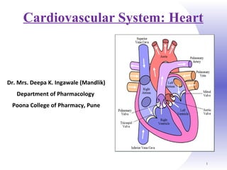
SlideShare a Scribd company logo
1 of 93
1
Cardiovascular System: Heart
Dr. Mrs. Deepa K. Ingawale (Mandlik)
Department of Pharmacology
Poona College of Pharmacy, Pune
Heart
 Cardiology: It is the branch of science that deals with study of
heart & disease of heart.
2
Heart Anatomy
Heart Anatomy
 Shape: Cone shaped
 Weight: 250 gm in adult females
300 gm in adult males
 Size: Approximately the size of closed
fist
 Location:
 Above the diaphragm
 Near the middle of thoracic cavity
 Between the lungs
 Dimensions: 12 cm long, 9 cm wide
& 6 cm thick
Parts:
Four chambers
2 Atria
2 Ventricles
Parts:
Four chambers
2 Atria
2 Ventricles
5
Coverings of Heart: Paricardium
 Pericardium – Double walled membrane that surrounds &
protects the heart
 It confines the heart to its position & allows sufficient freedom
of movement for contraction.
 It is composed of:
1. A superficial fibrous pericardium
2. A deep serous pericardium:
 Parietal layer
 Visceral layer
Diagram of Pericardium
Pericardium
 Fibrous pericardium:
 It is made of tough inelastic, dense irregular connective tissue.
 It prevents over stretching of heart, provides protection & holds the
heart at particular position.
 Serous pericardium:
 Is a thinner, more delicate membrane that forms double layer around
the heart.
 Outer parietal layer fused with fibrous pericardium.
 Inner visceral layer called as epicardium (external layer of heart
wall)
 Space between parietal & visceral layer is called as pericardial
cavity and filled with pericardial fluid. 7
Function of Pericardium
 The Function of the Pericardium:
 Protects & anchors the heart
 Prevents overfilling of the heart with blood
 Allows the heart to work in a relatively friction-free
environment
Heart Wall
 Wall of heart consists of 3 layers;
o Epicardium (External layer)
o Myocardium (Middle layer)
o Endocardium (Inner layer)
 Epicardium:
 Outermost, thin, transparent layer of heart wall.
 Also called as visceral layer of serous pericardium.
 Composed of delicate connective tissue that imparts smooth,
slippery texture to outer surface of heart.
Heart wall
 Myocardium:
 Middle layer, made up of cardiac muscle tissue, make up the bulk
of heart.
 Responsible for pumping action
 Endocardium:
 Inner layer of heart wall made up of endothelial cells
 Provides smooth lining for the chambers of heart & covers the
valve of heart.
Diagram of heart wall
 Vessels returning blood to the heart include:
1. Superior and inferior vena cava
2. Right and left pulmonary veins
 Vessels conveying blood away from the heart include:
1. Pulmonary trunk, which splits into right & left pulmonary
arteries
2. Ascending aorta (3 branches) –
a. Brachiocephalic artery
b. Left common carotid artery
c. Subclavian arteries
External Heart: Major Vessels of the Heart
External Heart: Anterior View
Blood Vessels
• 5 types of blood vessels
• Taking blood to the tissues & back
Arteries
Arterioles
Capillaries
Venules
Veins
Blood Vessels
 Arteries Arterioles carry blood away from the heart
Elastic Fibers
Smooth Muscle
 Capillaries – where gas exchange takes place.
One cell thick
Serves the Respiratory System
 Veins Venules moves blood towards the heart
One way valves
When they break - varicose veins form
The ARTERY
thick muscle and
elastic fibres
Arteries carry blood away from the heart.
the elastic fibres allow the
artery to stretch under
pressure
The VEIN
Veins carry blood towards the heart
thin muscle and
elastic fibres
veins have valves which
stop the blood from going
in wrong direction.
The CAPILLARY
Capillaries link Arteries with Veins
Wall of capillary
is only one cell thick
They exchange materials
between the blood and other
body cells.
artery vein
capillaries
body cell
The CAPILLARY
A collection of capillaries is known as a capillary bedcapillary bed.
The Vascular System
Blood Vessels: Anatomy
• Three layers (tunics)
• Tunic intima
• Endothelium
• Tunic media
• Smooth muscle
• Tunic externa
• Fibrous connective tissue
Artery/Vein differences
Arteries Veins
Direction of
flow
Blood Away from
Heart
Blood to Heart
Pressure Higher Lower
Walls THICKER: Tunica
media is thicker
THINNER: Tunica externa is
thinner
Lumen Smaller Larger
Valves No valves Valves
Gross Anatomy of Heart: Frontal Section
Chambers of Heart
Chambers of the Heart
 4 chambers of heart
 2 ventricles & 2 atria
 Right atrium (RA): collects blood from systemic circuit
 Right ventricle (RV): pumps blood to pulmonary circuit
 Left atrium (LA): collects blood from pulmonary circuit
 Left ventricle (LV): pumps blood to systemic circuit
Right Atrium (RA)
 Atria are the receiving chambers of the heart
 RA is roughly quadrangular in shape.
 Divided into 2 parts;
 Upper part
 Lower part
 Superior vena cava present at the upper part
 Inferior vena cava present at lower part
Right Ventricle (RV)
 Ventricles are the pumping chambers of the heart
 It is convex & forms large part of heart.
 The wall of RV is much thinner than LV.
Left Atrium (LA)
 Smaller in shape than RA.
 Roughly cuboidal in shape
 Four pulmonary veins open at the upper part of LA.
Left Ventricle (LV)
 It functions as a powerful pump operating at high pressure.
 The walls are three times more thicker as that of RV.
 Cone shaped, longer and narrower than RV
Myocardial Thickness &Function
Thickness of myocardium varies according to the function of chamber
Atria are thin walled, deliver blood to adjacent ventricles
Ventricle walls are much thicker and stronger
Thickness of Cardiac Walls
Pathway of Blood Through Heart & Lungs
 Right atrium  tricuspid valve  right ventricle
 Right ventricle  pulmonary semilunar valve  pulmonary
arteries  lungs
 Lungs  pulmonary veins  left atrium
 Left atrium  bicuspid valve  left ventricle
 Left ventricle  aortic semilunar valve  aorta
 Aorta  systemic circulation
Pathway of Blood Through Heart & Lungs
 The right side of heart pumps blood into the pulmonary
circuit:
 Blood returning from the body is relatively oxygen-poor and
carbon dioxide-rich
 Blood enters the right atrium and passes into the right
ventricle, which pumps it to the lungs via the pulmonary
arteries (conduct blood away from the heart)
 In the lungs, the blood unloads carbon dioxide and picks up
oxygen (oxygenated)
 The left side of the heart pumps blood into the systemic
circuit
Coronary Circulation
 Coronary circulation is blood supply to the heart
muscle itself
Arterial Supply Venous Supply
Heart Valves
 As each chamber of heart contracts, it pushes a portion of blood
into a ventricle or out of heart through an artery.
 To prevent back flow of blood, the heart has valve.
 Made up of dense connective tissue covered by endocardium
 2 types of valve
 Atrioventricular valve (AV valve)
 Tricuspid valve
 Bicuspid valve
 Semilunar valve (SL valve)
Atrioventricular (AV)
 Atrioventricular (AV) valves lie between the atria and ventricles
 AV valves prevent backflow of blood into the atria when
ventricles contract
 2 types
 Tricuspid valve
 Bicuspid valve
Atrioventricular (AV)
 Tricuspid valve:
 It is present between RA and RV is called as tricuspid valve
 Consist of 3 cusp (flaps)
 Septal cusp
 Anterior cusp
 Posterior cusp
 Bicuspid valve:
 It is present between LA and LV is called as bicuspid valve.
 Consist of 2 cusps.
 Also called as mitral valve.
Semilunar valves
 Semilunar valves prevent backflow of blood into the ventricles
 Aortic semilunar valve lies in the aorta
 Pulmonary semilunar valve lies in the pulmonary trunk
 Both the valves consist of 3 half moon shaped cusps.
 Permits blood flow in only one direction.
Heart Valves
Conducting system of heart
 A special system is available in the heart responsible for the
rhythmic contraction and conduction of impulses in the heart.
 Divided into 5 parts;
 SA Node or Sinoatrial Node
 AV Node or Atrioventricular Node
 AV Bundle (Bundle of His)
 Right & Left Bundle Branches
 Conduction Myofibers (Purkinje Fibers)
Conducting system of heart
 Sinoatrial (SA) node:
 It is located in the right atrial wall just below the opening of
superior vena cava.
 Cardiac excitation begins in the SA node,
 Each SA node impulse travels throughout the heart via the
conduction system
 Atrioventricular (AV) node:
 It is located in the septum between the two atria.
 The cardiac impulses spreads from SA node to AV node.
Conducting system of heart
 Atrioventricular bundle (bundle of His):
 From AV node, the impulse enters the Bundle of His, only
electrical connection between atria and ventricle.
 AV bundle splits into two pathways
1. Bundle branches carry the impulse toward the apex of the
heart
2. Purkinje fibers carry the impulse to the heart apex and
ventricular walls
Conducting system of heart
 Right & left bundle branches:
 From the bundle branches the impulses then enters the right &
left bundle branches that runs towards the apex of the heart.
 Perkinje Fibers:
 The impulse from right and left bundle branches enters into
Perkinje fibers.
 These conduct impulses to all parts of ventricles.
 Then the ventricles contracts pushing the blood upwards towards
the SA node.
Electrocardiogram
Electrocardiogram
 Conduction of action potential through heart generates electrical
currents that can be detected at the surface of the body.
 A recording of electrical changes during each cardiac cycle is
called as electrocardiogram (ECG).
 The instrument used to record the change is called as an
electrocardiograph.
 It consist of 3 waves;
 P wave
 QRS wave
 T wave
Electrocardiogram
 P wave:
 It is small upward wave.
 It represents atrial depolarization which spreads from SA node
throughout both atria.
 QRS wave:
 The complex represents 3 separate waves.
 Q wave, R wave and S wave
 The complex begins with downward deflection of Q wave,
continues as a large, upright, triangular defection of R wave &
ends as a downward deflection of S wave.
 The QRS complex represents ventricular depolarization.
Electrocardiogram
 T wave:
 It represents ventricular repolarisation.
 Third dome shaped upward deflection
 The T wave is small & more spread out than QRS complex
because repolarisation occurs more slow than the depolarisation.
 PQ or PR interval:
 The duration between beginning of P wave & beginning of QRS
wave is called as PQ interval.
 It is also called as PR interval because the Q wave is frequently
absent.
 It is interval between beginning of contarction of atria &
beginning of contraction of ventricles.
Electrocardiogram
 ST Segment:
 It begins at the end of S wave & starting of T wave.
 QT interval:
 The QT interval extends from the start of QRS complex to
the end of T wave.
 It is the time from beginning of ventricular depolarization
to the end of ventricular repolarization.
Electrocardiogram
 Following conclusions can be made with the altered ECG notes.
 Larger P wave: It indicates enlargement of atrium.
 Enlarged Q wave: It indicates myocardial infarction.
 Enlarged R wave: It indicated enlargement of ventricles.
 Flatter T wave: It indicates insufficient oxygen supply to
myocardium.
 Larger PQ interval: It indicates formation of scar tissue in heart
due to coronary artery disease.
 Larger ST segment: It indicates acute myocardial infarction
when elevated above the baseline & insufficient oxygen supply
to heart muscle when depressed below the baseline.
 Renin:
 It is secreted by the juxtaglomerular cells in Kidney
 Angiotensinogen:
 It is a glycoprotein synthesized by liver & secreted into the
bloodstream
 Aldosterone:
 It is a mineralocorticoid produced in the adrenal cortex
 It plays a central role in the regulation of blood pressure mainly
by acting on the distal tubules & collecting ducts of nephron
Renin-Angiotensin-Aldosterone System
 Angiotensin
 It is a peptide hormone that causes vasoconstriction
and a subsequent increase in blood pressure.
 Angiotensin-I
 Angiotensin-II
 Angiotensin converting enzyme (ACE):
 It converts angiotensin I to II (vasoconstrictor)
Renin-Angiotensin-Aldosterone System
Renin Angiotensin aldosterone System (RAA)
 Stimuli that initiate the renin–angiotensin–aldosterone
pathway include dehydration, Na+
deficiency, or hemorrhage.
 These conditions cause a decrease in blood volume.
 Decreased blood volume leads to decreased blood pressure.
 Lowered blood pressure stimulates certain cells of the
kidneys, called juxtaglomerular cells, to secrete the enzyme
renin.
 The level of renin in the blood increases.
 Renin converts angiotensinogen, a plasma protein produced
by the liver, into angiotensin I.
Renin Angiotensin aldosterone System (RAA)
 Blood containing increased levels of angiotensin I circulates
in the body.
 As blood flows through capillaries, particularly those of the
lungs, the enzyme angiotensin-converting enzyme (ACE)
converts angiotensin I into the hormone angiotensin II.
 Blood level of angiotensin II increases.
 Angiotensin II stimulates the adrenal cortex to secrete
aldosterone.
 Blood containing increased levels of aldosterone circulates
to the kidneys.
Renin Angiotensin aldosterone System (RAA)
 In the kidneys, aldosterone increases reabsorption of Na+
and
water so that less is lost in the urine. Aldosterone also stimulates
the kidneys to increase secretion of K+
and H+
into the urine.
 With increased water reabsorption by the kidneys, blood volume
increases.
 As blood volume increases, blood pressure increases to normal.
 Angiotensin II also stimulates contraction of smooth muscle in
the walls of arterioles. The resulting vasoconstriction of the
arterioles increases blood pressure and thus helps raise blood
pressure to normal.
 Besides angiotensin II, a second stimulator of aldosterone
secretion is an increase in the K+
concentration of blood (or
interstitial fluid). A decrease in the blood K+
level has the opposite
effect.
Pulse ratePulse rate
• Pulse: Means expansion and elongation of arterial walls by
the pressure changes during systole (contraction) and
diastole (relaxation)
Pulse rate is recorded for 1
minute
Normal ranges:
New born: 140 beats/minutes
Children: 100 beats/minutes
Adult human: 60-80 beats/minutes
Tachycardia: Increase in HR than normal
Bradycardia: Decrease in HR than normal
Blood PressureBlood Pressure
• Blood pressure: It is the pressure excreted by blood
on the wall of arteries.
• Systolic BP–Ventricular contraction
• Diastolic BP–Ventricles relaxation
• Normal BP = 120/80 mm Hg
• Pressure in blood vessels decreases as the distance
from the heart increases
• It is essential to record both BP’s as it gives
information regarding the status of working heart.
• BP varies from different physiological parameters like
age, sex, exercise, posture, sleep during
emotions, etc.
Methods of BP determination
 Oscillatory method
 Palpatory method
 Auscultatory method
Stethoscope
Sphygmomanometer
Blood pressure
It depends on the speed of blood coming into a vessel and width of
vessel itself.
Arteries
Speed: high
Width: medium
Pressure: high
Capillaries
Speed: medium
Width: narrow
Pressure: medium
Veins
Speed: low
Width: wide
Pressure: low
Blood pressure
An individual’s blood pressure is affected by a number of factors.
Age – It increases as you get older.
Gender – Men tend to have higher blood pressure than
women.
Stress - Can cause increase blood pressure.
Diet – Salt and saturated fats can increase blood pressure.
Exercise – Exercise lowers the blood pressure
Having high blood pressure puts stress on heart. It can lead to
angina, heart attacks and strokes.
Auscultatory method
Auscultatory method
 Initially the cuff is inflated to a level higher than the systolic pressure.
 Thus the artery is completely compressed, there is no blood flow, and
no sounds are heard.
 The cuff pressure is slowly decreased. At the point where the systolic
pressure exceeds the cuff pressure, the Korotkoff sounds are first
heard and blood passes in turbulent flow through the partially
constricted artery.
 Korotkoff sounds will continue to be heard as the cuff pressure is
further lowered.
 However, when the cuff pressure reaches diastolic pressure, the sounds
disappear.
 Now at all points in time during the cardiac cycle, the blood pressure is
greater than the cuff pressure, and the artery remains open.
 Auscultation – listening to heart sound via stethoscope
 Four heart sounds
 S1– “lubb” caused by the closing of the AV valves
 S2 – “dupp” caused by the closing of the semilunar valves
 S3 – a faint sound associated with blood flowing into the
ventricles
 S4 – another faint sound associated with atrial contraction
Heart sounds
Variations in Blood PressureVariations in Blood Pressure
• Human normal range is variable
• Normal
• 140–110 mm Hg systolic
• 80–75 mm Hg diastolic
• Hypotension
• Low systolic (below 110 mm HG)
• Often associated with illness
• Hypertension
• High systolic (above 140 mm HG)
• Can be dangerous if it is chronic
Control of BP
70
 Blood pressure is controlled in 2 ways:
 Short term control: Mainly involves the baroreceptor reflex,
chemoreceptor & circulating hormones
 Long term control: Involves regulation of blood volume by the kidneys
and RAA system
 The cardiovascular centre (CVC) is a collection of
interconnected neurons in the brain
 The CVC receives, integrates & coordinates inputs
from:
 Baroreceptors (pressure receptors)
 Chemoreceptor
 Higher centers in the brain
Baroreceptors
 These are nerve endings sensitive to pressure changes (stretch)
within the vessel, situated in the arch of the aorta
 Rise in B.P. in these arteries
 Stimulation of Baroreceptors
 Increasing their input to the CVC
 Increases parasympathetic nerve activity to heart
 Decreases HR & decreases FC
 Vasodilation
 Fall in systemic blood pressure
71
Conversely
 Fall in B.P. aortic arch and carotid sinuses
 Deactivation of Baroreceptors
 Decreasing their input to the CVC
 Increases sympathetic nerve activity to heart
 Increases HR & FC
 Vasoconstriction
 Rise in systemic blood pressure
Chemoreceptor
 These are nerve endings situated in the carotid and aortic
bodies.
 Involved in control of respiration.
 Sensitive to changes in the levels of carbon dioxide, oxygen &
the acidity of the blood (pH).
Hormonal regulation of BP
 Renin-angiotensin-Aldosterone System: Discussed above
 Epinephrine & Nor-epinephrine: Adrenal medulla releases
epinephrine and nor-epinephrine. These changes increases CO by
increase in the HR & FC.
 Antidiuretic hormone (ADH): It is produced by hypothalamus
causes vasoconstriction that increases BP.
 Hence, it is also called as vasopressin.
 Atrial natriuretic peptide (ANP): It is released by the cells in
the atria of heart. ANP lowers BP by causing vasodilation and by
promoting the loss of salt & water in urine which reduces blood
volume.
Auto regulation of blood pressure
 The ability of a tissue to automatically adjust its blood flow to
match its metabolic demands called as auto regulation.
 Two general types of stimuli cause auto regulatory
changes in blood flow.
 Physical change:
 Warming promotes vasodilation & cooling causes
vasoconstriction.
 Vasodialating & vasoconstricting chemicals:
Auto regulation of blood pressure
 Several types of cells such as WBC, Platelets, smooth muscle
fibers, macrophages, endothelial cells-release a wide variety of
chemicals that alters blood vessels diameter.
 Vasodialating chemicals released by metabolically active tissue
cells include K+
, H+
, lactic acid & adenosine (From ATP).
 Important vasodilator released by endothelial cell is NO named as
endothelium derived relaxation factor (EDRF).
Filling of Heart Chambers – Cardiac CycleFilling of Heart Chambers – Cardiac Cycle
Cardiac Cycle
 The event occurring in the heart from the beginning of one
heart beat to the beginning of other is called as cardiac cycle.
 In normal cardiac cycle the two atria contracts while the two
ventricles relax. Then, while the two ventricles contract, the two
atria relax.
 Cardiac cycle consists of systole and diastole of both the atria &
ventricles.
 Cardiac cycle refers to all events associated with blood flow
through the heart
 Systole – contraction of heart muscle
 Diastole – relaxation of heart muscle
Phases of the Cardiac Cycle
 Cardiac cycle is divided into 3 phases
 Ventricular filling
 Ventricular contraction
 Ventricular relaxation
Ventricular Filling
 During ventricular relaxation, large amount of blood collects in
the atria, as the AV valve are closed.
 This increases the pressure in the atria and AV valves get opens
and semilunar valve are closed.
 So, the blood flow rapidly into the ventricles.
 First 1/3th
time of ventricular filling is called as period of rapid
ventricular filling.
 Later on only small amount of blood flows into the ventricles.
 P wave on ECG indicates atrial depolarization
Ventricular contraction
 Period of isovolumetric contraction:
 Immediately after ventricular filling the pressure inside the
ventricles rises suddenly.
 This rise in pressure tries to push blood back to the atria and
due to this AV valves get closed.
 At this particular junction, the AV valves and SL valves are
closed and the volume inside the ventricles does not change
called as period of isovolumetric contraction.
Ventricular contraction
 Period of ventricular ejection:
 As further ventricles starts contracting the pressure inside rises
sharply.
 When the pressure rises above the aortic pressure and
pulmonary trunk pressure SL valve get opens.
 As the SL get opens the blood get ejected out of the ventricles.
This period is called as ventricular ejection.
 After this ventricular pressure falls, the period of ventricular
relaxation is repeated.
Ventricular relaxation
 Ventricles starts to relax at the end of heart beat.
 At this particular point all the chambers of heart are
relaxing.
 This represent T wave on ECG.
 As the ventricles starts relaxing pressure inside the
ventricles drops suddenly.
 This drop in pressure leads to back flow of blood from the
pulmonary trunk and aorta.
 This forceful back flow of blood closes the SL valves
suddenly.
Ventricular relaxation
 This pressure produces a bump called as dicrotic wave.
 At this particular point both the SL valve and AV valves are
closed.
 Due to this the ventricular volume does not change and
this period is called as isovolumetric relaxation.
 With the further relaxation of ventricles there is further fall
in pressure inside the ventricles.
 When this ventricular pressure drops below the atrial
pressure AV valves opens and ventricular filling begins.
Phases of the Cardiac Cycle
Cardiac Output (CO) and Reserve
 Cardiac Output is the amount of blood pumped by each
ventricle in one minute
 CO is the product of heart rate (HR) and stroke volume
(SV)
 HR is the number of heart beats per minute
 SV is the amount of blood pumped out by a ventricle
with each beat
 Cardiac reserve is the difference between resting and
maximal CO
 CO (ml/min) = HR (75 beats/min) x SV (70 ml/beat)
 CO = 5250 ml/min (5.25 L/min)
Stroke Volume
 SV = End diastolic V(EDV) - End systolic V (ESV)
 EDV = amount of blood collected in a ventricle
during diastole
 ESV = amount of blood remaining in a ventricle after
contraction
Factors Affecting Stroke Volume
 Afterload is the tension developed in the wall of the left ventricle
during ejection
 Preload is pressure that stretches the right or left ventricle of the
heart to its greatest geometric dimensions under variable
physiologic demand
89
Congestive Heart Failure (CHF)
 Congestive heart failure (CHF) is caused by:
 Coronary atherosclerosis
 Persistent high blood pressure
 Multiple myocardial infarcts
 Dilated cardiomyopathy (DCM) – main pumping
chambers of the heart are dilated and contract poorly
Chapter 18, Cardiovascular System 90
Congestive Heart Failure
 Causes of CHF
 coronary artery disease, hypertension, MI, valve disorders,
congenital defects
 Left side heart failure
 less effective pump so more blood remains in ventricle
 heart is overstretched & even more blood remains
 blood backs up into lungs as pulmonary edema
 suffocation & lack of oxygen to the tissues
 Right side failure
 fluid builds up in tissues as peripheral edema
Coronary Artery Disease
 Heart muscle receiving
insufficient blood supply
 narrowing of vessels---
atherosclerosis, artery
spasm or clot
 atherosclerosis--smooth
muscle & fatty deposits in
walls of arteries
 Treatment
 drugs, bypass graft,
angioplasty, stent
By-pass Graft
Chapter 18, Cardiovascular System 93
Artificial Heart

More Related Content

What's hot

Bloodvessels
BloodvesselsBloodvessels
Bloodvessels
scuffruff
 
Circulatory System.ppt
Circulatory System.pptCirculatory System.ppt
Circulatory System.ppt
Shama
 
Blood functions and characteristics
Blood functions and characteristicsBlood functions and characteristics
Blood functions and characteristics
stewart_j
 
Cardiovascular System
Cardiovascular SystemCardiovascular System
Cardiovascular System
000 07
 

What's hot (20)

THE CARDIOVASCULAR SYSTEM CHAPTER 4
THE CARDIOVASCULAR SYSTEM CHAPTER 4THE CARDIOVASCULAR SYSTEM CHAPTER 4
THE CARDIOVASCULAR SYSTEM CHAPTER 4
 
Bloodvessels
BloodvesselsBloodvessels
Bloodvessels
 
Pulmonary & systemic circulation
Pulmonary & systemic circulationPulmonary & systemic circulation
Pulmonary & systemic circulation
 
Blood Flow Through the Heart
Blood Flow Through the HeartBlood Flow Through the Heart
Blood Flow Through the Heart
 
Cardiovascular system
Cardiovascular systemCardiovascular system
Cardiovascular system
 
Circulatory System.ppt
Circulatory System.pptCirculatory System.ppt
Circulatory System.ppt
 
Respiratory system (Structure).pptx
Respiratory system (Structure).pptxRespiratory system (Structure).pptx
Respiratory system (Structure).pptx
 
Introduction of CVS
Introduction of CVSIntroduction of CVS
Introduction of CVS
 
Blood functions and characteristics
Blood functions and characteristicsBlood functions and characteristics
Blood functions and characteristics
 
Cardiovascular System
Cardiovascular SystemCardiovascular System
Cardiovascular System
 
2.1 blood circulation
2.1 blood circulation2.1 blood circulation
2.1 blood circulation
 
Anatomy and physiology of the digestive system
Anatomy and physiology of the digestive systemAnatomy and physiology of the digestive system
Anatomy and physiology of the digestive system
 
Blood vessels.2.
Blood vessels.2.Blood vessels.2.
Blood vessels.2.
 
Circulatory System Physiology [Zoo 403]
Circulatory System Physiology [Zoo 403]Circulatory System Physiology [Zoo 403]
Circulatory System Physiology [Zoo 403]
 
Structure of blood vessels
Structure of blood vesselsStructure of blood vessels
Structure of blood vessels
 
Anatomy of the heart
Anatomy of the heartAnatomy of the heart
Anatomy of the heart
 
Anatomy and physiology of the respiratory system
Anatomy and physiology of the respiratory systemAnatomy and physiology of the respiratory system
Anatomy and physiology of the respiratory system
 
Urinary System Notes
Urinary System NotesUrinary System Notes
Urinary System Notes
 
Types of bones
Types of bonesTypes of bones
Types of bones
 
PPT on Lymphatic system
PPT on Lymphatic systemPPT on Lymphatic system
PPT on Lymphatic system
 

Similar to Cardiovascular system

Introduction to the_cardiovascular_system
Introduction to the_cardiovascular_systemIntroduction to the_cardiovascular_system
Introduction to the_cardiovascular_system
Personal
 
Marina bozilovic heart medical terminology
Marina bozilovic heart medical terminologyMarina bozilovic heart medical terminology
Marina bozilovic heart medical terminology
mbozilovic
 
Anatomy Of The Heart
Anatomy Of The HeartAnatomy Of The Heart
Anatomy Of The Heart
shabeel pn
 

Similar to Cardiovascular system (20)

Introduction to the_cardiovascular_system
Introduction to the_cardiovascular_systemIntroduction to the_cardiovascular_system
Introduction to the_cardiovascular_system
 
Anatomy and physiology of the heart by Adeboye Oluwajuyitan
Anatomy and physiology of the heart by Adeboye OluwajuyitanAnatomy and physiology of the heart by Adeboye Oluwajuyitan
Anatomy and physiology of the heart by Adeboye Oluwajuyitan
 
Cardiovascular System.pptx
Cardiovascular System.pptxCardiovascular System.pptx
Cardiovascular System.pptx
 
heart anatomy pdf.pdf
heart anatomy pdf.pdfheart anatomy pdf.pdf
heart anatomy pdf.pdf
 
INTRODUCTION TO CVS PHYSIOLOGY-BSCN.pptx
INTRODUCTION TO CVS PHYSIOLOGY-BSCN.pptxINTRODUCTION TO CVS PHYSIOLOGY-BSCN.pptx
INTRODUCTION TO CVS PHYSIOLOGY-BSCN.pptx
 
Cardiovascular System.pptx
Cardiovascular System.pptxCardiovascular System.pptx
Cardiovascular System.pptx
 
CARDIOVASCULAR SYSTEM.pptx
CARDIOVASCULAR SYSTEM.pptxCARDIOVASCULAR SYSTEM.pptx
CARDIOVASCULAR SYSTEM.pptx
 
Cardiovascular System
Cardiovascular SystemCardiovascular System
Cardiovascular System
 
Heart.pptx
Heart.pptxHeart.pptx
Heart.pptx
 
circulatory system mammals.ppt
circulatory system mammals.pptcirculatory system mammals.ppt
circulatory system mammals.ppt
 
08 Heart.ppt
08 Heart.ppt08 Heart.ppt
08 Heart.ppt
 
Marina bozilovic heart medical terminology
Marina bozilovic heart medical terminologyMarina bozilovic heart medical terminology
Marina bozilovic heart medical terminology
 
Cardiovascular System Part 1
Cardiovascular System Part 1Cardiovascular System Part 1
Cardiovascular System Part 1
 
Chapter19 heartmarieb
Chapter19 heartmariebChapter19 heartmarieb
Chapter19 heartmarieb
 
CARDIOVASCULAR SYSTEM
CARDIOVASCULAR SYSTEMCARDIOVASCULAR SYSTEM
CARDIOVASCULAR SYSTEM
 
Cardiovascular system of goat
Cardiovascular system of goatCardiovascular system of goat
Cardiovascular system of goat
 
Cardiovascular System & Antihypertensives drugs.pptx
Cardiovascular System & Antihypertensives drugs.pptxCardiovascular System & Antihypertensives drugs.pptx
Cardiovascular System & Antihypertensives drugs.pptx
 
Circualtory system 6a.pptx
Circualtory system 6a.pptxCircualtory system 6a.pptx
Circualtory system 6a.pptx
 
Anatomy Of The Heart
Anatomy Of The HeartAnatomy Of The Heart
Anatomy Of The Heart
 
The cardiovascular system.pptx
The cardiovascular system.pptxThe cardiovascular system.pptx
The cardiovascular system.pptx
 

More from deepaingawale21 (12)

Introduction to hap
Introduction to hapIntroduction to hap
Introduction to hap
 
Urinary system
Urinary systemUrinary system
Urinary system
 
Special sense organs
Special sense organsSpecial sense organs
Special sense organs
 
Respiratory system
Respiratory systemRespiratory system
Respiratory system
 
Reproductive system
Reproductive systemReproductive system
Reproductive system
 
Nervous system
Nervous systemNervous system
Nervous system
 
Lymphatic system
Lymphatic systemLymphatic system
Lymphatic system
 
Cell and tissue
Cell and tissueCell and tissue
Cell and tissue
 
Muscular system
Muscular systemMuscular system
Muscular system
 
Health education ppt
Health education pptHealth education ppt
Health education ppt
 
Digestive system
Digestive systemDigestive system
Digestive system
 
Blood
BloodBlood
Blood
 

Recently uploaded

❤️Amritsar Escorts Service☎️9815674956☎️ Call Girl service in Amritsar☎️ Amri...
❤️Amritsar Escorts Service☎️9815674956☎️ Call Girl service in Amritsar☎️ Amri...❤️Amritsar Escorts Service☎️9815674956☎️ Call Girl service in Amritsar☎️ Amri...
❤️Amritsar Escorts Service☎️9815674956☎️ Call Girl service in Amritsar☎️ Amri...
Sheetaleventcompany
 
Cara Menggugurkan Kandungan Dengan Cepat Selesai Dalam 24 Jam Secara Alami Bu...
Cara Menggugurkan Kandungan Dengan Cepat Selesai Dalam 24 Jam Secara Alami Bu...Cara Menggugurkan Kandungan Dengan Cepat Selesai Dalam 24 Jam Secara Alami Bu...
Cara Menggugurkan Kandungan Dengan Cepat Selesai Dalam 24 Jam Secara Alami Bu...
Cara Menggugurkan Kandungan 087776558899
 
💚Chandigarh Call Girls Service 💯Piya 📲🔝8868886958🔝Call Girls In Chandigarh No...
💚Chandigarh Call Girls Service 💯Piya 📲🔝8868886958🔝Call Girls In Chandigarh No...💚Chandigarh Call Girls Service 💯Piya 📲🔝8868886958🔝Call Girls In Chandigarh No...
💚Chandigarh Call Girls Service 💯Piya 📲🔝8868886958🔝Call Girls In Chandigarh No...
Sheetaleventcompany
 
Hyderabad Call Girls Service ❤️ 7783825323 Independent Escort Service Hyderabad
Hyderabad Call Girls Service ❤️ 7783825323 Independent Escort Service HyderabadHyderabad Call Girls Service ❤️ 7783825323 Independent Escort Service Hyderabad
Hyderabad Call Girls Service ❤️ 7783825323 Independent Escort Service Hyderabad
Sheetaleventcompany
 
👉 Chennai Sexy Aunty’s WhatsApp Number 👉📞 7427069034 👉📞 Just📲 Call Ruhi Colle...
👉 Chennai Sexy Aunty’s WhatsApp Number 👉📞 7427069034 👉📞 Just📲 Call Ruhi Colle...👉 Chennai Sexy Aunty’s WhatsApp Number 👉📞 7427069034 👉📞 Just📲 Call Ruhi Colle...
👉 Chennai Sexy Aunty’s WhatsApp Number 👉📞 7427069034 👉📞 Just📲 Call Ruhi Colle...
rajnisinghkjn
 
Difference Between Skeletal Smooth and Cardiac Muscles
Difference Between Skeletal Smooth and Cardiac MusclesDifference Between Skeletal Smooth and Cardiac Muscles
Difference Between Skeletal Smooth and Cardiac Muscles
MedicoseAcademics
 
Ahmedabad Call Girls Book Now 9630942363 Top Class Ahmedabad Escort Service A...
Ahmedabad Call Girls Book Now 9630942363 Top Class Ahmedabad Escort Service A...Ahmedabad Call Girls Book Now 9630942363 Top Class Ahmedabad Escort Service A...
Ahmedabad Call Girls Book Now 9630942363 Top Class Ahmedabad Escort Service A...
Sheetaleventcompany
 

Recently uploaded (20)

❤️Call Girl Service In Chandigarh☎️9814379184☎️ Call Girl in Chandigarh☎️ Cha...
❤️Call Girl Service In Chandigarh☎️9814379184☎️ Call Girl in Chandigarh☎️ Cha...❤️Call Girl Service In Chandigarh☎️9814379184☎️ Call Girl in Chandigarh☎️ Cha...
❤️Call Girl Service In Chandigarh☎️9814379184☎️ Call Girl in Chandigarh☎️ Cha...
 
❤️Amritsar Escorts Service☎️9815674956☎️ Call Girl service in Amritsar☎️ Amri...
❤️Amritsar Escorts Service☎️9815674956☎️ Call Girl service in Amritsar☎️ Amri...❤️Amritsar Escorts Service☎️9815674956☎️ Call Girl service in Amritsar☎️ Amri...
❤️Amritsar Escorts Service☎️9815674956☎️ Call Girl service in Amritsar☎️ Amri...
 
Circulatory Shock, types and stages, compensatory mechanisms
Circulatory Shock, types and stages, compensatory mechanismsCirculatory Shock, types and stages, compensatory mechanisms
Circulatory Shock, types and stages, compensatory mechanisms
 
Low Cost Call Girls Bangalore {9179660964} ❤️VVIP NISHA Call Girls in Bangalo...
Low Cost Call Girls Bangalore {9179660964} ❤️VVIP NISHA Call Girls in Bangalo...Low Cost Call Girls Bangalore {9179660964} ❤️VVIP NISHA Call Girls in Bangalo...
Low Cost Call Girls Bangalore {9179660964} ❤️VVIP NISHA Call Girls in Bangalo...
 
Lucknow Call Girls Service { 9984666624 } ❤️VVIP ROCKY Call Girl in Lucknow U...
Lucknow Call Girls Service { 9984666624 } ❤️VVIP ROCKY Call Girl in Lucknow U...Lucknow Call Girls Service { 9984666624 } ❤️VVIP ROCKY Call Girl in Lucknow U...
Lucknow Call Girls Service { 9984666624 } ❤️VVIP ROCKY Call Girl in Lucknow U...
 
Call Girls Service Jaipur {9521753030 } ❤️VVIP BHAWNA Call Girl in Jaipur Raj...
Call Girls Service Jaipur {9521753030 } ❤️VVIP BHAWNA Call Girl in Jaipur Raj...Call Girls Service Jaipur {9521753030 } ❤️VVIP BHAWNA Call Girl in Jaipur Raj...
Call Girls Service Jaipur {9521753030 } ❤️VVIP BHAWNA Call Girl in Jaipur Raj...
 
Cara Menggugurkan Kandungan Dengan Cepat Selesai Dalam 24 Jam Secara Alami Bu...
Cara Menggugurkan Kandungan Dengan Cepat Selesai Dalam 24 Jam Secara Alami Bu...Cara Menggugurkan Kandungan Dengan Cepat Selesai Dalam 24 Jam Secara Alami Bu...
Cara Menggugurkan Kandungan Dengan Cepat Selesai Dalam 24 Jam Secara Alami Bu...
 
💚Chandigarh Call Girls Service 💯Piya 📲🔝8868886958🔝Call Girls In Chandigarh No...
💚Chandigarh Call Girls Service 💯Piya 📲🔝8868886958🔝Call Girls In Chandigarh No...💚Chandigarh Call Girls Service 💯Piya 📲🔝8868886958🔝Call Girls In Chandigarh No...
💚Chandigarh Call Girls Service 💯Piya 📲🔝8868886958🔝Call Girls In Chandigarh No...
 
Call Girls in Lucknow Just Call 👉👉 8875999948 Top Class Call Girl Service Ava...
Call Girls in Lucknow Just Call 👉👉 8875999948 Top Class Call Girl Service Ava...Call Girls in Lucknow Just Call 👉👉 8875999948 Top Class Call Girl Service Ava...
Call Girls in Lucknow Just Call 👉👉 8875999948 Top Class Call Girl Service Ava...
 
Lucknow Call Girls Just Call 👉👉8630512678 Top Class Call Girl Service Available
Lucknow Call Girls Just Call 👉👉8630512678 Top Class Call Girl Service AvailableLucknow Call Girls Just Call 👉👉8630512678 Top Class Call Girl Service Available
Lucknow Call Girls Just Call 👉👉8630512678 Top Class Call Girl Service Available
 
Cardiac Output, Venous Return, and Their Regulation
Cardiac Output, Venous Return, and Their RegulationCardiac Output, Venous Return, and Their Regulation
Cardiac Output, Venous Return, and Their Regulation
 
Hyderabad Call Girls Service ❤️ 7783825323 Independent Escort Service Hyderabad
Hyderabad Call Girls Service ❤️ 7783825323 Independent Escort Service HyderabadHyderabad Call Girls Service ❤️ 7783825323 Independent Escort Service Hyderabad
Hyderabad Call Girls Service ❤️ 7783825323 Independent Escort Service Hyderabad
 
👉 Chennai Sexy Aunty’s WhatsApp Number 👉📞 7427069034 👉📞 Just📲 Call Ruhi Colle...
👉 Chennai Sexy Aunty’s WhatsApp Number 👉📞 7427069034 👉📞 Just📲 Call Ruhi Colle...👉 Chennai Sexy Aunty’s WhatsApp Number 👉📞 7427069034 👉📞 Just📲 Call Ruhi Colle...
👉 Chennai Sexy Aunty’s WhatsApp Number 👉📞 7427069034 👉📞 Just📲 Call Ruhi Colle...
 
Difference Between Skeletal Smooth and Cardiac Muscles
Difference Between Skeletal Smooth and Cardiac MusclesDifference Between Skeletal Smooth and Cardiac Muscles
Difference Between Skeletal Smooth and Cardiac Muscles
 
7 steps How to prevent Thalassemia : Dr Sharda Jain & Vandana Gupta
7 steps How to prevent Thalassemia : Dr Sharda Jain & Vandana Gupta7 steps How to prevent Thalassemia : Dr Sharda Jain & Vandana Gupta
7 steps How to prevent Thalassemia : Dr Sharda Jain & Vandana Gupta
 
Bhawanipatna Call Girls 📞9332606886 Call Girls in Bhawanipatna Escorts servic...
Bhawanipatna Call Girls 📞9332606886 Call Girls in Bhawanipatna Escorts servic...Bhawanipatna Call Girls 📞9332606886 Call Girls in Bhawanipatna Escorts servic...
Bhawanipatna Call Girls 📞9332606886 Call Girls in Bhawanipatna Escorts servic...
 
Chennai ❣️ Call Girl 6378878445 Call Girls in Chennai Escort service book now
Chennai ❣️ Call Girl 6378878445 Call Girls in Chennai Escort service book nowChennai ❣️ Call Girl 6378878445 Call Girls in Chennai Escort service book now
Chennai ❣️ Call Girl 6378878445 Call Girls in Chennai Escort service book now
 
Ahmedabad Call Girls Book Now 9630942363 Top Class Ahmedabad Escort Service A...
Ahmedabad Call Girls Book Now 9630942363 Top Class Ahmedabad Escort Service A...Ahmedabad Call Girls Book Now 9630942363 Top Class Ahmedabad Escort Service A...
Ahmedabad Call Girls Book Now 9630942363 Top Class Ahmedabad Escort Service A...
 
Kolkata Call Girls Naktala 💯Call Us 🔝 8005736733 🔝 💃 Top Class Call Girl Se...
Kolkata Call Girls Naktala  💯Call Us 🔝 8005736733 🔝 💃  Top Class Call Girl Se...Kolkata Call Girls Naktala  💯Call Us 🔝 8005736733 🔝 💃  Top Class Call Girl Se...
Kolkata Call Girls Naktala 💯Call Us 🔝 8005736733 🔝 💃 Top Class Call Girl Se...
 
Call Girls Mussoorie Just Call 8854095900 Top Class Call Girl Service Available
Call Girls Mussoorie Just Call 8854095900 Top Class Call Girl Service AvailableCall Girls Mussoorie Just Call 8854095900 Top Class Call Girl Service Available
Call Girls Mussoorie Just Call 8854095900 Top Class Call Girl Service Available
 

Cardiovascular system

  • 1. 1 Cardiovascular System: Heart Dr. Mrs. Deepa K. Ingawale (Mandlik) Department of Pharmacology Poona College of Pharmacy, Pune
  • 2. Heart  Cardiology: It is the branch of science that deals with study of heart & disease of heart. 2
  • 4. Heart Anatomy  Shape: Cone shaped  Weight: 250 gm in adult females 300 gm in adult males  Size: Approximately the size of closed fist  Location:  Above the diaphragm  Near the middle of thoracic cavity  Between the lungs  Dimensions: 12 cm long, 9 cm wide & 6 cm thick Parts: Four chambers 2 Atria 2 Ventricles Parts: Four chambers 2 Atria 2 Ventricles
  • 5. 5 Coverings of Heart: Paricardium  Pericardium – Double walled membrane that surrounds & protects the heart  It confines the heart to its position & allows sufficient freedom of movement for contraction.  It is composed of: 1. A superficial fibrous pericardium 2. A deep serous pericardium:  Parietal layer  Visceral layer
  • 7. Pericardium  Fibrous pericardium:  It is made of tough inelastic, dense irregular connective tissue.  It prevents over stretching of heart, provides protection & holds the heart at particular position.  Serous pericardium:  Is a thinner, more delicate membrane that forms double layer around the heart.  Outer parietal layer fused with fibrous pericardium.  Inner visceral layer called as epicardium (external layer of heart wall)  Space between parietal & visceral layer is called as pericardial cavity and filled with pericardial fluid. 7
  • 8. Function of Pericardium  The Function of the Pericardium:  Protects & anchors the heart  Prevents overfilling of the heart with blood  Allows the heart to work in a relatively friction-free environment
  • 9. Heart Wall  Wall of heart consists of 3 layers; o Epicardium (External layer) o Myocardium (Middle layer) o Endocardium (Inner layer)  Epicardium:  Outermost, thin, transparent layer of heart wall.  Also called as visceral layer of serous pericardium.  Composed of delicate connective tissue that imparts smooth, slippery texture to outer surface of heart.
  • 10. Heart wall  Myocardium:  Middle layer, made up of cardiac muscle tissue, make up the bulk of heart.  Responsible for pumping action  Endocardium:  Inner layer of heart wall made up of endothelial cells  Provides smooth lining for the chambers of heart & covers the valve of heart.
  • 12.  Vessels returning blood to the heart include: 1. Superior and inferior vena cava 2. Right and left pulmonary veins  Vessels conveying blood away from the heart include: 1. Pulmonary trunk, which splits into right & left pulmonary arteries 2. Ascending aorta (3 branches) – a. Brachiocephalic artery b. Left common carotid artery c. Subclavian arteries External Heart: Major Vessels of the Heart
  • 14. Blood Vessels • 5 types of blood vessels • Taking blood to the tissues & back Arteries Arterioles Capillaries Venules Veins
  • 15.
  • 16. Blood Vessels  Arteries Arterioles carry blood away from the heart Elastic Fibers Smooth Muscle  Capillaries – where gas exchange takes place. One cell thick Serves the Respiratory System  Veins Venules moves blood towards the heart One way valves When they break - varicose veins form
  • 17. The ARTERY thick muscle and elastic fibres Arteries carry blood away from the heart. the elastic fibres allow the artery to stretch under pressure
  • 18. The VEIN Veins carry blood towards the heart thin muscle and elastic fibres veins have valves which stop the blood from going in wrong direction.
  • 19. The CAPILLARY Capillaries link Arteries with Veins Wall of capillary is only one cell thick They exchange materials between the blood and other body cells.
  • 20. artery vein capillaries body cell The CAPILLARY A collection of capillaries is known as a capillary bedcapillary bed.
  • 22. Blood Vessels: Anatomy • Three layers (tunics) • Tunic intima • Endothelium • Tunic media • Smooth muscle • Tunic externa • Fibrous connective tissue
  • 23. Artery/Vein differences Arteries Veins Direction of flow Blood Away from Heart Blood to Heart Pressure Higher Lower Walls THICKER: Tunica media is thicker THINNER: Tunica externa is thinner Lumen Smaller Larger Valves No valves Valves
  • 24. Gross Anatomy of Heart: Frontal Section
  • 26. Chambers of the Heart  4 chambers of heart  2 ventricles & 2 atria  Right atrium (RA): collects blood from systemic circuit  Right ventricle (RV): pumps blood to pulmonary circuit  Left atrium (LA): collects blood from pulmonary circuit  Left ventricle (LV): pumps blood to systemic circuit
  • 27. Right Atrium (RA)  Atria are the receiving chambers of the heart  RA is roughly quadrangular in shape.  Divided into 2 parts;  Upper part  Lower part  Superior vena cava present at the upper part  Inferior vena cava present at lower part
  • 28. Right Ventricle (RV)  Ventricles are the pumping chambers of the heart  It is convex & forms large part of heart.  The wall of RV is much thinner than LV.
  • 29. Left Atrium (LA)  Smaller in shape than RA.  Roughly cuboidal in shape  Four pulmonary veins open at the upper part of LA.
  • 30. Left Ventricle (LV)  It functions as a powerful pump operating at high pressure.  The walls are three times more thicker as that of RV.  Cone shaped, longer and narrower than RV
  • 31. Myocardial Thickness &Function Thickness of myocardium varies according to the function of chamber Atria are thin walled, deliver blood to adjacent ventricles Ventricle walls are much thicker and stronger
  • 33. Pathway of Blood Through Heart & Lungs
  • 34. Pathway of Blood Through Heart & Lungs  Right atrium  tricuspid valve  right ventricle  Right ventricle  pulmonary semilunar valve  pulmonary arteries  lungs  Lungs  pulmonary veins  left atrium  Left atrium  bicuspid valve  left ventricle  Left ventricle  aortic semilunar valve  aorta  Aorta  systemic circulation
  • 35. Pathway of Blood Through Heart & Lungs
  • 36. Pathway of Blood Through Heart & Lungs  The right side of heart pumps blood into the pulmonary circuit:  Blood returning from the body is relatively oxygen-poor and carbon dioxide-rich  Blood enters the right atrium and passes into the right ventricle, which pumps it to the lungs via the pulmonary arteries (conduct blood away from the heart)  In the lungs, the blood unloads carbon dioxide and picks up oxygen (oxygenated)  The left side of the heart pumps blood into the systemic circuit
  • 37. Coronary Circulation  Coronary circulation is blood supply to the heart muscle itself Arterial Supply Venous Supply
  • 38. Heart Valves  As each chamber of heart contracts, it pushes a portion of blood into a ventricle or out of heart through an artery.  To prevent back flow of blood, the heart has valve.  Made up of dense connective tissue covered by endocardium  2 types of valve  Atrioventricular valve (AV valve)  Tricuspid valve  Bicuspid valve  Semilunar valve (SL valve)
  • 39. Atrioventricular (AV)  Atrioventricular (AV) valves lie between the atria and ventricles  AV valves prevent backflow of blood into the atria when ventricles contract  2 types  Tricuspid valve  Bicuspid valve
  • 40. Atrioventricular (AV)  Tricuspid valve:  It is present between RA and RV is called as tricuspid valve  Consist of 3 cusp (flaps)  Septal cusp  Anterior cusp  Posterior cusp  Bicuspid valve:  It is present between LA and LV is called as bicuspid valve.  Consist of 2 cusps.  Also called as mitral valve.
  • 41. Semilunar valves  Semilunar valves prevent backflow of blood into the ventricles  Aortic semilunar valve lies in the aorta  Pulmonary semilunar valve lies in the pulmonary trunk  Both the valves consist of 3 half moon shaped cusps.  Permits blood flow in only one direction.
  • 44. Conducting system of heart  A special system is available in the heart responsible for the rhythmic contraction and conduction of impulses in the heart.  Divided into 5 parts;  SA Node or Sinoatrial Node  AV Node or Atrioventricular Node  AV Bundle (Bundle of His)  Right & Left Bundle Branches  Conduction Myofibers (Purkinje Fibers)
  • 45. Conducting system of heart  Sinoatrial (SA) node:  It is located in the right atrial wall just below the opening of superior vena cava.  Cardiac excitation begins in the SA node,  Each SA node impulse travels throughout the heart via the conduction system  Atrioventricular (AV) node:  It is located in the septum between the two atria.  The cardiac impulses spreads from SA node to AV node.
  • 46. Conducting system of heart  Atrioventricular bundle (bundle of His):  From AV node, the impulse enters the Bundle of His, only electrical connection between atria and ventricle.  AV bundle splits into two pathways 1. Bundle branches carry the impulse toward the apex of the heart 2. Purkinje fibers carry the impulse to the heart apex and ventricular walls
  • 47. Conducting system of heart  Right & left bundle branches:  From the bundle branches the impulses then enters the right & left bundle branches that runs towards the apex of the heart.  Perkinje Fibers:  The impulse from right and left bundle branches enters into Perkinje fibers.  These conduct impulses to all parts of ventricles.  Then the ventricles contracts pushing the blood upwards towards the SA node.
  • 49.
  • 50. Electrocardiogram  Conduction of action potential through heart generates electrical currents that can be detected at the surface of the body.  A recording of electrical changes during each cardiac cycle is called as electrocardiogram (ECG).  The instrument used to record the change is called as an electrocardiograph.  It consist of 3 waves;  P wave  QRS wave  T wave
  • 51. Electrocardiogram  P wave:  It is small upward wave.  It represents atrial depolarization which spreads from SA node throughout both atria.  QRS wave:  The complex represents 3 separate waves.  Q wave, R wave and S wave  The complex begins with downward deflection of Q wave, continues as a large, upright, triangular defection of R wave & ends as a downward deflection of S wave.  The QRS complex represents ventricular depolarization.
  • 52. Electrocardiogram  T wave:  It represents ventricular repolarisation.  Third dome shaped upward deflection  The T wave is small & more spread out than QRS complex because repolarisation occurs more slow than the depolarisation.  PQ or PR interval:  The duration between beginning of P wave & beginning of QRS wave is called as PQ interval.  It is also called as PR interval because the Q wave is frequently absent.  It is interval between beginning of contarction of atria & beginning of contraction of ventricles.
  • 53. Electrocardiogram  ST Segment:  It begins at the end of S wave & starting of T wave.  QT interval:  The QT interval extends from the start of QRS complex to the end of T wave.  It is the time from beginning of ventricular depolarization to the end of ventricular repolarization.
  • 54. Electrocardiogram  Following conclusions can be made with the altered ECG notes.  Larger P wave: It indicates enlargement of atrium.  Enlarged Q wave: It indicates myocardial infarction.  Enlarged R wave: It indicated enlargement of ventricles.  Flatter T wave: It indicates insufficient oxygen supply to myocardium.  Larger PQ interval: It indicates formation of scar tissue in heart due to coronary artery disease.  Larger ST segment: It indicates acute myocardial infarction when elevated above the baseline & insufficient oxygen supply to heart muscle when depressed below the baseline.
  • 55.  Renin:  It is secreted by the juxtaglomerular cells in Kidney  Angiotensinogen:  It is a glycoprotein synthesized by liver & secreted into the bloodstream  Aldosterone:  It is a mineralocorticoid produced in the adrenal cortex  It plays a central role in the regulation of blood pressure mainly by acting on the distal tubules & collecting ducts of nephron Renin-Angiotensin-Aldosterone System
  • 56.  Angiotensin  It is a peptide hormone that causes vasoconstriction and a subsequent increase in blood pressure.  Angiotensin-I  Angiotensin-II  Angiotensin converting enzyme (ACE):  It converts angiotensin I to II (vasoconstrictor) Renin-Angiotensin-Aldosterone System
  • 58. Renin Angiotensin aldosterone System (RAA)  Stimuli that initiate the renin–angiotensin–aldosterone pathway include dehydration, Na+ deficiency, or hemorrhage.  These conditions cause a decrease in blood volume.  Decreased blood volume leads to decreased blood pressure.  Lowered blood pressure stimulates certain cells of the kidneys, called juxtaglomerular cells, to secrete the enzyme renin.  The level of renin in the blood increases.  Renin converts angiotensinogen, a plasma protein produced by the liver, into angiotensin I.
  • 59. Renin Angiotensin aldosterone System (RAA)  Blood containing increased levels of angiotensin I circulates in the body.  As blood flows through capillaries, particularly those of the lungs, the enzyme angiotensin-converting enzyme (ACE) converts angiotensin I into the hormone angiotensin II.  Blood level of angiotensin II increases.  Angiotensin II stimulates the adrenal cortex to secrete aldosterone.  Blood containing increased levels of aldosterone circulates to the kidneys.
  • 60. Renin Angiotensin aldosterone System (RAA)  In the kidneys, aldosterone increases reabsorption of Na+ and water so that less is lost in the urine. Aldosterone also stimulates the kidneys to increase secretion of K+ and H+ into the urine.  With increased water reabsorption by the kidneys, blood volume increases.  As blood volume increases, blood pressure increases to normal.  Angiotensin II also stimulates contraction of smooth muscle in the walls of arterioles. The resulting vasoconstriction of the arterioles increases blood pressure and thus helps raise blood pressure to normal.  Besides angiotensin II, a second stimulator of aldosterone secretion is an increase in the K+ concentration of blood (or interstitial fluid). A decrease in the blood K+ level has the opposite effect.
  • 61. Pulse ratePulse rate • Pulse: Means expansion and elongation of arterial walls by the pressure changes during systole (contraction) and diastole (relaxation) Pulse rate is recorded for 1 minute Normal ranges: New born: 140 beats/minutes Children: 100 beats/minutes Adult human: 60-80 beats/minutes Tachycardia: Increase in HR than normal Bradycardia: Decrease in HR than normal
  • 62. Blood PressureBlood Pressure • Blood pressure: It is the pressure excreted by blood on the wall of arteries. • Systolic BP–Ventricular contraction • Diastolic BP–Ventricles relaxation • Normal BP = 120/80 mm Hg • Pressure in blood vessels decreases as the distance from the heart increases • It is essential to record both BP’s as it gives information regarding the status of working heart. • BP varies from different physiological parameters like age, sex, exercise, posture, sleep during emotions, etc.
  • 63. Methods of BP determination  Oscillatory method  Palpatory method  Auscultatory method Stethoscope Sphygmomanometer
  • 64. Blood pressure It depends on the speed of blood coming into a vessel and width of vessel itself. Arteries Speed: high Width: medium Pressure: high Capillaries Speed: medium Width: narrow Pressure: medium Veins Speed: low Width: wide Pressure: low
  • 65. Blood pressure An individual’s blood pressure is affected by a number of factors. Age – It increases as you get older. Gender – Men tend to have higher blood pressure than women. Stress - Can cause increase blood pressure. Diet – Salt and saturated fats can increase blood pressure. Exercise – Exercise lowers the blood pressure Having high blood pressure puts stress on heart. It can lead to angina, heart attacks and strokes.
  • 67. Auscultatory method  Initially the cuff is inflated to a level higher than the systolic pressure.  Thus the artery is completely compressed, there is no blood flow, and no sounds are heard.  The cuff pressure is slowly decreased. At the point where the systolic pressure exceeds the cuff pressure, the Korotkoff sounds are first heard and blood passes in turbulent flow through the partially constricted artery.  Korotkoff sounds will continue to be heard as the cuff pressure is further lowered.  However, when the cuff pressure reaches diastolic pressure, the sounds disappear.  Now at all points in time during the cardiac cycle, the blood pressure is greater than the cuff pressure, and the artery remains open.
  • 68.  Auscultation – listening to heart sound via stethoscope  Four heart sounds  S1– “lubb” caused by the closing of the AV valves  S2 – “dupp” caused by the closing of the semilunar valves  S3 – a faint sound associated with blood flowing into the ventricles  S4 – another faint sound associated with atrial contraction Heart sounds
  • 69. Variations in Blood PressureVariations in Blood Pressure • Human normal range is variable • Normal • 140–110 mm Hg systolic • 80–75 mm Hg diastolic • Hypotension • Low systolic (below 110 mm HG) • Often associated with illness • Hypertension • High systolic (above 140 mm HG) • Can be dangerous if it is chronic
  • 70. Control of BP 70  Blood pressure is controlled in 2 ways:  Short term control: Mainly involves the baroreceptor reflex, chemoreceptor & circulating hormones  Long term control: Involves regulation of blood volume by the kidneys and RAA system  The cardiovascular centre (CVC) is a collection of interconnected neurons in the brain  The CVC receives, integrates & coordinates inputs from:  Baroreceptors (pressure receptors)  Chemoreceptor  Higher centers in the brain
  • 71. Baroreceptors  These are nerve endings sensitive to pressure changes (stretch) within the vessel, situated in the arch of the aorta  Rise in B.P. in these arteries  Stimulation of Baroreceptors  Increasing their input to the CVC  Increases parasympathetic nerve activity to heart  Decreases HR & decreases FC  Vasodilation  Fall in systemic blood pressure 71
  • 72. Conversely  Fall in B.P. aortic arch and carotid sinuses  Deactivation of Baroreceptors  Decreasing their input to the CVC  Increases sympathetic nerve activity to heart  Increases HR & FC  Vasoconstriction  Rise in systemic blood pressure
  • 73. Chemoreceptor  These are nerve endings situated in the carotid and aortic bodies.  Involved in control of respiration.  Sensitive to changes in the levels of carbon dioxide, oxygen & the acidity of the blood (pH).
  • 74. Hormonal regulation of BP  Renin-angiotensin-Aldosterone System: Discussed above  Epinephrine & Nor-epinephrine: Adrenal medulla releases epinephrine and nor-epinephrine. These changes increases CO by increase in the HR & FC.  Antidiuretic hormone (ADH): It is produced by hypothalamus causes vasoconstriction that increases BP.  Hence, it is also called as vasopressin.  Atrial natriuretic peptide (ANP): It is released by the cells in the atria of heart. ANP lowers BP by causing vasodilation and by promoting the loss of salt & water in urine which reduces blood volume.
  • 75. Auto regulation of blood pressure  The ability of a tissue to automatically adjust its blood flow to match its metabolic demands called as auto regulation.  Two general types of stimuli cause auto regulatory changes in blood flow.  Physical change:  Warming promotes vasodilation & cooling causes vasoconstriction.  Vasodialating & vasoconstricting chemicals:
  • 76. Auto regulation of blood pressure  Several types of cells such as WBC, Platelets, smooth muscle fibers, macrophages, endothelial cells-release a wide variety of chemicals that alters blood vessels diameter.  Vasodialating chemicals released by metabolically active tissue cells include K+ , H+ , lactic acid & adenosine (From ATP).  Important vasodilator released by endothelial cell is NO named as endothelium derived relaxation factor (EDRF).
  • 77. Filling of Heart Chambers – Cardiac CycleFilling of Heart Chambers – Cardiac Cycle
  • 78. Cardiac Cycle  The event occurring in the heart from the beginning of one heart beat to the beginning of other is called as cardiac cycle.  In normal cardiac cycle the two atria contracts while the two ventricles relax. Then, while the two ventricles contract, the two atria relax.  Cardiac cycle consists of systole and diastole of both the atria & ventricles.  Cardiac cycle refers to all events associated with blood flow through the heart  Systole – contraction of heart muscle  Diastole – relaxation of heart muscle
  • 79. Phases of the Cardiac Cycle  Cardiac cycle is divided into 3 phases  Ventricular filling  Ventricular contraction  Ventricular relaxation
  • 80. Ventricular Filling  During ventricular relaxation, large amount of blood collects in the atria, as the AV valve are closed.  This increases the pressure in the atria and AV valves get opens and semilunar valve are closed.  So, the blood flow rapidly into the ventricles.  First 1/3th time of ventricular filling is called as period of rapid ventricular filling.  Later on only small amount of blood flows into the ventricles.  P wave on ECG indicates atrial depolarization
  • 81. Ventricular contraction  Period of isovolumetric contraction:  Immediately after ventricular filling the pressure inside the ventricles rises suddenly.  This rise in pressure tries to push blood back to the atria and due to this AV valves get closed.  At this particular junction, the AV valves and SL valves are closed and the volume inside the ventricles does not change called as period of isovolumetric contraction.
  • 82. Ventricular contraction  Period of ventricular ejection:  As further ventricles starts contracting the pressure inside rises sharply.  When the pressure rises above the aortic pressure and pulmonary trunk pressure SL valve get opens.  As the SL get opens the blood get ejected out of the ventricles. This period is called as ventricular ejection.  After this ventricular pressure falls, the period of ventricular relaxation is repeated.
  • 83. Ventricular relaxation  Ventricles starts to relax at the end of heart beat.  At this particular point all the chambers of heart are relaxing.  This represent T wave on ECG.  As the ventricles starts relaxing pressure inside the ventricles drops suddenly.  This drop in pressure leads to back flow of blood from the pulmonary trunk and aorta.  This forceful back flow of blood closes the SL valves suddenly.
  • 84. Ventricular relaxation  This pressure produces a bump called as dicrotic wave.  At this particular point both the SL valve and AV valves are closed.  Due to this the ventricular volume does not change and this period is called as isovolumetric relaxation.  With the further relaxation of ventricles there is further fall in pressure inside the ventricles.  When this ventricular pressure drops below the atrial pressure AV valves opens and ventricular filling begins.
  • 85. Phases of the Cardiac Cycle
  • 86. Cardiac Output (CO) and Reserve  Cardiac Output is the amount of blood pumped by each ventricle in one minute  CO is the product of heart rate (HR) and stroke volume (SV)  HR is the number of heart beats per minute  SV is the amount of blood pumped out by a ventricle with each beat  Cardiac reserve is the difference between resting and maximal CO  CO (ml/min) = HR (75 beats/min) x SV (70 ml/beat)  CO = 5250 ml/min (5.25 L/min)
  • 87. Stroke Volume  SV = End diastolic V(EDV) - End systolic V (ESV)  EDV = amount of blood collected in a ventricle during diastole  ESV = amount of blood remaining in a ventricle after contraction
  • 88. Factors Affecting Stroke Volume  Afterload is the tension developed in the wall of the left ventricle during ejection  Preload is pressure that stretches the right or left ventricle of the heart to its greatest geometric dimensions under variable physiologic demand
  • 89. 89 Congestive Heart Failure (CHF)  Congestive heart failure (CHF) is caused by:  Coronary atherosclerosis  Persistent high blood pressure  Multiple myocardial infarcts  Dilated cardiomyopathy (DCM) – main pumping chambers of the heart are dilated and contract poorly
  • 90. Chapter 18, Cardiovascular System 90 Congestive Heart Failure  Causes of CHF  coronary artery disease, hypertension, MI, valve disorders, congenital defects  Left side heart failure  less effective pump so more blood remains in ventricle  heart is overstretched & even more blood remains  blood backs up into lungs as pulmonary edema  suffocation & lack of oxygen to the tissues  Right side failure  fluid builds up in tissues as peripheral edema
  • 91. Coronary Artery Disease  Heart muscle receiving insufficient blood supply  narrowing of vessels--- atherosclerosis, artery spasm or clot  atherosclerosis--smooth muscle & fatty deposits in walls of arteries  Treatment  drugs, bypass graft, angioplasty, stent
  • 93. Chapter 18, Cardiovascular System 93 Artificial Heart