Meditation states and traits eeg erpand neuroimaging studies cahn polish2006
•
2 likes•1,949 views


Recommended
More Related Content
What's hot
What's hot (20)
Similar to Meditation states and traits eeg erpand neuroimaging studies cahn polish2006
Similar to Meditation states and traits eeg erpand neuroimaging studies cahn polish2006 (20)
Recently uploaded
Recently uploaded (20)
Meditation states and traits eeg erpand neuroimaging studies cahn polish2006
- 1. Psychological Bulletin 2006, Vol. 132, No. 2, 180 –211 Copyright 2006 by the American Psychological Association 0033-2909/06/$12.00 DOI: 10.1037/0033-2909.132.2.180 Meditation States and Traits: EEG, ERP, and Neuroimaging Studies B. Rael Cahn John Polich University of California, San Diego, and University of Zurich Hospital of Psychiatry The Scripps Research Institute Neuroelectric and imaging studies of meditation are reviewed. Electroencephalographic measures indicate an overall slowing subsequent to meditation, with theta and alpha activation related to proficiency of practice. Sensory evoked potential assessment of concentrative meditation yields amplitude and latency changes for some components and practices. Cognitive event-related potential evaluation of meditation implies that practice changes attentional allocation. Neuroimaging studies indicate increased regional cerebral blood flow measures during meditation. Taken together, meditation appears to reflect changes in anterior cingulate cortex and dorsolateral prefrontal areas. Neurophysiological meditative state and trait effects are variable but are beginning to demonstrate consistent outcomes for research and clinical applications. Psychological and clinical effects of meditation are summarized, integrated, and discussed with respect to neuroimaging data. Keywords: meditation, EEG, ERP, fMRI The word meditation is used to describe practices that selfregulate the body and mind, thereby affecting mental events by engaging a specific attentional set. These practices are a subset of those used to induce relaxation or altered states such as hypnosis, progressive relaxation, and trance-induction techniques (Vaitl et al., 2005). Given that regulation of attention is the central commonality across the many divergent methods (R. J. Davidson & Goleman, 1977), meditative styles can be usefully classified into two types—mindfulness and concentrative— depending on how the attentional processes are directed. Most meditative techniques lie somewhere on a continuum between the poles of these two general methods (Andresen, 2000; Shapiro & Walsh, 1984; B. A. Wallace, 1999). However, meditative traditions often do not characterize themselves according to this schema but rather place more emphasis on the benefits from the practice. Mindfulness practices involve allowing any thoughts, feelings, or sensations to arise while maintaining a specific attentional stance: awareness of the phenomenal field as an attentive and nonattached observer without judgment or analysis. Examples include Zen, Vipassana, and the Western adaptation to mindfulness meditation (Kabat-Zinn, 2003). Concentrative meditational techniques involve focusing on specific mental or sensory activity: a repeated sound, an imagined image, or specific body sensations such as the breath. Examples include forms of yogic meditation and the Buddhist Samatha meditation focus on the sensation of breath. Transcendental meditation (TM) fits somewhat within the concentrative forms, because practice centers on the repetition of a mantra, but the method places a primary emphasis on absence of concentrative effort and the development of a witnessing, thought-free “transcendental awareness.” The mantra is thought to eventually occupy awareness during meditative practice without concentrative effort, thereby possibly distinguishing the technique from other concentrative practices (Mahesh Yogi, 1963; Travis, Teece, Arenander, & Wallace, 2002). However, the development of a transcending observer’s perspective on their mental contents is an implicit or explicit goal of most meditative traditions (Goleman, 1996; Kabat-Zinn, Overview and Definitions Electroencephalographic (EEG) studies of meditative states have been conducted for almost 50 years, but no clear consensus about the underlying neurophysiological changes from meditation practice has emerged. Sensory evoked potential (EP) and cognitive event-related potential (ERP) assessments of meditative practice also reflect variegated results. Some reliable meditation-related EEG frequency effects for theta and alpha activity, as well as EEG coherence and ERP component changes, have been observed. Positron emission tomography (PET) and functional magnetic resonance imaging (fMRI) studies are beginning to refine the neuroelectric data by suggesting possible neural loci for meditation effects, although how and where such practice may alter the central nervous system (CNS) have not yet been well characterized. The current study reviews and summarizes the neuroelectric results in conjunction with neuroimaging findings. Toward this end, meditation terms and effects are defined, the results of neuroelectric meditation studies are collated, and the findings are related to other neuroimaging reports. B. Rael Cahn, Department of Neurosciences and Medical School, University of California, San Diego, and Laboratory for Psychopharmacology and Brain Imaging, University of Zurich Hospital of Psychiatry, Zurich, Switzerland; John Polich, Department of Neuropharmacology, The Scripps Research Institute, La Jolla, California. This work was supported by National Institute on Drug Abuse Grants DA14115, DA18262, and 3P50AA06420 to John Polich. B. Rael Cahn was supported in part by the Heffter Institute, the Fetzer Institute, and National Institute of General Medical Sciences Medical Scientist Training Grant T32 GM07198.This paper is 16434-NP from The Scripps Research Institute. We thank Arnaud Delorme and Lee Schroeder for helpful comments, and gratefully acknowledge the support and guidance of Mark Geyer and Franz Vollenweider. Correspondence concerning this article should be addressed to John Polich, Cognitive Electrophysiology Laboratory, Department of Neuropharmacology TPC-10, The Scripps Research Institute, 10550 North Torrey Pines Road, La Jolla, CA 92037. E-mail: polich@scripps.edu 180
- 2. MEDITATION STATES AND TRAITS 1990; Walsh, 1982). This distinction, if more thoroughly assessed across meditative traditions, might evolve as a second dimension for the state space into which different techniques could be categorized usefully. Although these perspectives make it difficult to classify a given meditative practice as purely mindfulness or concentrative meditation, the two styles overlap in their approach toward similar goals. The former requires the maintenance of attention in a state of open perceptivity, and the latter requires narrowing of attentional focus. Mindfulness-based practices tend to encourage a continual return to an attentive set that is characterized by open, nonjudgmental awareness of the sensory and cognitive fields and include a meta-awareness or observation of the ongoing contents of thought. Concentrative techniques incorporate mindfulness by allowing other thoughts and sensations to arise and pass without clinging to them and bringing attention back to a specific object of concentrative awareness to develop an internal “witnessing observer.” Thus, the methods used to elicit specific states differ across practices, but the results similarly produce reported trait changes in self-experience: eliciting shift toward expanded experience of self not centered on the individual’s body schema and mental contents (Mahesh Yogi, 1963; Naranjo & Ornstein, 1971; Ornstein, 1972; Wallace, 1999; West, 1987). An early theoretical model for understanding the neurophysiology of meditative states and traits used a continuum of autonomic arousal from parasympathetic (trophotropic) to sympathetic (ergotrophic) dominance (Fischer, 1971; Gellhorn & Kiely, 1972). Mystical experiences of consciousness can be considered related to ergotrophic states similar to those seen in psychiatric disturbance, ecstatic ritual, and hallucinogenic drug intoxication, but they also can be elicited through trophotropic meditative practice by means of a hypothetical rebound effect (Fischer, 1971). This framework has utility in reconciling the neurophysiological arousal of peak experiences in meditative states with the more commonly observed hypoarousal of meditative practice (J. M. Davidson, 1976). However, broad and encompassing statements about “the neurophysiology of meditation” are as yet unrealistic, because brain differences among meditative practices have not been well established (Dunn, Hartigan, & Mikulas, 1999; Lazar et al., 2003; Lehmann et al., 2001; Lou et al., 1999; Lutz, Greischar, Rawlings, Ricard, & Davidson, 2004). Some progress has been made to identify structure–function CNS relationships of meditative states and traits (Travis & Wallace, 1999); changes in arousal and attentional state involved in meditation are also related to hypnosis (Holroyd, 2003; Otani, 2003), drowsiness, sleep, and unconsciousness (Austin, 1998; Vaitl et al., 2005). Meditation States and Traits Measurement of the brain response to meditative practice is based on the premise that different conscious states are accompanied by different neurophysiological states and on reports that meditation practice induces distinct states and traits of consciousness. State refers to the altered sensory, cognitive, and selfreferential awareness that can arise during meditation practice, whereas trait refers to the lasting changes in these dimensions that persist in the meditator irrespective of being actively engaged in meditation (Austin, 1998; Shapiro & Walsh, 1984; West, 1987). 181 Regular meditation practice can produce relatively short-term states as well as long-term changes in traits. State changes from the meditative and religious traditions are reported to include a deep sense of calm peacefulness, a cessation or slowing of the mind’s internal dialogue, and experiences of perceptual clarity and conscious awareness merging completely with the object of meditation, regardless of whether a mantra, image, or the whole of phenomenal experience is the focal point (D. P. Brown, 1977; Wallace, 1999; West, 1987). A common experience of many meditative practices is a metacognitive shift in the relationship between thoughts and feelings; they come to be observed as arising phenomena instead of occupying full attention (Wallace, 1999; West, 1987). Also possible are “peak experiences,” characterized by blissful absorption into the current moment (e.g., Samadhi, nirvana, oneness); different traditions use specific names to describe the resulting ineffable states (Forman, 1990; Goleman, 1996; Mahesh Yogi, 1963; Wilber, 1977) that are affected by the extent of practice (Travis et al., 2002; Wallace, 1999). Although such peak–mystical states spurred the evolution of different meditation traditions, the practice is centered on trait effects (Dalai Lama & Cutler, 1998; Goleman, 1996, 2003; Kwon, Hahm, & Rhi, 1996), because peak experiences can occur under circumstances unrelated to meditation (James, 1902/1985; Maslow, 1964). Trait changes from long-term meditation include a deepened sense of calmness, increased sense of comfort, heightened awareness of the sensory field, and a shift in the relationship to thoughts, feelings, and experience of self. States of awareness sometimes referred to as “the witness” or “transcendental experience” are also claimed to ensue over time. This experience consists of contentless awareness that is independent of mental activities, can be present during deep sleep, and produces the perception of an altered self-identity wherein the separation perceived between the observer and the observed grows ever fainter (Austin, 2000; Forman, 1990; Travis et al., 2002; West, 1987). As the perceived lack of separation develops, the sense of self seems to shift from mental thought centered in the body to an impersonal beingness. This awareness is related to the essential emptiness of a separate and isolated self-identity. Studies to date have not been optimally designed to assess both meditation state and trait effects, in part because of the administrative challenge, difficulty in defining appropriate control groups and conditions, and complications arising from the synergistic association between meditative states and traits (Goleman, 1996; Travis, Arenander, & DuBois, 2004; Walsh, 1980; Wilber, 1977). Meditators consistently evince a witnessing awareness stance to their emotional and cognitive fields through their meditative practice and, therefore, cannot disengage this metacognitive shift. Hence, an observed state of meditation in a meditator may be a deeper reflection of the trait and may be observed in a meditator told to keep the mind busy with thoughts instead of meditating (Goleman, 1996; Mahesh Yogi, 1963). Moreover, nonmeditators simply cannot keep themselves in a state of physical immobility for the long lengths of time trained meditators can exhibit, making comparisons with the prolonged meditative state of a meditator practically impossible. Attempts to assess state versus trait effects have largely ignored these issues and used protocols that omit counterbalancing of meditation versus nonmeditation states, minimized the duration of nonmeditation simulations (Aftanas &
- 3. CAHN AND POLICH 182 Golocheikine, 2002; Hebert & Lehmann, 1977; Kwon et al., 1996; Wallace, 1970), or only compared meditators and controls at rest to measure trait effects (R. J. Davidson et al., 2003; Travis et al., 2002; Travis, Tecce, & Guttman, 2000). The developing field of neurophenomenology emphasizes the need to define the underlying neurophysiological correlates of conscious states and internal experience (Delacour, 1997; Gallagher, 1997; Jack & Roepstorff, 2002; Jack & Shallice, 2001; Lutz, Lachaux, Martinerie, & Varela, 2002; McIntosh, Fitzpatrick, & Friston, 2001; Varela, 1996). The goal is to use first-person reports to correlate internal experience with brain activity to guide neuroimaging analysis. For example, studies of TM states have begun to incorporate protocol methodology that marks the neurophysiological data with repeated reports from meditative participants to inform the neurophenomenological correlation (Mason et al., 1997; Travis, 2001; Travis & Pearson, 1999; Travis & Wallace, 1997); similar efforts are used for neuroimaging of hypnosis states (Rainville & Price, 2003). Collaborations between members of meditative traditions and neuroscientists have begun to distill the range of phenomenological changes from long-term contemplative practice (Goleman, 2003; Mason et al., 1997; Rapgay, Rinpoche, & Jessum, 2000; Travis et al., 2004). This approach is a necessary step to avoid the confound of meditation self-selection characteristics underlying the observed effects (Schuman, 1980; Shapiro & Walsh, 1984; West, 1980a), with trait measured using longitudinal prospective studies of meditative practice compared with nonmeditating controls (R. J. Davidson et al., 2003). One common parameter of internal experience secondary to meditative practices is the expansiveness in the experience of self, which includes agency, autobiographical memory referencing, and psychiatric or drug-induced changes in self-experience and depersonalization phenomena (Farrer et al., 2003; Farrer & Frith, 2002; Kircher & David, 2003; MacDonald & Paus, 2003; Mathew et al., 1999; Sierra, Lopera, Lambert, Phillips, & David, 2002; Simeon et al., 2000; Vollenweider, 1998; Vollenweider & Geyer, 2001; Vollenweider et al., 1997). However, neurophysiological studies of the altered self-experience from meditative practice are largely absent because of the difficulty in quantifying self-experience. Psychometric state and trait measures have been constructed (Dittrich, 1998; Friedman, 1983; Friedman & MacDonald, 1997; Vaitl et al., 2005), and some studies have begun use this approach to amplify meditation CNS findings (Lehmann et al., 2001; Travis et al., 2002, 2004). EEG and Meditation Continuous EEG The EEG signal generated by alpha (8 –12 Hz) activity was first described by Hans Berger in 1929, when he demonstrated that closing the eyes decreased sensory input and increased alpha power over the occipital scalp (Berger, 1929). EEG studies have used these methods to limn the neurophysiological changes that occur in meditation. Although the neuroelectric correlates of meditative altered consciousness states are not yet firmly established, the primary findings have implicated increases in theta and alpha band power and decreases in overall frequency (for reviews, see Andresen, 2000; J. M. Davidson, 1976; Delmonte, 1984b; Fenwick, 1987; Pagano & Warrenburg, 1983; Schuman, 1980; Sha- piro, 1980; Shapiro & Walsh, 1984; Shimokochi, 1996; West, 1979, 1980a; Woolfolk, 1975). The association between alpha changes and cortical activation has been assessed with combined EEG and fMRI–PET studies, with increased alpha power related to decreased blood flow in inferior frontal, cingulate, superior temporal, and occipital cortices (Goldman, Stern, Engel, & Cohen, 2002; Sadato et al., 1998). Stimulation of the sensory systems or by attentional focusing is associated with decreases in alpha power from the corresponding sensory area as well (Basar, Schurmann, Basar-Eroglu, & Karakas, ¸ ¸ ¸ 1997; Niedermeyer & Lopes da Silva, 1999; Schurmann & Basar, ¸ 2001). Results suggest a positive correlation between thalamic activity and alpha power at some but not all locations (Schreckenberger et al., 2004). Although an integrated model of the neural generators for alpha and other frequencies has not yet been established (Basar, Basar-Eroglu, Karakas, & Schurmann, 2001; Nied¸ ¸ ¸ ermeyer, 1997), alpha appears to be a dynamic signal with diverse properties that is sensitive to stimulus presentation and expectation (Schurrmann & Basar, 2001; Steriade, 2000). ¸ Table 1 summarizes the findings from EEG meditation studies. Alpha power increases are often observed when meditators are evaluated during meditating compared with control conditions (Aftanas & Golocheikine, 2001; Anand, Chhina, & Singh, 1961; Arambula, Peper, Kawakami, & Gibney, 2001; Banquet, 1973; Deepak, Manchanda, & Maheshwari, 1994; Dunn et al., 1999; Echenhofer, Coombs, & Samten, 1992; Ghista et al., 1976; Kamei et al., 2000; Kasamatsu & Hirai, 1966; Khare & Nigam, 2000; Lee et al., 1997; Litscher, Wenzel, Niederwieser, & Schwarz, 2001; Saletu, 1987; Taneli & Krahne, 1987; Wallace, 1970; Wallace, Benson, & Wilson, 1971; Wenger & Bagchi, 1961), and this band is stronger at rest in meditators compared with nonmeditator controls (Aftanas & Golocheikine, 2001, 2005; Corby, Roth, Zarcone, & Kopell, 1978; Deepak et al., 1994; Elson, Hauri, & Cunis, 1977; Kasamatsu & Hirai, 1966; Khare & Nigam, 2000; Satyanarayana, Rajeswari, Rani, Krishna, & Rao, 1992; Travis, 1991; Travis et al., 2002), suggesting that both state and trait alpha changes emerge from meditation practice (Delmonte, 1984a; Fenwick, 1987; West, 1980a). This outcome has been related to early biofeedback studies in which greater levels of alpha activity were found to be correlated with lower levels of anxiety and feelings of calm and positive affect (B. B. Brown, 1970a, 1970b; Hardt & Kamiya, 1978; Kamiya, 1969). However, subsequent reports suggested that the apparent increased alpha trait effect could be correlated with relaxation and selection bias for those who choose to meditate or stay with the practice, and not all meditation studies show an alpha state effect (Aftanas & Golocheikine, 2001; Benson, Malhotra, Goldman, Jacobs, & Hopkins, 1990; Drennen & O’Reilly, 1986; Hebert & Lehmann, 1977; G. D. Jacobs, Benson, & Friedman, 1996; Kwon et al., 1996; Pagano & Warrenburg, 1983; Schuman, 1980; Travis & Wallace, 1999). In sum, alpha power increases are associated with relaxation, which is observed in some individuals when meditating compared with baseline (Morse, Martin, Furst, & Dubin, 1977). What is much less clear is whether and how meditation practices produce increased alpha beyond that obtained from reducing general arousal, which may become apparent only when fine-grained topographic mapping is combined with other neuroimaging methods. Studies using counterbalanced control relaxation conditions (text continues on page 186)
- 4. MEDITATION STATES AND TRAITS 183 Table 1 Summary of Meditation Studies Using Electroencephalographic (EEG) Methods Study Meditation type N 7 Experimental design Das & Gastaut (1957) Kriya yoga Advanced yogic meditators Rest 3 meditation 3 rest Wenger & Bagchi (1961) Yoga Anand et al. (1961) Raj yoga Kasamatsu & Hirai (1966) Zen 70 Meditators vs. controls EEG during eyes-open rest or meditation R. K. Wallace (1970) TM 15 Rest vs. meditation Some photic and auditory stimuli R. K. Wallace et al. (1971) TM 36 Rest vs. meditation Banquet (1973) TM 24 Rest vs. meditation, with repeated sessions Some photic and auditory stimuli Williams & West (1975) TM 19 Photic stimulation during rest Younger et al. (1975) TM 8 Tebecis (1975) TM, self-hypnosis 14 Rest vs. meditation 6 Rest vs. meditation 42 Meditation-4 sessions per participant Rest 3 meditation/self hypnosis, meditation/selfhypnosis 3 rest Pagano et al. (1976) TM 5 Napping vs. meditation, 5 conditions per individual Ghista et al. (1976) Ananda Marga 5 Before, during, and after meditation Bennett & Trinder (1977) TM 32 Meditators vs. controls, each participant had 2 analytic tasks and 2 spatial tasks; tasks and meditation-relaxation order counterbalanced Hebert & Lehmann (1977) TM 13 Meditators vs. controls Morse et al. (1977) TM, hypnosis, PR 2 48 Rest 3 meditation 3 rest Randomized order induction of various relaxation states Findings State: alpha activity decrease, frequency increase; Samadhi with increased amplitude fast beta activity; no alpha blocking to stimuli; resting alpha with increased amplitude and wider distribution after meditation vs. before Trait: NA State: Alpha activity increase, no alpha blocking Trait: NA State: increased alpha power during Samadhi; no alpha blocking to visual, auditory, or painful stimuli during meditation Trait: high alpha amplitude at rest, beginners with higher alpha showed greater zeal to continue State: increased alpha amplitude 3 decreased alpha frequency 3 alpha activity spreading frontally 3 theta bursts (3 alpha persists in eyes open rest state), nonhabituating alpha blocking Trait: increased alpha amplitude State: decreased alpha frequency and increased in alpha amplitude; alpha blocking with no habituation Trait: NA State: increased alpha (8–10 Hz) amplitude, some participants with theta bursts Trait: NA State: decreased alpha frequency 3 theta activity in some; deep meditation states with generalized fast frequencies at 20 and 40 Hz, persistent alpha activity after meditation, no alpha blocking, no statistics Trait: none reported State: NA Trait: more early alpha induction, less alpha blocking during rest session; maintenance of low-arousal state without progression toward sleep State: 40% in sleep Stages I or II (range 0–70%) Trait: NA State: none Trait: higher theta power in meditators and selfhypnosis State: 40% time in meditation met criteria for sleep Stages II–IV Trait: NA State: increases in alpha and theta power Trait: NA State: trend toward less variation in asymmetry during meditation compared with control relaxation; only alpha asymmetry assessed Trait: greater left asymmetry on analytical tasks, right asymmetry on spatial tasks State: frontal-central theta bursts more common during meditation, associated with peaceful “drifting,” not drowsy Trait: more theta burst subjects (30% vs. 0%) State: all relaxation methods induced equal alpha in some but not all participants Trait: none reported (table continues)
- 5. CAHN AND POLICH 184 Table 1 (continued ) Study Meditation type N Experimental design Rest 3 meditate 3 rest Fenwick et al. (1977) TM Elson et al. (1977) Ananda Marga yoga 22 Rest 3 meditate 3 rest Corby et al. (1978) Ananda Marga yoga 30 LTM vs. STM vs. controls Rest 3 breath focus 3 mantra Warrenburg et al. (1980) TM, PR 27 LTM vs. PR vs. controls Lehrer et al. (1980) Passive meditation 32 Novices with 5 week passive meditation vs. PR vs. controls; tones Stigsby et al. (1981) TM 26 Meditators vs. controls Becker & Shapiro (1981) TM, Zen, yoga 50 Different groups vs. attend and ignore controls Dillbeck & Bronson (1981) TM 15 Beginning meditators, longitudinal study: 2 weeks of relaxation vs. 2 weeks TM Orme-Johnson & Haynes (1981) TM 22 Meditators vs. controls at rest Farrow & Hebert (1982) TM 1 Pagano & Warrenburg (1983) TM 48 Persinger (1984) TM 1 Badawi et al. (1984) TM 11 Dillbeck & Vesely (1986) TM 5 22 Heide (1986) TM 34 Meditators vs. controls Tones presented Taneli & Krahne (1987) TM 10 Repeated rest and meditation recordings, subjective reports Saletu (1987) TM 4 Experienced meditators Rest 3 meditation Advanced TM meditator LTM vs. STM vs. PR practitioners vs. controls Rest 3 meditation 3 PR 3 rest Case study Meditation with subjective reports EEG during a cognitive learning task Findings State: theta bursts in some; meditation indistinguishable from stage-onset sleep; meditation appeared as drowsiness that does not descend to sleep as in rest periods Trait: NA State: nondescending alpha-theta (Stage I like) controls descend to Stage II during relaxation, increased alpha-theta in eyes-open rest after meditation; very advanced practitioner in nondescending alpha-theta with eyes open had highest theta Trait: higher alpha-theta power State: theta power proportional to proficiency, experts with lowest percentage of sleep scores during meditation or relaxation Trait: higher theta and alpha power State: none Trait: increased theta State: increased frontal alpha after auditory stimulation Trait: NA State: decreased mean frequency in left frontal area, intra- and interhemispheric values stable, alpha-theta power for TM was between wakefulness and drowsiness and remained stable Trait: slower mean frequency in meditators (1 Hz) State: no effect of meditation on alpha blocking Trait: NA State: increase in frontal alpha coherence Trait: NA State: NA Trait: increased alpha coherence State: increased alpha power and coherence during reported thought-free pure conscious experiences Trait: NA State: increased theta, decreased alpha, no hemispheric asymmetry Trait: increased theta in long-term practitioners State: during TM practice peak experience accompanied by right temporal lobe delta wave-dominant seizure Trait: NA State: thought-free respiratory suspension with increased delta, theta, alpha, and beta coherence; increased theta power Trait: NA State: NA Trait: Increased alpha coherence State: no changes in alpha blocking or habituation Trait: NA State: increased alpha amplitude Trait: NA State: increased alpha and theta power Trait: NA
- 6. MEDITATION STATES AND TRAITS 185 Table 1 (continued ) Study Meditation type N Experimental design Ikemi (1988) SRM 12 Before vs. during SRM vs. during drowsiness, novices J. Z. Zhang et al. (1988) Qigong 32 LTM vs. STM vs. controls Rest 3 meditation Gaylord et al. (1989) TM 83 TM vs. PR vs. controls Longitudinal study, novices Jacobs & Lubar (1989) Autogenic training 28 Longitudinal 7 weeks Relaxed listening to radio vs. autogenic training Benson et al. (1990) Tibetan Buddhist “gTum-mo” (heat generating) Travis (1991) TM Satyanarayana et al. (1992) Santhi Kriya yoga meditation Echenhofer et al. (1992) Tibetan Buddhism Deepak et al. (1994) Mantra 3 20 8 6 20 Comparative study of rest meditation, stabilization meditation, g Tum-mo LTM vs. STM Before and after meditation on Training Days 1, 10, 20, 30 LTM vs. STM Meditation 3 rest Epileptic patients in 1-year clinical trial using EEG Pan et al. (1994) Concentrative vs. nonconcentrative Qigong 73 Two groups of Qigong meditators vs. controls, Rest 3 meditation Jacobs et al. (1996) Relaxation response (mantra based) 20 Novices guided meditation tape vs. listening to talk radio Kwon et al. (1996) Traditional Korean meditation 11 Rest 3 meditation 3 rest Mason et al. (1997) TM 31 LTM vs. STM vs. controls, sleep records Lee et al. (1997) Qigong 13 Rest 3 meditation Travis & Wallace (1999) TM 20 10-min rest and meditation counterbalanced order Dunn et al. (1999) Breath-focused Concentrative vs. Mindfulness 10 Relaxation and 2 meditation conditions counterbalanced, each practiced for 15 min Kamei et al. (2000) Yoga 8 Rest 3 yoga with postures 3 yogic breathing 3 yogic meditation Khare & Nigam (2000) Yogic meditation, TM 40 Yogic vs. TM meditators vs. controls, rest 3 meditation Findings State: increased theta power, decreased beta power Trait: NA State: LTM increased alpha power frontally and decreased occipitally, decreased alpha frequency Trait: not reported State: increased alpha and theta coherence Trait: none State: with training increased theta power, increased theta power (35%), decreased alpha power (41%) Trait: none observed State: increased beta activity, greater asymmetries (right sided), increased finger and toe temperature of up to 8.3 °C Trait: NA State: NA Trait: increased alpha power and alpha coherence at rest State: none Trait: increased occipital and prefrontal alpha power State: increased theta-alpha (6–12 Hz) power Trait: none reported State: patients, no effects, LTM increased alpha Trait: increased alpha with decreased seizure frequency State: concentrative Qigong associated with increased frontal midline theta activity Trait: none reported State: decreased frontal beta power Trait: NA State: variable; 6 participants with signs of drowsiness, 5 with highly individual patterns Trait: NA State: NA Trait: increased 6–10 Hz spectral power in Stage III–IV sleep with increased meditation and reports of awareness during sleep State: increased alpha power Trait: NA State: increased intrahemispheric frontal-central and interhemispheric frontal alpha (8–10 Hz) coherence Trait: NA State: meditation vs. relaxation (increased beta and posterior alpha, decreased delta and theta power; mindfulness vs. concentrative meditation), increased anterior theta, centralposterior alpha, and beta power Trait: NA State: increased alpha power and decreased serum cortisol, inverse correlation between alpha power and cortisol levels Trait: NA State: increased alpha power and coherence Trait: increased alpha power (table continues)
- 7. CAHN AND POLICH 186 Table 1 (continued ) Study Meditation type N Experimental design Arambula et al. (2001) Kundalini yoga 1 Rest 3 meditation 3 rest Litscher et al. (2001) Qigong 2 Qigong masters, meditation 3 mentally recite poem Travis (2001) TM Lehmann et al. (2001) Tibetan Buddhist practices Travis et al. (2002) TM 30 1 51 Meditation with periodic bell rings eliciting subjective reports Five different meditative practices in succession for 2 min, with each repeated LTM vs. STM vs. controls recorded during cognitive task Aftanas & Golocheikine (2001, 2002, 2003) Sahaja yoga 27 STM vs. LTM Rest 3 meditation Davidson et al. (2003) Mindfulness based stress reduction 32 Before and after meditation training intervention, EEG at rest and to emotional films Lutz et al. (2003) Tibetan Buddhist 11 LTM vs. controls Rest 3 meditation 3 rest 3 meditation Hebert & Tan (2004) TM 30 LTM vs. controls Faber et al. (2004) Zen 1 Repeated measures, 4 days with one control and 3 meditation scans Lutz et al. (2004) Tibetan Buddhist nonreferential love-compassion 18 LTM vs. controls Rest 3 meditation 3 rest 3 meditation Murata et al. (2004) Zen 22 Novice meditators Takahashi et al. (2005) Zen 20 Novice meditators Aftanas & Golocheikine (2005) Sahaja yoga 50 LTM vs. controls Findings State: increased alpha power (P4-O2 electrodes) Trait: NA State: increased alpha power Trait: NA State: increased theta-alpha (6–12 Hz) power and anterior-posterior coherence with pure conscious experiences Trait: NA State: different gamma (35–44 Hz) power increases associated with each practice Trait: NA State: NA Trait: increased theta-alpha (6–10 Hz) power and increased frontal coherence across all bands during cognitive CNV task State: increased theta and alpha power (frontalcentral), increased theta coherence, decreased dimensional complexity Trait: decreased alpha frequency, increased thetaalpha power State: NA Trait: leftward shift of frontal asymmetry State: increased gamma power, different gamma coherence patterns among practices Trait: none reported State: NA Trait: increase in anterior-posterior alpha coherence State: increased theta coherence, decreased gamma coherence except increased gamma coherence temporally Trait: NA State: increased gamma power ratio, increased absolute gamma power, increased gamma synchrony Trait: increased gamma power ratio correlated with length of meditative training State: increased frontal alpha coherence Trait: NA State: increased frontal theta and low alpha Trait: NA State: NA Trait: increased theta and low alpha power at rest, decreased left hemispheric laterality in temporoparietal areas at rest, decreased induction of frontal gamma synchrony to aversive movie viewing Note. NA ϭ not applicable; TM ϭ transcendental meditation; PR ϭ progressive relaxation; LTM ϭ long-term meditators; STM ϭ short-term meditators; SRM ϭ self-regulation method. consistently have found a lack of alpha power increases or even decreases in a comparison of relaxation and meditation for both TM and yogic meditation (Corby et al., 1978; Hebert & Lehmann, 1977; G. D. Jacobs & Lubar, 1989; Lehrer, Schoicket, Carrington, & Woolfolk, 1980; Lehrer, Woolfolk, Rooney, McCann, & Carrington, 1983; Lou et al., 1999; Pagano & Warrenburg, 1983; Tebecis, 1975; Travis & Wallace, 1999). How- ever, some forms of meditation may affect alpha selectively, because a highly accomplished Kundalini yoga meditator was reported to produce a fivefold increase in alpha during meditative practice; only moderate increases in theta were found after the meditation period (Arambula et al., 2001). Further, advanced, but not beginner, Qigong meditators increased alpha power selectively over frontal cortex; decreases in alpha power over occipital cortex and
- 8. MEDITATION STATES AND TRAITS concomitant decreases in peak alpha frequency were observed (J. Z. Zhang, Li, & He, 1988). Meditation appears to affect the EEG frequency distribution within the alpha band as both a state and a trait effect; a staterelated alpha band slowing was observed in conjunction with increases in power (Banquet, 1973; Hirai, 1974; Kasamatsu & Hirai, 1966; Taneli & Krahne, 1987). A group of epileptics who were taught a yogic concentrative meditation and who were assessed at baseline and at 1 year demonstrated a decrease in the 1to 8-Hz band and an increase in the 8- to 12-Hz band (Deepak et al., 1994). TM meditators produced an overall 1-Hz slower mean frequency relative to controls (Stigsby, Rodenberg, & Moth, 1981), and a 0.8-Hz trait-related alpha frequency difference between novices and long-term Sahaja yoga meditators of the same age was observed (Aftanas & Golocheikine, 2001). A number of reports have suggested that increased theta (4 – 8 Hz) rather than increases in alpha power during meditation may be a specific state effect of meditative practice (Aftanas & Golocheikine, 2001, 2002; Anand et al., 1961; Banquet, 1973; Corby et al., 1978; Elson et al., 1977; Fenwick et al., 1977; Hebert & Lehmann, 1977; Hirai, 1974; G. D. Jacobs & Lubar, 1989; Pagano & Warrenburg, 1983; Travis et al., 2002; Wallace et al., 1971; Warrenburg, Pagano, Woods, & Hlastala, 1980). Some studies of yogic meditative practice found increases in theta to be associated with proficiency in meditative technique (Aftanas & Golocheikine, 2001; Corby et al., 1978; Elson et al., 1977; Kasamatsu & Hirai, 1966), and early investigations with Zen meditation indicate theta increases to be characteristic of only the more advanced practitioners (Kasamatsu & Hirai, 1966). Long-term meditators relative to nonmeditator controls exhibit trait higher theta and alpha power, perhaps related to the specific meditative technique and a slower baseline EEG frequency (Aftanas & Golocheikine, 2005; Andresen, 2000; J. M. Davidson, 1976; Delmonte, 1984a; Jevning, Wallace, & Beidebach, 1992; Schuman, 1980; West, 1979, 1980a; Woolfolk, 1975). However, self-selection effects cannot be ruled out, because EEG slowing is a typical finding for both state and trait meditation effects (Corby et al., 1978; Elson et al., 1977; J. Z. Zhang et al., 1988). In addition, there are some findings of alpha power decreases instead of increases for meditators (G. D. Jacobs & Lubar, 1989; Pagano & Warrenburg, 1983), with other suggestions of no systematic EEG change related to meditation state (Kwon et al., 1996; Tebecis, 1975; Travis & Wallace, 1999). This variability may stem from technical environments that impair relaxation or focus before or during a meditative session as well as participant– experimenter interactions and expectation influences during psychophysiological recordings (Cuthbert, Kristeller, Simons, Hodes, & Lang, 1981; Delmonte, 1985). Theta power increases for meditative practice have been widely reported (Aftanas & Golocheikine, 2001; Ghista et al., 1976; Kasamatsu & Hirai, 1966; Kasamatsu et al., 1957; Lehmann et al., 2001; Lou et al., 1999; Pagano & Warrenburg, 1983; Schacter, 1977; Tebecis, 1975; R. K. Wallace, 1970; West, 1980b). Increased frontal midline theta power during meditation also has been observed (Aftanas & Golocheikine, 2002; Hebert & Lehmann, 1977; Kubota et al., 2001; Pan, Zhang, & Xia, 1994), although a similar activation occurs in non-meditation-related studies of sustained attention (Asada, Fukuda, Tsunoda, Yamaguchi, & Tonoike, 1999; Gevins, Smith, McEvoy, & Yu, 1997; Ishii et al., 1999; Mizuki, Tanaka, Isozaki, Nishijima, & Ianaga, 1980). 187 Attempting to relate the frontal midline theta to the concentrative aspect of meditational practices, Qigong practitioners of two different forms were assayed. One form of Qigong is a concentrationbased practice, and the other is more mindfulness based (Pan et al., 1994). Even though the level of expertise in the two groups was equal, the concentrative Qigong technique produced frontal midline theta activity in practitioners, whereas the other more passive form did not. Although mindfulness-based practices have been assessed with EEG less often than concentrative practices, a comparative study found that mindfulness meditation produced greater frontal theta than concentrative meditation (Dunn et al., 1999). This is an odd outcome given the presumed association between frontal theta and focused concentration. Moreover, novice meditators were assessed, and global theta was shown to be higher during resting relaxation than either of the two meditative conditions, thereby implicating drowsiness as the source of the theta activity in this study. Frontal midline theta activity is generated by anterior cingulate cortex, medial prefrontal cortex, or dorsolateral prefrontal cortex (Asada et al., 1999; Ishii et al., 1999). This activity is correlated with attention-demanding tasks (Gevins et al., 1997; Mizuki et al., 1980), and individuals exhibiting greater theta activity tend to have lower state and trait anxiety scores (Inanaga, 1998). Hence, increased frontal theta for both state and trait effects in meditation is associated with reported decreases in anxiety level resulting from practice (Shapiro, 1980; West, 1987), a finding that may be associated with the feelings of peace or blissfulness and low thought content that have been correlated with theta burst occurrence (Aftanas & Golocheikine, 2001; Hebert & Lehmann, 1977). Hypnotic states also appear associated with frontal midline theta and anterior cingulate cortex activation (Holroyd, 2003; Rainville, Duncan, Price, Carrier, & Bushnell, 1997; Rainville, Hofbauer, Bushnell, Duncan, & Price, 2002; Rainville et al., 1999), which has been observed during autonomic self-regulation as assessed by galvanic skin response biofeedback (Critchley, Melmed, Featherstone, Mathias, & Dolan, 2001, 2002). The scalp topography of the theta meditation effect is an important issue (e.g., Gevins et al., 1997), because most early reports used only a few parietal or occipital electrodes, so that claims for frontal midline theta may be unwarranted. Indeed, assessment of a relaxation-focused yogic nidra meditation with 16 electrodes found increases in theta power for all electrodes, suggesting that this type of practice may produce generalized rather than frontal-specific theta activity increases (Lou et al., 1999). EEG coherence refers to the squared cross-correlation between EEG power from two scalp locations within a frequency band and indexes the functional covariation of activity among different cortical areas (Gevins, Bressler, et al., 1989; Gevins, Cutillo, et al., 1989; Nunez et al., 1997, 1999; Thatcher, Krause, & Hrybyk, 1986). Increased alpha–theta range coherence among recording sites has been observed intra- and interhemispherically for state effects during meditation (Aftanas & Golocheikine, 2001; Badawi, Wallace, Orme-Johnson, & Rouzere, 1984; Dillbeck & Bronson, 1981; Faber, Lehmann, Gianotti, Kaelin, & Pascual-Marqui, 2004; Farrow & Hebert, 1982; Gaylord, Orme-Johnson, & Travis, 1989; Hebert & Tan, 2004; Travis, 2001; Travis & Pearson, 1999; Travis & Wallace, 1999); similar trait effects were found in long-term meditators at rest or engaged in cognitive tasks (Dillbeck & Vesely, 1986; Hebert & Tan, 2004; Orme-Johnson & Haynes,
- 9. 188 CAHN AND POLICH 1981; Travis, 1991; Travis et al., 2002). Interpreting coherence requires consideration of methodological issues; false-positive results from different electrode configurations may color the interpretation of early coherence reports (Fenwick, 1987; Shaw, 1984). EEG measures of phasic states during meditation have been described across studies, but the lack of a standardized phenomenological description compounds the problem: One mediator’s ecstasy may not have much in common with another’s pure conscious event, bliss, or absolute unitary being (d’Aquili & Newberg, 2000; Newberg et al., 2001). Some assessments of meditators in subjectively reported deep states of meditation found alpha desynchronization with fast beta rhythms predominant (Anand et al., 1961; Banquet, 1973; Das & Gastaut, 1955; Elson, 1979; Elson et al., 1977; Lo, Huang, & Chang, 2003). Other investigations have found increased activity in the temporal lobes for absorptive states of meditative ecstasy (Persinger, 1983, 1984). These activity patterns are similar to temporal lobe epilepsy and reports of profound ecstasy and spiritual, mystical, or religious experience from seizures (Asheim Hansen & Brodtkorb, 2003; Cirignotta, Todesco, & Lugaresi, 1980; Dewhurst & Beard, 1970; Foote-Smith & Smith, 1996; Persinger, 1993). Given the infrequent number of ecstatic states assayed, temporal involvement in peak experiences may occur, but the evidence is unclear. Studies of TM have indicated increases of alpha coherence and respiratory suspension during episodes of thoughtless awareness or transcendent experiences (Badawi et al., 1984; Farrow & Hebert, 1982; Travis, 2001). A report of yogic meditation found respiratory suspension but no observable EEG changes for the experience of “near Samadhi” (Corby et al., 1978). These discrepancies may originate from the focus on affectively neutral pure consciousness events and thoughtless awareness as the main phenomenological correlate in the TM studies, whereas the assayed yogic states were characterized by blissful affect and unity of awareness (Travis & Pearson, 1999). Although meditative practice can influence EEG measures, how meditation affects cognitive states and alters CNS traits is unclear. Some techniques may change alpha power as a trait effect toward the beginning of meditation training (Aftanas & Golocheikine, 2003; Deepak et al., 1994; Elson, 1979; Elson et al., 1977; Glueck & Stroebel, 1975; Khare & Nigam, 2000; Satyanarayana et al., 1992; Stigsby et al., 1981; Takahashi et al., 2005; Travis, 1991; Travis et al., 2002; Vassiliadis, 1973). Because baseline alpha levels equilibrate at higher power, theta power or theta–alpha coherence state effects might be manifested (Aftanas & Golocheikine, 2001; Corby et al., 1978; Travis & Wallace, 1999). A major limitation to date is the lack of sufficient topographic information, because most studies have used relatively few recording sites with little consistency of location (frontal, parietal, temporal, or occipital). Evaluation of different meditation techniques to characterize possible attentional and psychological set variation also is needed (R. J. Davidson & Goleman, 1977). Lateralized EEG Measures Following early theories of hemispheric specialization, the hypothesis developed that meditation practice was associated with right-hemispheric activity (Ornstein, 1972; West, 1987). State effects sometimes have been found: right-hemisphere relative to left-hemisphere decreases in alpha activity for meditators meditat- ing compared with resting (Ehrlichman & Wiener, 1980; Fenwick, 1987). Trait effects were observed, suggesting that, compared with nonmeditators, meditators demonstrated greater lateralized EEG alpha for hemispheric analytical versus spatial discrimination tasks (Bennett & Trinder, 1977). Further, an assessment of lateralization trait differences in long-term Sahaja yoga meditators versus controls found no hemispheric lateralization in the meditator group and greater right- than left-hemispheric power over temporal and parietal cortices, suggesting relatively greater left-sided activation in the control group (Aftanas & Golocheikine, 2005). However, no general difference in hemispheric functioning has been found during meditation (Bennett & Trinder, 1977; Pagano & Warrenburg, 1983; Schuman, 1980). A randomized controlled trial involving an 8-week training course in mindfulness meditation produced increases in right-sided alpha power at baseline and in response to emotion-inducing stimuli, an effect that was strongest at the medial central (C3 and C4) lateral recording sites (R. J. Davidson et al., 2003). Antibody titers to a flu shot also increased in the meditation group relative to controls, and the titer increase correlated with the degree of leftward lateralization observed in hemispheric cortical activity (cf. Smith, 2004; Travis & Arenander, 2004). These outcomes may reflect the relative activation of left and right prefrontal cortices, which indexes emotional tone and motivation such that greater left than right alpha power is associated with greater right frontal hemisphere activation (Coan & Allen, 2004; R. J. Davidson, 1988, 2003). In this framework, appetitive and approach-oriented emotional styles are characterized by a left-over-right prefrontal cortical activity, whereas avoidance and withdrawal-oriented styles are characterized by right-over-left prefrontal cortical dominance (R. J. Davidson, 1992; R. J. Davidson, Ekman, Saron, Senulis, & Friesen, 1990; R. J. Davidson & Irwin, 1999). Normal variation of positive versus negative affective states suggests left dominance for happier states and traits; left-over-right frontal hemispheric dominance is primarily related to the approach–withdrawal spectrum of emotion and motivation (R. J. Davidson, Jackson, & Kalin, 2000; Harmon-Jones, 2004; HarmonJones & Allen, 1998; Wheeler, Davidson, & Tomarken, 1993). In sum, meditation practice may alter the fundamental electrical balance between the cerebral hemispheres to modulate individual differences in affective experience; additional studies are warranted to assess this possibility. Sleep and Meditation After initial reports advocating a fourth state of consciousness originating from TM (R. K. Wallace, 1970; R. K. Wallace et al., 1971), several EEG meditation studies reported sleeplike stages during meditation with increased alpha and then theta power (Pagano, Rose, Stivers, & Warrenburg, 1976; Younger, Adriance, & Berger, 1975). Subsequent studies also seemed to suggest that meditation was a physiological twilight condition between waking and sleep, although this viewpoint did little to explain meditation state other than to indicate that it is not waking or sleeping as normally experienced (Fenwick et al., 1977; Williams & West, 1975). However, the ability to stay suspended between normal sleep and waking influenced meditation state assessment; EEG differences were found among meditation, baseline, and sleep (Corby et al., 1978; Elson et al., 1977; Stigsby et al., 1981;
- 10. MEDITATION STATES AND TRAITS Williams & West, 1975). These results contributed to the perspective that meditation training affects conscious awareness at a level similar to sleep Stage I, with marked increased alpha–theta power and a suspension of hypnagogic effects in a manner not reported by nonmeditators (Fenwick, 1987; Fenwick et al., 1977; Schuman, 1980; Stigsby et al., 1981; Tebecis, 1975; Young & Taylor, 1998). Meditators may stay suspended in a physiological state similar to the brief period of Stage I, in which theta predominates before transitioning to Stage II in normal individuals; such an explanation may account for increased theta levels observed in proficient meditators (Elson et al., 1977). Early reports attempted to distinguish between meditative state and Stage I sleep by presenting auditory stimuli. It was found that during meditation theta desynchronization occurred, whereas during Stage I sleep alpha activity was induced (Banquet, 1973; Kasamatsu & Hirai, 1966). Differential EEG band patterns are observed in meditation compared with Stage I sleep: Meditationrelated increases in theta are accompanied by stable or increased alpha power (Lou et al., 1999), whereas the increased theta power in sleep Stage I is accompanied by about a 50% decrease in alpha power (Rechtschaffen & Kales, 1968). Relative to relaxed but alert wakefulness, alpha coherence decreases are observed in drowsiness (Cantero, Atienza, Salas, & Gomez, 1999). In contrast, increases in theta and alpha coherence above baseline resting wakefulness are commonly found during meditation, further dissociating meditation from drowsiness and early sleep stages (Aftanas & Golocheikine, 2003; Faber et al., 2004; Travis, 1991; Travis et al., 2002; Travis & Wallace, 1999). Increases in overall cerebral blood flow during meditation have been observed, whereas decreases are characteristic of sleep (Jevning, Anand, Biedebach, & Fernando, 1996). This outcome may be related to findings of increased melatonin levels in meditators at baseline and increased levels in meditators during sleep on nights after meditating (Harinath et al., 2004; Solberg et al., 2004; Tooley, Armstrong, Norman, & Sali, 2000). These results support subjective reports that meditation and sleep are not equivalent states (Aftanas & Golocheikine, 2001; Banquet & Sailhan, 1974; Corby et al., 1978; Delmonte, 1984b; Hebert & Lehmann, 1977; Ikemi, 1988; Naveen & Telles, 2003; Paty, Brenot, Tignol, & Bourgeois, 1978; Stigsby et al., 1981). The effects of meditation on sleep also have been assessed. An early study comparing sleep in TM meditators with controls reported higher levels of alpha activity for the meditators during sleep Stages III and IV (Banquet & Sailhan, 1974). Accomplished TM meditators who reported maintaining witnessing awareness throughout their sleep cycles demonstrated greater amounts of fast theta and slow alpha (6 –10 Hz) power during sleep Stages III and IV (when such activity is at a minimum) relative to controls. Long-term meditators not reporting awareness throughout the sleep cycle also exhibited increased theta and alpha activity during deep sleep but of smaller amplitude (Mason et al., 1997). These findings have been hypothesized to reflect the development of a transcendental consciousness that persists during waking, dreaming, and deep sleep. Meditation experience may, therefore, produce neurophysiological changes during sleep that correspond to a progression along a continuum from being totally unconscious to totally conscious during deep sleep (Varela, 1997). 189 Alpha Blocking and Alpha Habituation An initial conceptualization of meditation effects proposed that deautomization was induced, such that each stimulus occurrence was perceived as fresh under mindfulness, open-awareness meditative states relative to rest conditions (Deikman, 1966; Kasamatsu & Hirai, 1966). A possible measure of this process is EEG alpha blocking, which is defined as a decrease in ongoing alpha (8 –12 Hz) power when comparing prestimulus to poststimulus activity. Prototypical alpha blocking occurs when alpha power is reduced after closed eyes are opened and is most pronounced in the occipital cortex, reflecting the association between alpha activity and decreases in cortical processing (Basar et al., 1997; Nieder¸ meyer, 1997). Alpha blocking also is observed when a series of discrete stimuli are presented, such that small alpha power decreases are obtained between pre- and poststimulus alpha activity. This effect habituates over the course of a stimulus train after 10 to 20 stimuli, and an absence of alpha decrement from stimulus presentations is typical (Barlow, 1985; Morrell, 1966). In addition, increased alpha activity is induced when normal individuals are aroused from drowsiness or sleep by stimuli (Niedermeyer, 1997). Field recordings of meditating Indian yogis found no alpha blocking in response to both auditory and physical stimuli such as hands placed into ice water (Anand et al., 1961; Das & Gastaut, 1957; Wenger & Bagchi, 1961). However, subsequent studies of Japanese Zen monks reported alpha blocking to auditory stimuli that did not habituate (Hirai, 1974; Kasamatsu & Hirai, 1966). Similar early studies of TM practitioners while meditating yielded conflicting results; one found an absence of alpha blocking, and another indicated that most participants demonstrated no alphablocking habituation to auditory stimuli (Banquet, 1973; R. K. Wallace, 1970). Both Zen and TM meditators, however, produced theta activity during meditation that was associated with states of consciousness different than those observed for drowsiness, because auditory stimuli produced a general EEG desynchronization compared with the alpha induction found in drowsy nonmeditator controls (Blake & Gerard, 1937; Morrell, 1966). These early findings suggest that specific meditation practices might produce EEG measures that reflect baseline levels, stimulus reactivity, and brain state differences. EEG studies of meditation in response to stimuli have attempted to characterize state and trait effects for alpha reactivity. Longterm TM meditators were instructed to “just rest” with eyes closed as photic stimulator light flashes were presented (Williams & West, 1975). The major findings for meditators compared with controls were as follows: (a) Alpha activity during the prestimulus interval was greater, (b) alpha induction occurred earlier with more regularity, and (c) alpha blocking continued throughout the stimulus train (i.e., less habituation was observed). These results suggested that the TM individuals in a resting state demonstrated substantially less EEG shifting along the wake– drowsy continuum. A subsequent study assessed TM, Zen, and yoga mantra meditation techniques in advanced practitioners; separate nonmeditator “attend” and “ignore” control groups were included (Becker & Shapiro, 1981). The attend group was told to “pay strong attention” to each click, notice all of its sound qualities and subtleties, and count the number of clicks; the ignore group was told to “try not to let the clicks disturb your relaxed state.” Pre- and poststimulus amplitude measures indicated comparable alpha
- 11. 190 CAHN AND POLICH blocking and habituation among groups. Another study of TM meditators likewise found no effects of meditation on alpha blocking (Heide, 1986). Thus, comparison of well-defined meditating and appropriate nonmeditating controls failed to produce the previously reported findings on alpha blocking and habituation to auditory stimuli. Variation in meditation experience, recording environments, and methodological details may have contributed to the differences between the initial field and later laboratory findings. The early studies demonstrated that yogic (toward the extreme of concentrative-based) practice was characterized by the absence of alpha blocking and Zen (toward the extreme of mindfulness-based) practice was characterized by a lack of alpha-blocking habituation. These outcomes are consistent with the reported subjective states of being deeply immersed and removed from sensory experience during yogic practices, even while being more present to the ongoing moment-to-moment sensory experiences during Zen. Hence, literature reviews that highlight different meditative techniques have accepted the differential effects for the two techniques as fact (Andresen, 2000; Jevning et al., 1992; West, 1980a; Woolfolk, 1975). The lack of replication for these effects may reflect an absence of adequate control conditions or the challenge in finding sufficiently trained meditators (Becker & Shapiro, 1981). Additional early meditation studies have shown relatively increased alpha power after aversive stimuli. Comparison of meditation intervention and a progressive relaxation training intervention in controls found greater frontal alpha power in response to loud stimuli for the meditation group (Lehrer et al., 1980). In experiments with affectively arousing name calling, highly experienced Zen practitioners showed no alpha blocking (Kinoshita, 1975). Subsequent assessment of highly experienced Tibetan Buddhist monks indicated that dramatically reduced alpha blocking could occur, because an accomplished monk engaged in an openawareness meditative technique yielded a complete lack of startle response, a finding consistent with a possible underlying lack of alpha blocking (Goleman, 2003). In sum, the effects of different meditative practices and induced states on EEG alpha responsiveness to stimuli are still unclear with respect to both state and trait effects. Advanced EEG Meditation Studies Specificity of neuroelectric measures in meditation has been increased by assessment of EEG coherency and high-frequency gamma band (30 – 80 Hz) in attempts to characterize mechanisms of conscious awareness and perceptual binding (Croft, Williams, Haenschel, & Gruzelier, 2002; Engel & Singer, 2001; Llinas & Ribary, 1993; Meador, Ray, Echauz, Loring, & Vachtsevanos, 2002; Rodriguez et al., 1999; Sauve, 1999; Sewards & Sewards, 2001; Uchida et al., 2000). The low-resolution electromagnetic tomography algorithm (LORETA) of EEG signals selects the smoothest of all possible three-dimensional current distributions to localize scalp signals in a manner compatible with fMRI localization obtained in conjunction with simultaneous EEG and intracranial measurements (Lantz et al., 1997; Pascual-Marqui, Michel, & Lehmann, 1994; Vitacco, Brandeis, Pascual-Marqui, & Martin, 2002). A single highly experienced meditation teacher was evaluated using LORETA across four meditative states—visualization, mantra, self-dissolution, and self-reconstruction—in a case study with repeated elicitation of the meditative states but no resting condition (Lehmann et al., 2001). Gamma activity was the only band demonstrating differential spatial distributions for the various meditations; gamma power increased during the visualization and verbalization meditations in the right posterior occipital and left central–temporal regions, respectively. Increased gamma activity also was observed during the self-dissolution meditation in the right superior frontal gyrus, a brain area linked to an altered sense of self from cannabinoid-induced depersonalization and cognitive self-detachment from lesions (Mathew et al., 1999; B. L. Miller et al., 2001). These findings are consistent with right frontal involvement in the experience of agency, self-awareness, and selfreferenced memory (Keenan, Nelson, O’Connor, & PascualLeone, 2001; Keenan, Wheeler, Gallup, & Pascual-Leone, 2000; Wheeler, Stuss, & Tulving, 1997). Highly experienced Tibetan Buddhist meditators and novices who practiced the method for just 1 week were compared while engaged in three separate techniques: one-pointed concentration on an object, attention without object, and a state of nonreferential love and compassion (Lutz et al., 2004; Lutz, Greischar, Ricard, & Davidson, 2003). Large increases in 40-Hz gamma power were recorded in the meditators for the meditative state compared with the rest state. Different synchrony patterns between the two groups and among the meditative states were observed that imply changes in both state and trait effects in the gamma band. Another study of advanced Tibetan Buddhist meditators using ambiguous bistable visual stimuli found different effects for concentrative compared with compassion meditation, thereby supporting the idea that these forms of practice lead to distinct mind– brain states (Carter et al., 2005). For the nonreferential love state, some meditators demonstrated greater average gamma power over frontal areas during meditation than alpha power; an absence of similar spectral changes was found in the nonmeditator controls. Further, the ratio of gamma to theta power was larger in the meditators at baseline; increases were observed during the meditative practice. A significant increase in gamma synchrony also was found in the meditator but not the control group during meditation. These findings indicate that, at least for meditative practices involving affective regulation, gamma activity may play a prominent role. Sahaja yoga meditators, who practiced daily for 5 years, were compared with a group with less than 6 months experience (Aftanas & Golocheikine, 2001, 2002, 2003). The long-term meditators relative to novices exhibited slower mean frequency and greater theta–alpha power at rest, widespread increases in theta and early alpha power, and enhanced theta coherence at frontal– central locations. Theta coherence was most pronounced in the left frontal pole, and the theta power increases correlated positively with self-reported blissful affect and negatively with thought appearance rates. As EEG frequencies for long-term meditators were slowed, alpha frequency was defined individually with early alpha at 5.6 to 7.5 Hz, which most previous studies would have attributed to theta activity. To date, this is the only meditation study to define individual alpha frequencies before analysis, and the results may help account for the variegated previous findings. Decreased chaotic dimensional complexity over midline frontal and central cortical regions also was observed and may reflect decreased information processing mediated by frontal midline theta exerting an inhibitory influence on the normally automatic processing of association cortices. A related report assessing trait effects found that
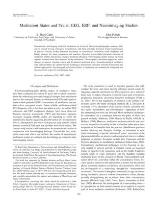
- 12. MEDITATION STATES AND TRAITS long-term Sahaja yoga meditators differed from controls in their lack of frontal gamma power increases to emotionally aversive movie clips (Aftanas & Golocheikine, 2005). These findings are intriguing because it has long been claimed that one of the primary benefits from meditative training is greater emotional stability for challenging life events (Kabat-Zinn, 1990). Conclusions From EEG Meditation Studies It is difficult to draw specific inferences from these studies other than the fact that theta and alpha band activity seems affected by meditation (state), which may alter the long-term neuroelectric profile (trait). The effects suggest that meditation practice is related to increased power in theta and alpha bands and decreased frequency at least in the alpha band, with overall slowing and alteration of coherence and gamma effects. Several factors could contribute to the observed variability. First, the word meditation includes many different techniques, and the specific practices may lead to different state and trait changes. Second, within a specific meditation tradition, individuals can vary in their degree of meditative practice, and their self-selection for participating in EEG studies could affect state and especially trait measurement outcome; that is, how constitutional variables such as affective valence, introversion versus extroversion, and anxiety level affect these measures is unknown. Third, neurophysiological markers of meditative states could alter baseline EEG patterns, such that clear 191 within-group meditation effects are obscured (e.g., overall large spectral power would mask pre- versus postmeditation state changes). Fourth, how EEG measures might be affected by meditator age has not been determined despite the neuroelectric changes that occur from early to middle age adulthood in humans (Polich, 1997). Fifth, methodological difficulties limit the generalizability of early recordings and analysis, especially when stimuli were used to elicit different alpha activity levels. ERPs and Meditation Figure 1 schematically illustrates brain potentials that can be elicited after a stimulus is presented. EPs are evoked automatically with repetitive sensory stimulation, whereas ERPs are elicited with cognitive task processing (Hall, 1992; Picton & Hillyard, 1974; Picton, Hillyard, Krausz, & Galambos, 1974). Auditory stimuli produce the auditory brainstem response and middle latency response. The longer latency auditory EPs are thought to reflect the activation of primary auditory cortex (Polich & Starr, 1983; Wood & Wolpaw, 1982). Visual and somatosensory EPs also can be evoked; standard clinical procedures are now well defined (Chiappa, 1996). The P300 component is usually elicited by assigning individuals a stimulus discrimination task and can be obtained across modalities (Donchin, 1981; Johnson, 1988; Picton, 1992; Polich, 2003, 2004). Figure 1. Schematic illustration of evoked and event-related brain potentials from auditory stimuli. Logarithmic scales for amplitude and latency are used for illustrative purposes only. MMN ϭ Mismatch Negativity. From “Human Auditory Evoked Potentials: I. Evaluation of Components,” by T. W. Picton, S. A. Hillyard, H. I. Drausz, and R. Galambos, 1974, Electroencephalography and Clinical Neurophysiology, 36, p. 181. Copyright 1974 by Elsevier Scientific Publishing Company. Adapted with permission.
- 13. CAHN AND POLICH 192 Table 2 Summary of Meditation Studies Using Evoked Potential (EP) or Event-Related Potential (ERP) Methods Study Meditation type N Experimental design EP/ERPs Paty et al. (1978) TM 25 Meditators vs. controls, before vs. after meditation/relaxation CNV Barwood et al. (1978) TM 8 Before, during, and after meditation; sleeping AEP Corby et al. (1978) Tantric yoga Ananda Marga 30 LTM vs. STM vs. controls, before vs. breath-focused vs. mantra meditation EEG, passive auditory oddball task Banquet & Lesevre (1980) ´ Yoga 20 Meditators vs. controls, before vs. after meditation or rest Visual oddball task McEvoy et al. (1980) TM-Siddhi 5 Meditators vs. controls, before vs. after meditation ABR Becker & Shapiro (1981) TM, Zen, yoga 50 Different meditation groups; attend and ignore control groups AEP and EEG Ikemi (1988) SRM 12 Before vs. during SRM vs. during drowsiness, beginning meditators CNV Goddard (1989) TM 26 Elderly meditators vs. elderly controls Auditory and visual oddball task Liu et al. (1990) Qigong 21 Before, during, and after meditation ABR, MLR, AEP Cranson et al. (1990) TM 39 LTM vs. STM vs. controls Auditory oddball task Goddard (1992) Gordeev et al. (1992) TM Yogic 32 29 Elderly meditators vs. elderly controls vs. young meditators vs. young controls Meditators vs. controls Visual oddball task VEPs, SEP Trait: none reported Findings State: increased CNV amplitude after meditation, decreased amplitude after sleeplike relaxation control period Trait: NA State: nonsignificant decrease in N1 latency during meditation Trait: NA State: no findings; all groups showed equivalent decreases in component amplitudes across sessions Trait: NA State: after meditation, increased P300 amplitude; after rest, decreased P300 amplitude Trait: shorter RT, fewer mistakes, increased N120 and P200 amplitudes State: Wave V latency increased at 45–50 dB and decreased at 60–70 dB; intensity-latency relationship increased in slope from 45–70 dB, central transmission time (Wave V-Wave I) increased at 50 dB Trait: NA State: AEP, no effect of meditation on average N1, P2, P3, early larger N1 amplitude that habituated to the mean in yoga and TM groups Trait: NA State: during SRM, decreased CNV amplitude, error rate; during drowsiness, decreased CNV amplitude, increased RT, error rate Trait: NA State: NA Trait: visual P300 latencies shorter in meditators, no auditory P300 effects State: ABR Waves I-V amplitudes increased, MLR Na-Pa amplitude decrease; AEP P2 amplitude decrease Trait: NA State: NA Trait: P300 latency inversely correlated with length of meditation practice: none Ͼ short Ͼ long State: NA Trait: P300 latencies longer in elderly than young; elderly meditators vs. elderly controls had shorter P300 latencies and longer RTs; dissociation of P300 latency and RT State: amplitude of intermediate and late components of VEPs and SEPs diminished 2–4 fold; SEP early components decreased in amplitude in hemisphere ipsilateral to stimulation only
- 14. MEDITATION STATES AND TRAITS 193 Table 2 (continued ) Study Telles & Desiraju (1993) Meditation type Om mantra meditation N Experimental design 14 Meditators vs. controls, before vs. during meditation technique EP/ERPs MLR W. Zhang et al. (1993) 2 types of Qigong 48 Two groups of LTM vs. STM vs. controls Flash VEP Telles et al. (1994) Om mantra meditation 18 Meditators vs. controls, baseline vs. om meditation vs. one meditation MLR Travis & Miskov (1994) TM 11 Before vs. after meditation vs. after rest Auditory oddball task Murthy et al. (1997, 1998) Kriya yoga, 3month training 45 Patients: depressed vs. dysthymic vs. controls Auditory oddball task Panjwani et al. (2000) Travis et al. (2000) Sahaja yoga TM 34 41 Epilepsy patients: yoga group vs. sham yoga group vs. controls Three groups varying in TM experience, frequency of transcendent experiences ABR, MLR, VCS CNV, simple; CNV, distraction task Findings State: NA Trait: Nb latency decrease in meditator group but no effect seen in controls, small effect size State: VEP amplitude increase in one form of Qigong and decreased in the other Trait: NA State: Na amplitude increased in meditators and decreased in nonmeditators during om; Na amplitude decreased in meditators during one Trait: NA State: decreased latency P300 after TM but not rest; trend toward higher amplitude P300 after TM Trait: NA State: NA Trait: improvement in depressive symptoms and increase of P300 amplitude in novice meditators; effect perhaps from arousal due to alleviation of depression State: NA Trait: ABR, no effects; MLR, increased Na-Pa amplitude at 6 months in meditation group, VCS increased State: NA Trait: CNV amplitude proportional to TM practice and frequency of transcendental experiences; distraction effects (decreases in CNV amplitude) inversely proportional to frequency of transcendent experiences Travis et al. (2002) TM 51 LTM vs. STM vs. controlCNV, simple CNV, choice task State: NA Trait: simple CNV amplitude proportional-choice CNV amplitude inversely proportional to frequency of transcendental experiences and TM practice Note. TM ϭ transcendental meditation; CNV ϭ contingent negative variation; NA ϭ not applicable; AEP ϭ auditory evoked potential (long latency); LTM ϭ long-term meditators; STM ϭ short-term meditators; EEG ϭ electroencephalographic; RT ϭ response time; ABR ϭ auditory brain stem response; SRM ϭ self-regulation method; MLR ϭ middle latency response; VEP ϭ visual evoked potential (flash stimulus); SEP ϭ somatosensory evoked potential; VCS ϭ visual contrast sensitivity. Table 2 summarizes the major EP and ERP meditation studies. The meditation effects are reviewed next for the sensory and cognitive domains. A summary of studies using contingent negative variation (CNV) is then presented. The rationale for these investigations is derived from the early EEG studies outlined previously. Meditators sometimes produced altered amplitudes and shorter potential latencies when stimuli were presented and EEG was recorded, thereby suggesting increased attentional control and CNS quiescence (Banquet & Lesevre, 1980). This interpretation is ´ consonant with results from the 1970s in normal individuals that selective attention and later cognitive processing were reflected by different ERP components. Advanced concentrative meditation
- 15. 194 CAHN AND POLICH practitioners seemed to demonstrate decreased amplitude and latency for several sensory EPs (e.g., Anand et al., 1961; Gordeev, Baziian, & Liubimov, 1992), whereas mindfulness-based practices sometimes induced a decrease in habituation (e.g., Banquet, 1973; Kasamatsu & Hirai, 1966). Thus, these methods were used to characterize sensory and cognitive information processing in meditation as has been done with behavioral measures indicating enhanced perceptual acuity (D. P. Brown, Forte, & Dysart, 1984a, 1984b; Panjwani et al., 2000). Auditory Stimulus Potentials Brainstem potentials. Auditory brainstem responses occur within 10 ms after stimulus presentation and reflect initial sensory processing. Assuming that meditation practice affects attentional mechanisms, these potentials should not be influenced by either meditation state or trait. Auditory brainstem responses were obtained from practitioners of TM–Siddhi meditation supposed to augment normal hearing by using attention to special mantras constructed to sensitize the auditory system and lead to awareness of subtle inner sounds not normally perceived (Mahesh Yogi, 1963; McEvoy, Frumkin, & Harkins, 1980). Binaural click stimuli were presented at 5 to 70 dB in different conditions to elicit auditory brainstem responses before and after meditation. As stimulus intensity increased, Wave V latencies were differentially affected by meditation. At threshold intensities of 45 to 50 dB, Wave V latency increased, whereas at intensities of 65–70 dB latencies decreased relative to baseline, thereby leading to an increased intensity–latency relationship between 45 and 70 dB after meditation. Wave V–I latency differences (central transmission time) also increased after meditation for 50 dB but not other intensities. Background noise is 40 to 50 dB, so that meditation may attenuate the sensitivity to these intensities, thereby enhancing sounds at threshold (5– 40 dB) and speech (60 –70 dB) intensity levels. Middle latency potentials. Middle latency response potentials are generated post-brain stem and reflect initial cortical auditory processing occurring between 10 and 80 ms. Recordings of meditators using the traditional mantra om were made before and during meditation; a nonmeditator control group was comparably assessed while resting quietly at two different times (Telles & Desiraju, 1993). The meditators produced a small but reliable decrease in Nb component latency after meditating relative to the preceding rest period, whereas for the control group no changes were found. In a subsequent study involving novice and expert mantra meditators, Telles, Nagarathna, Nagendra, and Desiraju (1994) compared middle latency response measures before and after meditating on the syllable om versus the word one. Novice meditators demonstrated a decrease in Na amplitude in the om condition; expert meditators demonstrated an increase in Na amplitude for the om condition but an amplitude decrease for the one condition. Brahmakumaris Raja yoga meditators were assessed before and during meditation; a decrease in Na peak latency was found (Telles & Naveen, 2004). The Na potential is thought to be generated at the midbrain–thalamic level, so that concentrative mantra meditation may affect early thalamic sensory processes. Sahaja yoga emphasizes adopting the witness posture toward thoughts instead of flowing with them during meditation and is, therefore, very close to the mindfulness end of the meditational spectrum. This method was assessed in three groups of young adult epileptic patients (Panjwani et al., 2000). One group practiced Sahaja meditation, another group sat quietly in sham meditation, and a control patient group had no meditation instruction. Auditory brainstem response and middle latency response measures were obtained before the meditation intervention and again 3 months and 6 months later. No auditory brainstem response effects were obtained, but the Sahaja yoga group demonstrated an increase in middle latency response Na–Pa amplitude at 6 months. Although Sahaja yoga meditation in normal controls was not assessed, this outcome also suggests the influence of meditation on initial cortical auditory processing. Qigong is a distinct meditation technique that emphasizes becoming aware of the Qi or subtle energy in the body, and consciously manipulating it by means of intentionality, physical postures, and movements (McCaffrey & Fowler, 2003). Several different types of brain potentials were observed before, during, and after a Qigong meditation session in a within-subject design (Liu, Cui, Li, & Huang, 1990). Auditory brainstem response Waves I through V increased in amplitude 55% to 76%, whereas middle latency response Na and Pa amplitudes decreased 50% to 73% during Qigong meditation relative to the before and after conditions. The authors hypothesize that the brainstem may be synergistically released from descending inhibition to produce the auditory brainstem response amplitude increase when the initial cortical activity indexed by middle latency response potentials decreases during meditation. Long latency potentials. TM meditators presented with auditory tones (1/s) demonstrated decreased P1, N1, P2, and N2 component latencies for meditators at baseline and meditation–rest states compared with nonmeditator control group values (Wandhofer, Kobal, & Plattig, 1976). Another study used 50 tones (1-s duration) presented in three blocks to TM meditators before, during, and after meditation in a within-subject design; additional recordings were made during sleep. Although N1 latency was longer in the before control condition relative to the meditation condition, this effect was unreliable, and no other condition differences were found for any of the auditory long-latency potential components (Barwood, Empson, Lister, & Tilley, 1978). Ananda Marga meditative practice focuses initially on withdrawing from external orientation by means of breath-focused concentration, which is then followed by mantra meditation and, therefore, lies toward the concentrative meditation end of the spectrum. Experienced meditators were compared with novice meditators and nonmeditating controls (Corby et al., 1978). Each individual was exposed to a series of tones presented at a rate of 1/s for 20 min, with the inclusion of an oddball tone (1/15) in each of three conditions: baseline rest, breath-focused awareness, and mantra meditation. Nonmeditating controls mentally repeated a randomly chosen two-syllable word; all groups were instructed to ignore the tones. For the experienced meditators compared with other subjects, EEG theta and alpha power was higher in both the baseline and meditative conditions. For all three groups, infrequent tones elicited smaller N1 amplitudes and a positive potential occurring at approximately 250 ms (dubbed “P2-3” but likely a P2). Auditory long-latency potential components during the baseline rest were similar to the meditation conditions for both tones, but during meditation P2-3 amplitude decreased for infrequent tones and increased for frequent tones. Condition order was not counterbalanced, so it is likely that habituation effects produced
- 16. MEDITATION STATES AND TRAITS the amplitude decrements. The reason for the P2-3 amplitude increase to the frequent tones is unclear. Auditory long-latency potentials were obtained from five groups of participants—Zen, TM, yoga, and two groups of nonmeditator controls—who were instructed either to attend or ignore loud click stimuli (115 dB) presented at 15-s intervals (Becker & Shapiro, 1981). As noted, no differential alpha blocking was found among the five groups when meditators meditated and controls applied their instructed attentional focus at rest. No auditory long-latency potential components demonstrated any differences other than the production of larger passive P300 amplitudes in the attend group as observed previously (Becker & Shapiro, 1980). N100 amplitude for the TM and yoga meditation participants was increased over the first 30 stimulus presentations and then reduced to the same size as the other groups after 40 to 50 stimulus presentations. The authors suggested that, given the mantras used by both groups, the attentional state of the TM and yoga meditators may have been attuned to inner sounds, which could have contributed to a greater sensitivity for the auditory stimulus input, even above that of the control attend group specifically instructed to pay full attention to the auditory input. Qigong meditators were assessed by presenting 10-ms tones and recording before, during, and after a 30-min Qigong meditation session (Liu et al., 1990). P200 amplitude decreased 44% from the baseline to the meditation state and returned to baseline after meditation. This outcome suggests that later auditory long-latency potential measures may be sensitive to meditation state. Auditory P300. TM practice was studied using a passive auditory paradigm listening study with variable interstimulus intervals (1– 4 s) between identical tone stimuli (Cranson, Goddard, & Orme-Johnson, 1990). The participants were nonmeditator controls, novice, and highly experienced TM meditators (mean age ϭ 20, 28, and 41 years, respectively). IQ scores did not differ among the groups. Passive P300 potential latency was shorter for the two meditation groups; the long-term meditators showed the shortest P300 latency regardless of age (cf. Polich, 1996). These results imply that auditory long-latency potentials might reflect meditation trait differences. An auditory oddball task was used with eyes closed to assess experienced TM meditators at pretest baseline, after 10 min of rest, and after 10 min of TM practice; conditions were counterbalanced across participants (Travis & Miskov, 1994). P300 latency decreased at Pz after TM practice relative to no change after the rest condition. Sudarshan Kriya yoga is a meditation system that emphasizes breathing techniques. This technique was used as an intervention to assess dysthymic, dysthymic with melancholy, and unaffected control groups (Murthy, Gangadhar, Janakiramaiah, & Subbakrishna, 1997, 1998). At 3 months, P300 amplitude increased to control levels in the patient groups after initial values at baseline (7.5 V) and 1 month (10.4 V) that were well below normal values (14.4 V) at both time points. These reports suggest the possibility of some meditation effects on the P300 component. Visual Stimulus Potentials Visual EPs. Sensory potentials evoked by a light flashes were used to compare four populations: (a) long-term Qigong meditation practitioners, (b) long-term Nei Yang Gong practitioners (a variant of the older Qigong method), (c) beginning Nei Yang Gong meditators, and (4) nonmeditating controls (W. Zhang, Zheng, Zhang, Yu, & 195 Shen, 1993). Visual flash potentials were obtained under eyes-open conditions before, during, and after the meditative practice or analogously for a rest period in controls. The flash potentials were classified as early (N80-P115-N150) and late (N150-P200-N280) components, with peak-to-peak amplitudes measured. The long-term traditional Qigong practitioners demonstrated marginally significant decreased amplitude for the early and later flash potentials during meditation. However, the Nei Yang Gong practitioners demonstrated increased amplitudes for both the early and late flash potentials. No effects of meditation were reported for the beginning Nei Yang Gong or control groups. The authors concluded that the two types of Qigong meditative practice produce opposite effects on the relative excitability of the visual cortex, such that the more traditional Qigong leads to cortical inhibition and reduced flash potential amplitudes (Cui & Lui, 1987). Visual P300. ERPs obtained before and after a 30-min meditation period for experienced yogic meditators or a 30-min rest period for matched nonmeditator controls were compared (Banquet & Lesevre, 1980). A go/no-go task visually presented 450 ´ letters, with 10% randomly omitted. Participants were instructed to respond to each stimulus and to refrain from responding whenever they detected an omitted stimulus, so that state and trait effects could be evaluated under response and nonresponse conditions. For the meditators, P300 amplitude increased postmeditation; for the controls P300 amplitude decreased after rest. The meditators also demonstrated shorter response time (RT) and greater accuracy before and after the meditation period relative to controls; RT was shorter than P300 latency for the meditators but longer than P300 latency for the controls in both the pre- and postconditions. For the meditators compared with controls, P200 amplitudes from both the go and no-go stimuli were larger in the pre- and postmeditation– rest conditions, and N120 amplitude increased in the post-no-go task but decreased in latency in pre- and postconditions for the go task. The authors suggest that long-term meditative practice could increase selective attention capacity, which improves vigilance level to affect ERP measures. Such state effects also are consistent with meditation affecting deautomization of stimulus processing. Meditative practice and aging in TM meditators were evaluated relative to nonmeditating controls (66 years) with visual ERPs elicited by female and male names in a button-press task (Goddard, 1989, 1992). P300 latency was shorter in meditators than controls (543 vs. 703 ms). The same individuals also performed an auditory oddball task, but neither P300 latency nor RT differed between the groups. The results were interpreted as indicating that trait effects of long-term TM practice are observed only if mental processing demands are increased with more difficult visual tasks. A visual oddball task used to compare four groups of young (20 years) and older (69 years) meditators and controls found that P300 latency and RT increased as the discriminability of the targets was made more difficult for all groups (Goddard, 1992). P300 latencies were longer in older participants in all conditions, whereas RTs were shorter only as task difficulty increased. Further, P300 latencies were shorter in the older meditators versus nonmeditators. These results suggest the possibility of primarily P300 latency trait effects for meditating relative to nonmeditating older individuals. Somatosensory Potentials Somatosensory potentials are often evoked using mild electric shocks applied to the median nerve, with a series of potentials
- 17. 196 CAHN AND POLICH indexing transmission of the signal from the periphery to the cortex (Chiappa, 1996). TM meditators with 2 years practice demonstrated increased amplitudes of early components relative to controls (Petrenko, Orlova, & Liubimov, 1993). Yogic concentrative meditators with a 10- to 12-year practice history evinced amplitude decreases in the later components (Lyubimov, 1999). Similar yogic meditators produced somatosensory EP amplitude decreases when instructed to block out the sensory stimuli, whereas the controls produced no effects. Further, the early components decreased only on the recording sites ipsilateral to stimulation side, but late components decreased bilaterally (Gordeev et al., 1992). This outcome implies that some concentrative meditation practices states can block sensory input at a subcortical level. CNV CNV is elicited by presenting two stimuli in succession such that the first serves as an indicator for an impending second stimulus to which a response is required (Walter, Cooper, Aldridge, McCallum, & Winter, 1964). This negative-going waveform was one of the first reported cognitive ERPs and consists of an early deflection related to CNS orienting followed by a later deflection that is maximal before the imperative stimulus and thought to reflect stimulus expectancy (Gaillard, 1977; Irwin, Knott, McAdam, & Rebert, 1966; Rohrbaugh et al., 1997). An early study found meditation-induced state effects of increased CNV amplitudes following TM practice (Paty et al., 1978). The self-regulation method is a meditation technique combining aspects of Zen practice and autogenic training (Ikemi, 1988; Ikemi, Tomita, Kuroda, Hayashida, & Ikemi, 1986). After a 5-week training course, EEG and CNV assessments were carried out before and during practice as well as during a drowsy state. CNV was obtained with a choice task to the imperative second stimulus. During meditation accuracy increased and shorter RTs were observed, whereas during drowsiness accuracy decreased and longer RTs occurred. EEG demonstrated increased theta and decreased beta power for meditation, but during both meditation and drowsiness reduced CNV amplitudes for the choice-task were found. CNV processes, therefore, may be sensitive to meditation state. Groups of age-matched TM meditators who differed in length of practice and frequency of self-reported transcendental (defined as experiences of pure consciousness, devoid of thought, and marked by awareness of awareness itself) perceptions (Ͻ 1/year, 10 –20/ year, every day) were evaluated using simple and distracter CNV tasks (Travis et al., 2000). No group effects were observed for the earlier orienting CNV component, but greater negativity for the later expectancy wave was obtained as the frequency of reported transcendental experiences increased across groups and tasks (simple RT, distraction stimuli). The decrement in CNV amplitude induced by the distracting stimuli was inversely related to transcendental experience frequency. The findings implied that transcendental feelings may modulate cortical functioning by activating processing resources to facilitate greater attentional resource capacity and thereby increased CNV amplitude. A follow-up study assessed groups of older individuals who varied in TM background and reported varied transcendental experience levels: (a) no meditation practice (mean age ϭ 39.7 years), (b) TM practice and occasional transcendental events (mean age ϭ 42.5 years, mean years meditating ϭ 7.8), (c) long-term TM practice (mean age ϭ 46.5 years, mean years meditating ϭ 24.5) and continuous coexistence of transcendent experience in waking and sleeping states (Travis et al., 2002). For the simple RT task, CNV amplitudes were greater for individuals with more TM practice and greater frequency of transcendent experience. For the choice task, smaller CNV amplitudes were associated with more TM practice and transcendent experience frequency. These findings were interpreted as indicating that the brain of the meditators efficiently waited for the second stimulus information rather than automatically committing attentional resources to the imperative event. EEG recording during CNV task performance demonstrated increased theta–alpha (6 –12 Hz) power across the groups. Frontal broadband coherence values (6 –12 Hz, 12–25 Hz, and 25– 45 Hz) also were increased as meditation practice increased across groups. These effects suggested that development of transcendental awareness was a meditation trait. Follow-up psychometric assessment of these individuals indicated that greater meditation experience was also related to increased inner directedness, higher moral reasoning, lower anxiety, and more emotional stability (Travis et al., 2004). How self-selection bias of individuals choosing to meditate for long time periods may contribute to these outcomes is unknown. The CNV findings imply that meditation reduces choice-task CNV amplitude for state (Ikemi, 1988) and trait (Travis et al., 2002). In the simple CNV tasks, an increase in amplitude has been observed as both a state effect and a trait effect of meditation (Paty et al., 1978; Travis et al., 2000, 2002). One finding that may be related to these results is the inverse correlation between states of greater sympathetic activation and CNV amplitude, modifiable by autonomic biofeedback procedures (Nagai, Goldstein, Critchley, & Fenwick, 2004). Thus, CNV appears to be affected by meditative practice in a manner related to changes in attentional resource allocation and possibly autonomic activity. Conclusions From ERP Meditation Studies Sensory EP and cognitive ERP meditation assessments have produced a variety of effects. The major difficulties in many studies are a lack of methodological sophistication, no replication of critical conditions, and inconsistency of task and study populations. Some intriguing hints of meditation changing early cortical auditory processing appear reliable, with suggestions that P300 also can be affected by meditation practice. Possible stimulus modality differences in assessing meditation have not been systematically ascertained. Simple CNV tasks yield an increase in amplitude for both state and trait effects of meditation, such that CNV effects may reflect changes in attentional resource allocation. Brain Imaging and Meditation Table 3 summarizes the findings from other neuroimaging studies of meditation (reviewed next). The results complement and extend the neuroelectric findings presented previously. PET A PET study measured regional cerebral metabolic rate of glucose (rCMRGlc) in yoga meditation by comparing an eyes-
- 18. MEDITATION STATES AND TRAITS 197 Table 3 Summary of Meditation Studies Using Neuroimaging Methods Study Meditation type Herzog et al. (1990) Yoga meditation, eyes open Jevning et al. (1996) TM N Experimental design Method 8 Meditation vs. resting thought, separate days PET 34 Meditators vs. controls, rest 3 meditation Rheoencephalography Lou et al. (1999) Yoga nidra (guided) 9 Rest 3 meditation PET Lazar et al. (2000) Kundalini yoga mantra 5 Meditation vs. control periods silently generating number lists fMRI Khushu et al. (2000) Raja yoga 11 Rest 3 meditation fMRI Baerentsen (2001) Mindfulness 5 Rest 3 meditation fMRI Newberg et al. (2001) Tibetan Buddhist imagery-meditation 8 Meditators vs. controls, rest 3 meditation (self-reported peak) SPECT Azari et al. (2001) Psalm 23 recitation 12 Religious vs. nonreligious participants, rest, reading, and reciting Psalm 23 vs. versus nursery rhyme vs. phone book PET Kjaer et al. (2002) Yoga nidra (guided) 5 Separate days for meditation and baseline PET-11C-raclopride binding, EEG Ritskes et al. (2003) Zen Newberg et al. (2003) Christian prayer Lazar et al. (2003) Mindfulness vs. Kundalini yoga 11 3 33 Interleaved periods of meditation and rest fMRI Franciscan nuns, rest 3 prayer SPECT Mindfulness vs. Kundalini yoga meditators vs. controls, meditation vs. random number generation fMRI Findings (state effects) Increase: frontal-parietal and frontaloccipital activation ratios, lowresolution analysis Decrease: slight for posterior-anterior ratios Increase: frontal, occipital Decrease: none, low-resolution analysis Increase: anterior parietal (postcentral gyrus), fusiform gyrus, occipital cortex Decrease: dorsolateral orbital, cingulate, temporal, caudate, thalamus, pons, cerebellum Increase: DLPFC, ACC, parietal, hippocampus, temporal, striatum, hypothalamus, pre-post central gyri Decrease: 20% globally Increase: PFC Decrease: none, low-resolution analysis Increase: DLPFC, ACC Decrease: occipital Increase: cingulate, inferior-orbital, DLPFC, bilateral thalamus, midbrain, sensorimotor Decrease: PSPL; increases in left DLPFC correlated with decreases in left PSPL Increase: right and left DLPFC, right medial parietal dorsomedial prefrontal (pre-supplemental motor area), cerebellum Decrease: none reported Increase: EEG theta Decrease: raclopride binding in ventral striatum, indicating increase dopamine binding Increase: DLPFC (R Ͼ L), basal ganglia Decrease: right anterior superior occipital gyrus, ACC Increase: PFC, inferior parietal lobes, inferior frontal lobes Decrease: PSPL Increase: both showed cingulate activation, right temporal lobe (Vipassana only) Decrease: none reported; different distribution of activated networks in the two groups Note. PET ϭ positron emission tomography; TM ϭ transcendental meditation; fMRI ϭ functional magnetic imaging; DLPFC ϭ dorsolateral prefrontal cortex; ACC ϭ anterior cingulate cortex; PFC ϭ prefrontal cortex; SPECT ϭ single photon emission computed tomography; PSPL ϭ posterior superior parietal lobe; EEG ϭ electroencephalographic; R ϭ right; L ϭ left. open meditation and a control condition in which the participants were instructed to think of daily affairs (Herzog et al., 1990). The meditators reported feeling relaxed, at peace, and detached during meditation but not during the control condition. Half of the participants showed an overall increase, and half showed an overall decrease in general cerebral metabolic rate during meditation. This outcome may have resulted from the necessity of recording the two sessions on different days, so that differential practice effects could underlie arousal differences between the groups. No statistically reliable meditation effects on rCMRGlc for any brain regions were