1. AMYLOIDOSIS IN TUBERCULOSIS
M. S. A GNIHOTRI AND S. R ASTOGI
(From K.G.’s Medical College, Lucknow)
The term amyliodpsis is used to describe a male who was emaciated, the subcutaneous fat
hyaline material which accumulates between being scanty. Examination of the thoracic
parenchymatous cells and in connective tissue cavity revealed pleural adhsions on both sides.
of organs. Association of amyloidosis and Cut surface of the lungs revealed cavities of
tuberculosis is well known. Tuberculosis is a sizes varying from 2-4 cm in diameter. Their
common cause of secondary amyloidosis walls were smooth. The surrounding lung
(Anderson 1957; Mathur and Jhala 1964; parenchyma showed focal areas of caseation,
Chitkara et al. 1965; Reddy and Parvathi, 1-2 mm in diameter. The tracheobronchial
1968). Reddy et al. (1970) reported that in-
cidence of amyloidosis in tuberculosis was
19% in autopsies. Studies from western coun-
tries have reported higher incidence of amy-
loidosis in tuberculosis (Cohen 1943; Kozello
1965; Yoshizumi, 1962). Recently, studies
demonstrating changed pattern of amyloidosis
in treated patients of tuberculosis have appeared
in literature. Yoshizumi (1962) reported a
sharp decline in incidence of amyloidosis in
tuberculosis since the use of chemotherapy.
Maltchik (1959) observed that amyloidosis in-
volves lesser number of organs when it occurs
in chronic form of tuberculosis.
We are reporting a treated patient of
bilateral pulmonary tuburculosis with secon-
dary amyloidosis and miliary tuberculosis.
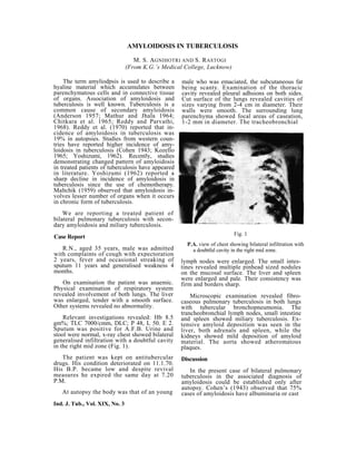
Fig. 1
Case Report
P.A. view of chest showing bilateral infiltration with
R.N., aged 35 years, male was admitted a doubtful cavity in the right mid zone.
with complaints of cough with expectoration
2 years, fever and occasional streaking of lymph nodes were enlarged. The small intes-
sputum 11 years and generalised weakness 4 tines revealed multiple pinhead sized nodules
months. on the mucosal surface. The liver and spleen
were enlarged and pale. Their consistency was
On examination the patient was anaemic. firm and borders sharp.
Physical examination of respiratory system
revealed involvement of both lungs. The liver Microscopic examination revealed fibro-
was enlarged, tender with a smooth surface. caseous pulmonary tuberculosis in both lungs
Other systems revealed no abnormality. with tubercular bronchopneumonia. The
trancheobronchial lymph nodes, small intestine
Relevant investigations revealed: Hb 8.5 and spleen showed miliary tuberculosis. Ex-
gm%; TLC 7000/cmm, DLC; P 48, L 50. E 2. tensive amyloid deposition was seen in the
Sputum was positive for A.F.B. Urine and liver, both adrenals and spleen, while the
stool were normal, x-ray chest showed bilateral kidneys showed mild deposition of amyloid
generalised infiltration with a doubtful cavity material. The aorta showed atheromatous
in the right mid zone (Fig. 1). plaques.
The patient was kept on antitubercular Discussion
drugs. His condition deteriorated on 11.1.70.
His B.P. became low and despite revival In the present case of bilateral pulmonary
measures he expired the same day at 7.20 tuberculosis in the associated diagnosis of
P.M. amyloidosis could be established only after
autopsy. Cohen’s (1943) observed that 75%
At autopsy the body was that of an young cases of amyloidosis have albuminuria or cast
Ind. J. Tub., Vol. XIX, No. 3
2. AMYLOIDOSIS IN TUBERCULOSIS 113
and therefore, their presence in the urine loidosis either before or after chemotherapy.
should make one suspect amyloidosis. Clinically Yoshizumi (1962) demonstrated a sharp
in our case urine examination revealed no decline in incidence of amyloidosis with the
abnormalities. Cango red test and gum biopsy advent of chemotherapy. It is not possible for
were also not done because of lack of clinical us to comment on incidence but the lower
diagnosis of amyloidosis. Autopsy of the incidence of amyloidosis in our country as
patient on 12.1.71 confirmed the diagnosis of compared to western countries, may be due to
bilateral pulmonary tuberculosis. The patient the lack of diagnosis. As in our case diagnosis
was also found to be suffering from generalised of amyloidosis could only be made after
miliary tuberculosis and amyloidosis. Secondary autopsy which is a rare diagnostic procedure
amyloidosis was seen in liver, spleen, kidney in our country.
and adrenal, whereas, miliary tuberculosis was
observed in intestine lymph node and spleen. Summary
Anderson (1957) reported that liver, kidney,
spleen and adrenal are commonly involved in A case with fibrocaseous pulmonary tuber-
secondary amyloidosis. Olekhnovich (1958) culosis with generalised secondary amyloidosis
reported that in 42 cases of tuberculosis with of adrenal, liver, spleen and kidney and miliary
amyloidosis, kidney was involved 42 times, tuberculosis of spleen, intestines and glands,
spleen 40 times and liver 30 times. In our is reported. Importance of autopsy in diagno-
case spleen was the only organ which demons- sis of amyloidosis is emphasised.
trated the presence of both amyloidosis and
miliary tuberculosis. Anderson (1957) reports REFERENCES
that the spleen is most commonly involved
organ in amyloidosis as well as it is one of the 1. Anderson, W.A. (1957), Pathology, C.V. Mostry
Co., St. Louis, p. 73.
organs involved at the earliest stages. Miliary
involvement of intestine, lymph node, spleen 2 Chitkara, N.L., Chugh T.D., Chuttani, P.N.
in our patient of fibrocaseous pulmonary tuber- and Chugh, K.S. (1965), Ind. J. Path. Bact. 8,
culosis was possibly due to haematogenous 285-293.
dissemination in preterminal stages of broncho-
genie tuberculosis, as our patient died within 3. Cohen, S., (1943), Ann. Int. Med. 19, 990-1002.
two weeks of admission in the hospital. Our 4. Kozello, N.A., (1963), Klin. Med. Mask. 41,
case also demonstrates that tuberculous involve- 79-85.
ment of an organ is not a pre-requisite for
amyloidosis. Furthermore, amyloid deposites 5. Maltchik, F.A., (1959), Probl. Tuberk, 97, 37.
in an organ do not favour development of
tuberculosis because both amyloidosis and 6. Mathur, B.B.L. and Jhala, C.I. (1964), Ind. J.
tuberculosis involved various organs in Path. Bact.l, 133-145.
the present case. But both amyloidosis and
tuberculosis simultaneously involved only 7. Reddy, C.R.R.M., and Parvathi, G. (1968),
spleen. Maltchik (1959) observation that amy- Ind. Jour. Med.Sci. 22, 770-774.
loidosis involves lesser number of organs when
it occurs in chronic forms of tuberculosis is 8. Reddy, C.R.R M., Sulochana, G., Rama Rao
contradictory to our case finding as in the T., Devi, C.S., (1970), 17, 70-71.
present case amyloidosis involved organs like 9. Olekhnovich, L.I. (1958), Probl. Tuberk. 96, 36.
liver, spleen, adrenal and lungs. Yoshizumi
(1962) reported that there is no difference in 10. Yoshizumi, M. and Lt, T.G. (1962), Amer, Rev,
pathologic involvement of organs by amy- Resp. Dis. 85, 432-435,
Ind. J. Tub., Vol. XIX, No. 3