4.diabetes basic
•Download as PPTX, PDF•
15 likes•11,721 views

The document discusses the gastrointestinal system and its organs including the mouth, stomach, pancreas, small intestine, and liver. It then describes the liver's functions of producing proteins and bile, storing vitamins and minerals, converting and utilizing fats and carbohydrates, and removing waste. The document notes that carbohydrates provide 60% of the body's energy, with proteins and fats each contributing around 10-12% and 30%, respectively. It outlines the journey of glucose from food to different body parts and its utilization and storage. Key steps in glucose utilization are its entry into cells, phosphorylation, and energy release.

Recommended
More Related Content
What's hot
What's hot (20)
Viewers also liked
Viewers also liked (20)
Similar to 4.diabetes basic
Similar to 4.diabetes basic (20)
More from Ashok Moses
Recently uploaded
Recently uploaded (20)
4.diabetes basic
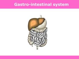
- 2. Gastrointestinal system • Mouth • Stomach and duodenum • Pancreas • Small intestine • Liver and other organs
- 3. Liver functions • Production – plasma proteins, blood clotting proteins, bile pigments • Storage – vitamins, minerals, fat, glucose as glycogen • Conversion/utilization – fats, carbohydrates, proteins • Removal – aged blood cells, drugs or toxins, waste products
- 4. Energy source for body • Proteins: 10-12% • Fats: 30% • Carbohydrates: 60%
- 5. Journey of Glucose Food Carbohydrates Formation formed of glucose Glucose Glucose Glucose enters enters reaches different blood Cell body parts Mediated by Insulin Glucose used for various functions Extra glucose stored in a different form
- 6. Steps in utilization of glucose Entry of glucose in cell Phosphorylation of glucose Release of energy
- 7. • Facilitates the transport of glucose into muscle and adipose cells • Facilitates the conversion of glucose to glycogen for storage in the liver and muscle. • Decreases the breakdown and release of glucose from glycogen by the liver Insulin-Carbohydrate Metabolism
- 8. • Stimulates protein synthesis • Inhibits protein breakdown; diminishes gluconeogenesis Insulin - Protein Metabolism
- 9. • Stimulates lipogenesis- the transport of triglycerides to adipose tissue • Inhibits lipolysis – prevents excessive production of ketones or ketoacidosis Insulin - Fat Metabolism
- 10. Pancreas • Exocrine function – Digestive enzymes • Pancreatic amylase • Pancreatic lipase • Trypsin • Chymotrypsin • Carboxypolypeptidase • Nuclease • Endocrine function – Hormones • Insulin • Glucagon • somatostatin
- 11. Endocrine function • Hormones act on target tissues to exert its effect • Produced by the islet of Langerhans – number of cells in each islet vary from several hundred to millions • Glucagon – alpha cells – Secreted when blood glucose levels fall • Insulin – beta cells – Secreted when blood glucose levels rise • Somatostatin – delta cells – Secreted in response to any kind of food intake – suppresses both insulin and glucagon and may extend the period of nutrient absorption and utilization
- 13. What is insulin? • A hormone – (from Greek - "to set in motion") is a chemical messenger from one cell (or group of cells) to another. • Insulin is the protein hormone produced by cells in the pancreas that regulate levels of glucose and regulate metabolism in glucose, fats, and proteins. • Insulin is composed of 51 amino acids. • Amino acids are the basic structural building units of proteins • Its formula is C254 H377 N65 O75 S6.
- 14. Role of Insulin • Hormone secreted by beta cells of pancreas • Controls the rate of entry of glucose inside the cell • Increases glucose utilization rate in the cell • Increases rate of glucose transport in the cell by more than 10 times. Hexamer of insulin
- 16. Regulation of Hormone Secretion • Non-hormonal – Control of release dependent upon concentration of other non-hormonal substance (i.e., glucose)
- 17. Few Important Definitions... • Glycolysis: Breakdown of glucose to release energy • Glycogenesis: Formation of glycogen for storage from unutilized glucose • Glycogenolysis: breakdown of stored glycogen into glucose • Gluconeogenesis: formation of glucose from sources other than carbohydrate (fat/protein) to meet energy requirement
- 18. C-peptide • Insulin manufactured and stored as proinsulin (86 AA) • C-peptide (31 AA) ensures correct folding of protein • Enzymatic cleavage (4 AA lost) and equal amount released along with insulin (51 AA) • C-peptide levels measured to assess insulin production • No physiological role – used as an insulin marker
- 19. GLUCOSE INSULIN CELL Energy How the body uses Food
- 20. Insulin Biosynthesis in the Beta Cell Insulin gene codes for pre-proinsulin Release by exocytosis Proinsulin C-peptide Glucose Insulin storage in vesicles
- 21. Physiological Effects of Insulin • Major target organs: – Liver: insulin increases storage of glucose as glycogen – Muscle: insulin stimulates glycogen and protein synthesis. – Adipose: insulin stimulates triglyceride storage
- 22. Date :12th Mar 09 Valid: 11th Mar 10 Arrest of K+ release Ca2+ Opening of Ca2+ channel Ca2+ K+ K+ K+ ATP Metabolism Glucokinase Glucose Glucose Glucose GLUT 2 Insulin release ATP Glucose Insulin release in non - diabetics BETA CELL
- 23. Date :12th Mar 09 Valid: 11th Mar 10 Insulin receptors Glucose Glycogen Metabolism GLUT4 Dephosphorylation Translocation Intracellular vesicle pGLUT4 ATP The non - diabetic peripheral cell
- 24. Insulin secretion • Insulin secretion increases almost 10 folds within 5 to 10 minutes of food intake. • Insulin secretion is stimulated by glucose
- 25. The Basal/Bolus Insulin Concept • Basal insulin – Continuous, constant, low level secretion for 24 hours – Suppresses glucose production between meals and overnight – 40% to 50% of daily needs • Bolus insulin (mealtime)- 2 phases – Limits hyperglycemia after meals – Immediate rise and sharp peak at 1 hour – 10% to 20% of total daily insulin requirement at each meal
- 26. 0 10 20 30 40 50 0 2 4 6 8 10 12 14 16 18 20 22 24 Time (Hours) Meal Meal Meal Basal Insulin Needs Bolus insulin needs Seruminsulin(mU/L)
- 27. Phases of insulin release • First phase – Release starts as soon as food comes to the stomach – Preformed stored insulin is released – 10-fold increase in levels within 3-5 minutes – Speeds up the use of glucose – Within 5-10 minutes, insulin secretion decreases by half • Second phase – Rising glucose levels send signals to the beta cell nucleus DNA produces mRNA mRNA produces more insulin – Causes a less acute rise in insulin levels – Reaches a plateau in 2-3 hours
- 28. Insulin release
- 29. glycogen synthesis glycogenolysis triglyceride synthesis ketogenesis gluconeogenesis glucose uptake protein synthesis protein degradation glycogen synthesis glycogenolysis glucose uptake triglyceride storage lipolysis Stimulates Inhibits Liver Skeletal Muscle Adipose tissue Promotes anabolic processes Inhibits catabolic processes Effects of insulin:
- 31. • 1500BC – Egyptians recorded diabetes as polyuria • 1st Century AD – diabetes described as “the melting down of flesh and limbs into urine” • 20th Century – children with Type 1 diabetes had life expectancy of 2 years – Hypothesized that liver and pancreas were involved in some way, although cause unknown • 1922 – Frederick Banting & Co. successfully isolate insulin extract for diabetes Type 1 • 2011 – Type 2 diabetes comprises roughly 90% of all diagnosed cases, likely due to increased obesity and inactivity levels History of Diabetes
- 32. Diabetes Mellitus Derived from Greek roots dia – through bainein – to go To go through – meaning syphon Mellitus – Latin „mel‟ for „honey‟ Diabetes Mellitus – Sweet syphon
- 33. Diabetes – Definition Diabetes Mellitus is a metabolic disorder caused by reduced availability or diminished effectiveness of insulin, characterized by hyperglycemia with or without glycosuria.
- 34. Diabetes Mellitus • Chronic medical condition • Inability to properly utilize glucose • Diabetes can cause acute medical emergencies –Too much glucose (hyperglycemia) –Too little glucose (hypoglycemia)
- 36. Diabetes Mellitus Types of Diabetes • Type 1 Diabetes • Type 2 Diabetes • Gestational Diabetes
- 37. Action of Insulin on the Cell Metabolism
- 38. • Low or absent endogenous insulin • Dependent on exogenous insulin for life • Onset generally < 30 years • 5-10% of cases of diabetes • Onset sudden – Symptoms: 3 P‟s: polyuria, polydypsia, polyphagia Type I Diabetes
- 39. Type I Diabetes Cell
- 40. • Genetic component to disease Type I Diabetes
- 41. • Insulin levels may be normal, elevated or depressed – Characterized by insulin resistance, – diminished tissue sensitivity to insulin, – and impaired beta cell function (delayed or inadequate insulin release) • Often occurs >40 years Type II Diabetes
- 42. Type II Diabetes
- 43. • Risk factors: family history, sedentary lifestyle, obesity and aging • Controlled by weight loss, oral hypoglycemic agents and or insulin Type II Diabetes
- 44. TYPE I + TYPE II Type I Diabetes Type II Diabetes GLUCOSE GLUCOSE INSULIN CELL CELL
- 45. DM – Type I / II
- 46. Pathogenesis of DM • Insulin resistance • Impaired insulin secretion • Excessive hepatic glucose production
- 48. Pathogenesis of DM (Contd.) Insulin Resistance: • Decreased ability of insulin to act effectively on peripheral tissue • This resistance is relative, since increased levels of insulin will normalize the Pl. glucose level Mechanism – exact not known • Decrease in insulin receptors or post receptor defect
- 49. • Major defect in individuals with type 2 diabetes1 • Reduced biological response to insulin1–3 • Strong predictor of type 2 diabetes4 • Closely associated with obesity5 IR 1American Diabetes Association. Diabetes Care 1998; 21:310–314. . J Clin Invest 1994; 94:1714–1721. 3Bloomgarden ZT. Clin Ther 1998; 20:216–231. 4Haffner SM, et al. Circulation 2000; 101:975–980. 5Boden G. Diabetes 1997; 46:3–10. Insulin Resistance 1American Diabetes Association. Diabetes Care 1998; 21:310–314. 2Beck-Nielsen H & Groop LC. J Clin Invest 1994; 94:1714–1721. 3Bloomgarden ZT. Clin Ther 1998; 20:216–231. 4Haffner SM, et al. Circulation 2000; 101:975–980. 5Boden G. Diabetes 1997; 46:3–10.
- 51. Pathogenesis of DM (Contd.) Impair insulin secretion • Initially insulin secretion increase in response to insulin resistance to maintain normal blood glucose levels • In later stage beta cell failure develops due to lipo and glucotoxicity - low insulin levels
- 52. Pathogenesis of DM (Contd.) Increased hepatic glucose production • Liver maintain Plasma glucose level during fasting state by glycogenolysis and gluconeogenesis (A.A., F.A., glycerol) • In DM because of insulin resistance, insulin fails to suppress gluconeogenesis which leads to increased blood glucose levels.
- 53. Diagnosis of Diabetes Polyuria – Increased micturition Polydipsia – Increased thirst Polyphagia – Increased hunger Fatigue Skin infections Impotence Tingling, numbness
- 54. Diagnosis of diabetes Symptoms + Elevated blood glucose level OR Elevated blood glucose levels on two occasions
- 55. Diagnostic criteria for type 2 diabetes Blood Glucose Parameter HbA1c FPG PPPG Normal < 6.5% < 100 mg/dl < 140 mg/dl Pre-diabetes ≥ 6.5 - 7% 100-125 mg/dl (IFG) 140-199 mg/dl (IGT) Diabetes ≥ 7% 126 mg/dl or above 200 mg/dl or above
- 56. Definitions IGT impaired glucose tolerance – – 2hr plasma glucose is between 7.8mmol/l (140mg/dl) and 11.0mmol/l (200mg/dl) IFG impaired fasting glucose – – Fasting plasma glucose is 6.1–6.9mmol/l (100–125mg/dl) Diabetes- – Confirmed fasting plasma glucose is ≥7.0mmol/l (126mg/dl) – 2hr plasma glucose is ≥11.0mmol/l (200mg/dl)
- 57. Prediabetes & Diabetes diabetesprediabetesnormoglycemic 100mg/dl 125 mg/dl 140 mg/dl 199 mg/dl Fasting glucose 2hr Plasma glucose Prediabetes is a condition in which the blood sugar level is higher than normal, but not high enough to be classified as diabetes
- 58. Insulin resistance Glucose output Glucose uptake Glucose uptake Hyperglycemia Liver Muscle Adipose tissue IR Insulin Resistance – Reduced response to circulating insulin
- 59. Chronic Hyperglycemia Over secretion of insulin to compensate for insulin resistance1,2 High circulating free fatty acids Glucotoxicity2 Pancreas Lipotoxicity3 -cell dysfunction . Eur J Clin Invest 2002; 32:14–23., 2Kaiser N, et al. J Pediatr Endocrinol Metab 2003; 16:5– 22.,3Finegood DT & Topp B. Diabetes Obes Metab 2001; 3 (Suppl. 1):S20–S27. Why does the -cell fail? 1Boden G & Shulman GI. Eur J Clin Invest 2002; 32:14–23. 2Kaiser N, et al. J Pediatr Endocrinol Metab 2003; 16:5–22. 3Finegood DT & Topp B. Diabetes Obes Metab 2001; 3 (Suppl. 1):S20–S27.
- 60. What is -cell dysfunction? • Major defect in individuals with type 2 diabetes • Reduced ability of -cells to secrete insulin in response to hyperglycemia DeFronzo RA, et al. Diabetes Care 1992; 15:318–354.
- 61. Insulin Resistance Genetic susceptibility, Obesity, Sedentary lifestyle Type 2 diabetes IR -cell dysfunction Core defects in T2DM Rhodes CJ & White MF. Eur J Clin Invest 2002; 32 (Suppl. 3):3–13.
- 63. Évolution of diabetes Normal Compensation phase Diabetes DeFronzo R.A. et al., Diabetes Care (1998) Insulin Resistance & Insulin Deficiency: 2 strongly linked mechanisms At the time of diagnosis, both defects are already combined Insulin resistance Fasting blood glucose Insulin secretion
- 64. Type 2 diabetes Years from diagnosis 0 5-10 -5 10 15 Pre-diabetes Onset Diagnosis Insulin secretion Insulin resistance Postprandial glucose Macrovascular complications Adapted from Ramlo-Halsted BA, Edelman SV. Prim Care. 1999;26:771-789; Nathan DM. N Engl J Med. 2002;347:1342-1349 Fasting glucose Microvascular complications Natural History of Type 2 Diabetes
- 65. Abnormal glucose tolerance Hyperinsulinemia, then -cell failure Normal IGT* Type 2 diabetes PPPG Insulin resistance Increased insulin resistance FPG Hyperglycemia Insulin secretion *IGT = impaired glucose tolerance Role of IR and -cell dysfunction in T2DM International Diabetes Center (IDC), Minneapolis, 2000.
- 66. -12 -6 0 6 12 0 20 40 60 80 100 Years Diagnosis UKPDS 16 diabetes 1995, 44:1249-1258 Progressive -cell Failure in Type 2 Diabetes
- 67. Insulin resistant; low insulin secretion (54%) Insulin resistant; good insulin secretion (29%) Insulin sensitive; good insulin secretion (1%) Insulin sensitive; low insulin secretion (16%) 83% More IR patients are progressing to T2DM Haffner SM, et al. Circulation 2000; 101:975–980.
- 68. Date :12th Mar 09 Valid: 11th Mar 10 Arrest of K+ release Ca2+ Opening of Ca2+ channel Ca2+ K+ K+ K+ ATP Metabolism Glucokinase Glucose Glucose Glucose GLUT 2 Insulin release ATP Glucose Insulin release in non-diabetics
- 69. Date :12th Mar 09 Valid: 11th Mar 10 Partial Arrest of K+ release Opening of Ca2+ channel Ca2+ K+ K+ K+ ATP Metabolism Glucokinase Less Glucose GLUT 2 Insulin release ATP Glucose Glucose Glucose Insulin release in diabetics K+
- 70. Date :12th Mar 09 Valid: 11th Mar 10 Insulin receptors Glucose Glycogen Metabolism GLUT4 Dephosphorylation Translocation Intracellular vesicle pGLUT4 ATP The non-diabetic peripheral cell
- 71. Date :12th Mar 09 Valid: 11th Mar 10 Insulin receptors Glucose Glycogen Metabolism GLUT4 Dephosphorylation Translocation Intracellular vesicle pGLUT4 ATP The diabetic peripheral cell
- 73. Absolute Insulin Deficiency Relative Insulin Deficiency No Glucose oxidation (lack of energy) Excessive Hunger (polyphagia) More Glucose production (from glycogen, amino acids and Glycerol) Kidney retains (up to 160-180 mg%) Above 160-180 mg % Glycosuria Polyuria Osmosis Water loss Polydipsia What goes wrong in diabetes?
- 74. Definition, Diagnosis and Classification of Diabetes Mellitus and its Complications. Department of Noncommunicable Disease Surveillance, World Health Organization, Geneva 1999. Available at: http://www.diabetes.org.uk/infocentre/carerec/diagnosi.doc Symptoms of Diabetes
- 75. Definition, Diagnosis and Classification of Diabetes Mellitus and its Complications. Department of Noncommunicable Disease Surveillance, World Health Organization, Geneva 1999. Available at: http://www.diabetes.org.uk/infocentre/carerec/diagnosi.doc Diagnosis of Diabetes Polyuria – Increased micturition Polydipsia – Increased thirst Polyphagia – Increased hunger Fatigue Skin infections Impotence Tingling, numbness
- 76. Definition, Diagnosis and Classification of Diabetes Mellitus and its Complications. Department of Noncommunicable Disease Surveillance, World Health Organization, Geneva 1999. Available at: http://www.diabetes.org.uk/infocentre/carerec/diagnosi.doc Diagnosis: Long Term Control
- 77. Definition, Diagnosis and Classification of Diabetes Mellitus and its Complications. Department of Noncommunicable Disease Surveillance, World Health Organization, Geneva 1999. Available at: http://www.diabetes.org.uk/infocentre/carerec/diagnosi.doc Glycated Hb (Hb A1c) • Increased blood glucose level leads to an increase in non enzymatic glycation of Hb. • It reflects glycaemic control over past 2-3 months. • Normal = < 6%
- 78. Definition, Diagnosis and Classification of Diabetes Mellitus and its Complications. Department of Noncommunicable Disease Surveillance, World Health Organization, Geneva 1999. Available at: http://www.diabetes.org.uk/infocentre/carerec/diagnosi.doc Chronic Complication of Diabetes Mellitus Microvascular Macrovascular Eye Disease Retinopathy (nonproliferative/proliferative) Macular edema Cataracts Glaucoma Neuropathy Sensory ,motor & Autonomic Nephropathy Coronary artery disease Peripheral vascular disease Cerebrovascular disease Other Gastrointestinal (gastroparesis, diarrhea) Genitourinary (uropathy/sexual dysfunction) Dermatologic
- 79. Definition, Diagnosis and Classification of Diabetes Mellitus and its Complications. Department of Noncommunicable Disease Surveillance, World Health Organization, Geneva 1999. Available at: http://www.diabetes.org.uk/infocentre/carerec/diagnosi.doc Diabetes – Chronic complications
- 80. Definition, Diagnosis and Classification of Diabetes Mellitus and its Complications. Department of Noncommunicable Disease Surveillance, World Health Organization, Geneva 1999. Available at: http://www.diabetes.org.uk/infocentre/carerec/diagnosi.doc
- 84. Underlying cause: AGE formation Increased Free Radicals Lipid Peroxidation Diabetic Retinopathy
- 85. Underlying cause: AGE formation inside Arterioles of Glomerulus Diabetic Nephropathy
- 86. Underlying cause Insufficient blood supply to nerves connecting peripheral parts. Microvascular AGE Diabetic Neuropathy
- 88. Stroke
- 90. Underlying cause: Plaque formation and Thrombogenesis. LDL oxidation and Lipid imbalance Coronary Artery Disease
- 91. Underlying cause: Disturbed or altered Systolic and Diastolic Blood-Pressure Hypertension
- 92. Bonora E, et al. Diabetes Care 2002; 25:1135–1141. Insulin Resistance is as strong a risk factor for Cardio Vascular Disease. Hanley AJ, et al. Diabetes Care 2002; 25:1177–1184. Bonora E, et al. Diabetes Care 2002; 25:1135–1141. 0.6 0.8 1.0 1.2 1.4 1.6 1.8 Age Smoking Total cholesterol: HDL cholesterol Insulin resistance
- 93. Present in > 80% of people with type 2 diabetes1 Approximately doubles the risk of a cardiac event2 Implicated in almost half of CHD events in individuals with type 2 diabetes2 Insulin Resistance IR 1Haffner SM, et al. Circulation 2000; 101:975–980.,2Strutton D, et al. Am J Man Care 2001; 7:765–773. Insulin Resistance is closely linked to Cardio Vascular Disease 1Haffner SM, et al. Circulation 2000; 101:975–980. 2Strutton D, et al. Am J Manag Care 2001; 7:765–773.
- 94. Atherosclerosis Hyperglycemia Dyslipidemia Hypertension Damage to blood vessels Clotting abnormalities Inflammation Insulin Resistance IR Insulin Resistance is linked to a range of Cardio Vascular risk factors Zimmet P. Trends Cardiovasc Med 2002; 12:354–362.
- 95. Obesity as a risk factor World Health Organization, 2005. http://www.who.int/dietphysicalactivity/publications/facts/obesity
- 96. Definition, Diagnosis and Classification of Diabetes Mellitus and its Complications. Department of Noncommunicable Disease Surveillance, World Health Organization, Geneva 1999. Available at: http://www.diabetes.org.uk/infocentre/carerec/diagnosi.doc Goals of Therapy TO MAINTAIN BLOOD GLUCOSE AT NEAR-NORMAL LEVELS (70-120MG/DL) REDUCE THE RISK OF COMPLICATIONS
- 97. Definition, Diagnosis and Classification of Diabetes Mellitus and its Complications. Department of Noncommunicable Disease Surveillance, World Health Organization, Geneva 1999. Available at: http://www.diabetes.org.uk/infocentre/carerec/diagnosi.doc Management Strategies SELF MEDICATIONS MONITORING EDUCATION
- 98. Definition, Diagnosis and Classification of Diabetes Mellitus and its Complications. Department of Noncommunicable Disease Surveillance, World Health Organization, Geneva 1999. Available at: http://www.diabetes.org.uk/infocentre/carerec/diagnosi.doc Management of Diabetes Diet Exercise Weight Management OHA Insulin
- 99. Definition, Diagnosis and Classification of Diabetes Mellitus and its Complications. Department of Noncommunicable Disease Surveillance, World Health Organization, Geneva 1999. Available at: http://www.diabetes.org.uk/infocentre/carerec/diagnosi.doc Treatment of Type 2 Diabetes • Monotherapy with oral agent • Combination therapy with oral agents • Insulin +/- oral agent –insulin required in 20-30% of patients With duration of the disease, more intensive therapy is required to maintain glycemic goals
- 100. Diabetes : Pathogenesis Circulatory System Pancreas Defective Insulin Secretion FFA Liver Adipose Muscle Circulatory System Glucose FFA
- 101. Address the underlying pathophysiology, including treatment of insulin resistance and Beta cell function Del Prato S, et al. Int J Clin Pract 2005; 59:1345–1355. How can diabetes care and outcomes be improved?
- 102. • By 2030, India will become the Diabetic Capital of the World • DM is the leading cause of blindness, End Stage Renal Disease and Amputations • Over 60% of ESRD is due to Diabetes • 70 % Diabetics die of – CHD, CVD • Leading cause of non traumatic LL amputation • So, screen all for Diabetes and for risk factors India Diabetes Fast Facts
- 103. Diabetes Medications • Biguanides Ex:Metformin • Sulfonylureas Ex:Tolbutamide, Glipizide, Glimepiride • Meglitinides Ex:Repaglinide, Nateglinide • Alpha Glucosidase Inhibitors Ex:Acarbose, Miglitol, Voglibose • Thiazolidinediones Ex:Pioglitazone • DPP4 inhibitors Ex:Sitagliptin, Vildagliptin, Saxagliptin • GLP-1 analogs Ex:Exanatide, Liraglutide • Insulin
- 104. Glucose output Insulin resistance Biguanides Insulin secretion Sulfonylureas/ meglitinides Carbohydrate breakdown/ absorption -glucosidase inhibitors Insulin resistance Thiazolidinediones Primary sites of action of Oral Anti-diabetic agents 1Kobayashi M. Diabetes Obes Metab 1999; 1 (Suppl. 1):S32–S40. 2Nattrass M & Bailey CJ. Baillieres Best Pract Res Clin Endo. Metab 1999; 13:309–329.
- 105. • Biguanides (Metformin) lowers the production of glucose made in the liver • Well accepted as the drug of first choice in Type II • Major side effects are GI • Lactic acidosis rare but serious side effect Biguanides:(Glyciphage-Metformin)
- 106. Metformin MOA
- 107. Myocardial infarction All-cause mortality Sulfonylureas/Insulin Myocardial infarction Significant All-cause mortality Significant Metformin 21% 8% 39% 36% UK Prospective Diabetes Study (UKPDS) Group. Lancet 1998; 352:854–865. Decreasing Insulin Resistance Decrease Macro Vascular complications UK Prospective Diabetes Study (UKPDS) Group. Lancet 1998; 352:854–865.
- 108. 12-monthcombinedeventrate(%) 0 10 20 30 40 Non-sensitizers Sensitizers 50 60 Kao JA, et al. J Am Coll Cardiol 2004; 43:37A. Insulin sensitizers reduce CV events in T2DM J Am Coll Cardiol 2004; 43:37A.
- 109. • Oldest of oral medecine • Until 1995 the only meds available • 1st gen- Tolbutamide • 2nd gen-Glipizide, Glibenclamide, Gliclazide • 3rd gen- Glimeperide • Stimulate the pancreas to release more insulin, hypoglycemia can be side effect Sulfonylureas
- 110. Glimepiride MOA Forced closure of K+ATP Channel by Glimepiride Glucose metabolism & Increase in ATP Decreased K efflux Depolarization of Membrane Voltage-gated Ca Channels open Translocation of Granules and Exocytosis Insulin release Glucose entry into cells Closes ATP-dep. K Channel
- 111. • Ex: Repaglinide, Nateglitinide • Stimulate insulin secretion when there is glucose present in the blood stream • Used with meals Meglitinides
- 112. • Example: Acarbose, Miglitol, Voglibose • Delay the conversion of carbohydrates into glucose during digestion • Major side effect gas/bloating limits use Alpha-Glucosidase Inhibitors
- 113. Blood glucose control GI tract Ingestion of food Villi of Small Intestine Voglibose Alpha Glucosidase Enzyme Glucose Prolongs glucose absorption due to reversible inhibition of enzyme Retards sudden absorption of glucose Voglibose
- 114. Class Mechanism Advantages TZDs (Pioglitazone) • PPAR-g activator • insulin sensitivity • No hypoglycemia • Durability • TGs, HDL-C, CVD Pioglitazone
- 115. Pioglitazone MOA Improved Insulin Sensitivity Thiazolidinedione
- 116. + HbA1c Insulin Resistance IR -cell function Lebovitz HE, et al. J Clin Endocrinol Metab 2001; 86:280–288. The dual action of TZDs 1Lebovitz HE, et al. J Clin Endocrinol Metab 2001; 86:280–288. 2Rosenblatt S, et al. Coron Artery Dis 2001; 12:413–423.
- 117. • Dipepityl Peptidase 4 inhibitor-slows the inactivation of GLP-1 and GIP (glucose-dependent insulinotropic polypeptide) • Example: Sitagliptin, Saxagliptin, Vildagliptin • Very minimal side effects, weight neutral • Most effective when used with metformin DPP-4 Inhibitors
- 118. • Exenatide-originally isolated from the saliva of Gila monster Lizard • Shares several of the coregulatory effects of the incretin glucagon- like peptide-1(GLP-1) • Improves glucose dependent insulin secretion • Restores first phase insulin response • Suppresses inappropriate glucagon secretion • Slows rate of gastric emptying • Increases satiety • BID injection, main side effect nausea/weight loss Incretin Mimetics
- 119. • Rapid Acting • Intermediate Acting • Long Acting • Premixed Insulin Insulin
- 120. PG = plasma glucose Diabetes Care, Diabetologia. 19 April 2012
- 121. ANTI-HYPERGLYCEMIC THERAPY Glycemic targets - HbA1c < 7.0% (mean PG 150-160 mg/dl [8.3-8.9 mmol/l]) - Pre-prandial PG <130 mg/dl (7.2 mmol/l) - Post-prandial PG <180 mg/dl (10.0 mmol/l) - Individualization is key: Tighter targets (6.0 - 6.5%) - younger, healthier Looser targets (7.5 - 8.0%+) - older, comorbidities, hypoglycemia prone, etc. PG = plasma glucose Diabetes Care, Diabetologia. 19 April 2012 ADA-EASD Position Statement: Management of Hyperglycemia in T2DM
- 122. Diabetes Care, Diabetologia., 19 April 2012 [Epub ahead of print]
- 123. American Diabetes Assoc. Goals HbA1C < 7.0% (individualization) Preprandial glucose 70-130 mg/dL (3.9-7.2 mmol/l) Postprandial glucose < 180 mg/dL Blood pressure < 130/80 mmHg Lipids LDL: < 100 mg/dL (2.59 mmol/l) < 70 mg/dL (1.81 mmol/l) (with overt CVD) HDL: > 40 mg/dL (1.04 mmol/l) > 50 mg/dL (1.30 mmol/l) TG: < 150 mg/dL (1.69 mmol/l) Guidelines for Glycemic, BP, & Lipid Control ADA. Diabetes Care. 2012;35:S11-63 HDL = high-density lipoprotein; LDL = low-density lipoprotein; PG = plasma glucose; TG = triglycerides.
- 124. Thank You
- 125. 126 Figure – Normal insulin action pathway Insulin Actions
Editor's Notes
- Ultimately, more intensive insulin regimens may be required (see Figure 3.)Dashed arrow line on the left-hand side of the figure denotes the option of a more rapid progression from a 2-drug combination directly to multiple daily insulin doses, in those patients with severe hyperglycaemia (e.g. HbA1c ≥10.0-12.0%). Consider beginning with insulin if patient presents with severe hyperglycemia (≥300-350 mg/dl [≥16.7-19.4 mmol/l]; HbA1c ≥10.0-12.0%) with or without catabolic features (weight loss, ketosis, etc).