Respiratory Pathology and Pathophysiology: Disease Presentation and Clinical Implications
•
64 likes•6,328 views

Respiratory Pathology and Pathophysiology and Clinical Presentation

Recommended
More Related Content
What's hot
What's hot (20)
Viewers also liked
Viewers also liked (10)
Similar to Respiratory Pathology and Pathophysiology: Disease Presentation and Clinical Implications
Similar to Respiratory Pathology and Pathophysiology: Disease Presentation and Clinical Implications (20)
More from Imhotep Virtual Medical School
More from Imhotep Virtual Medical School (20)
Recently uploaded
Recently uploaded (20)
Respiratory Pathology and Pathophysiology: Disease Presentation and Clinical Implications
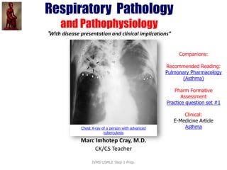
- 1. Respiratory Pathology and Pathophysiology “With disease presentation and clinical implications” Companions: Recommended Reading: Pulmonary Pharmacology (Asthma) Pharm Formative Assessment Practice question set #1 Clinical: e-Medicine Article Asthma Prepared and presented by Marc Imhotep Cray, M.D. BMS/CK Teacher Chest X-ray of a person with advanced tuberculosis
- 2. 2 Pulmonary Pathology Sections of the WebPath images are available for viewing by organ system. Each section consists of a series of images demonstrating gross and microscopic pathologic findings for a variety of disease processes. A short description accompanies each image. Internet Pathology Laboratory for Medical Education http://library.med.utah.edu/WebPath/webpath.html#MENU IVMS USMLE Step 1 Prep. Towards Understanding the Basic Medical Sciences Foundation of Clinical Medicine IVMS teaching philosophy is based on the integration of basic and clinical sciences...Learn More
- 3. Objectives • Understand presenting symptoms suggestive of pulmonary disease • Understand pathophysiology, pathology, disease presentation, implications, and treatment of major pulmonary diseases including, COPD, Restrictive lung disease, asthma, Cystic Fibrosis, and lung cancers IVMS USMLE Step 1 Prep. 3
- 4. Presenting Symptoms • Cough – Acute: viral or bacterial bronchitis, URI, or pneumonia – Chronic: asthma, postnasal drip, bronchitis, GERD • Hemoptysis – Ask the patient to estimate the amount of blood – Distinguish between epistaxis, hematemesis, and hemoptysis IVMS USMLE Step 1 Prep. 4
- 5. Presenting Symptoms (2) • Dyspnea – Timing, acuity of onset, exacerbating and alleviating factors, degree of functional impairment – Acute (p.e.) vs chronic (COPD) – Exertional or resting, episodic or continuous – Paroxysmal nocturnal dyspnea – Orthopnea IVMS USMLE Step 1 Prep. 5
- 6. Presenting Symptoms (3) • Chest pain – Many causes (cardiac, pulmonary, GI, musculoskeletal, etc) – Pulmonary causes: pleural disease, pulmonary vascular disease, musculoskeletal • lung parenchyma has no pain fibers – Pleuritic chest pain: sharp or stabbing pain on inspiration that can be positional IVMS USMLE Step 1 Prep. 6
- 7. Other important history • Cigarette smoking – Quantified as # of packs smoked/d X # of cumulative years (60pk year = 1 ppd X 60yrs) – Risk of lung disease is directly related to # of pack- years exposure and inversely to age at onset of smoking • Other environmental exposures, travel • Family history (CF, alpha-1 antitrypsin deficiency) IVMS USMLE Step 1 Prep. 7
- 8. Physical Exam • Watch the patient breath • RR, use of accessory muscles, paradoxical abdominal breathing, ability to speak in full sentences • Shape of the patient’s chest cavity – AP diameter suggestive of COPD • Auscultation –Rhonchi, rales, wheezing, rub • Clubbing IVMS USMLE Step 1 Prep. 8
- 9. Respiratory Infections • Upper respiratory infection – Most are viral: common cold, pharyngitis, etc • Lower respiratory infection – Frequently viral – Bronchitis: cough, wheezing, dyspnea – Pneumonia: cough, fever, rapid resp, dyspnea IVMS USMLE Step 1 Prep. 9
- 10. Pneumonias IVMS USMLE Step 1 Prep. 10 Compare the diffuse, patchy bilateral infiltrates of “atypical” interstitial pneumonia (A) with the localized, dense lesion of lobar pneumonia (B) Source: First Aid for the USMLE Step 1 2008, pg. 435
- 11. Pneumonias (2): Classification IVMS USMLE Step 1 Prep. 11 Source: First Aid for the USMLE Step 1 2008, pg. 468
- 12. Pneumonias (3): Gross and histopathology •Lung, bronchopneumonia, gross [XRAY] •Lung, bronchopneumonia, gross •Lung, bronchopneumonia, gross •Lung, lobar pneumonia, gross •Lung, empyema, gross •Lung, abscesses, gross •Lung, abscesses, gross •Lung, abscessing bronchopneumonia, gross •Lung, bronchopneumonia, low power microscopic •Lung, bronchopneumonina, high power microscopic •Lung, bronchopneumnia, high power microscopic •Lung, abscessing pneumonia, low power microscopic •Lung, abscessing pneumonia, high power microscopic •Lung, aspiration pneumonia, low power microscopic •Lung, aspiration pneumonia, high power microscopic •Lung, chronic abscess, gross IVMS USMLE Step 1 Prep. 12
- 13. Pulmonary Tuberculosis 13 http://emedicine.medscape.com/article/230802-overview Chandrasoma P, Taylor CR. Concise Pathology, 3rd ed. Stamford, CT: Appleton & Lange, 1998: 523 IVMS USMLE Step 1 Prep.
- 14. Pulmonary Tuberculosis (2) 14 http://upload.wikimedia.org/wikipedia/commons/2/2f/Tuberculosis_symptoms.svg IVMS USMLE Step 1 Prep.
- 15. Pulmonary Tuberculosis (3) • Caused by Mycobacterium tuberculosis • Major global problem; Seen in pts with HIV, other immunocompromised states, developing countries, etc • Contracted by inhalation Diagnosis suggested by: • chronic cough, • hemoptysis, • weight loss, • fevers, • night sweats M. tuberculosis bacterial colonies http://upload.wikimedia.org/wikipedia/co mmons/0/0a/TB_Culture.jpg Scanning electron micrograph of Mycobacterium tuberculosis IVMS USMLE Step 1 Prep. 15
- 16. Pulmonary TB (4) • Diagnosis: confirmed by CXR, PPD, sputum smears and culture Chest X-ray of a person with advanced tuberculosis http://upload.wikimedia.org/wikipedia/commons/9/9c/Tub erculosis-x-ray-1.jpg Mycobacterium tuberculosis Ziehl-Neelsen stain IVMS USMLE Step 1 Prep. 16 Treatment: 4 drug therapy See Tuberculosis Treatment & Management http://emedicine.medscape.com/article/230802- treatment
- 17. Obstructive Lung Disease General • Obstruction of air flow through airways • Major causes: – asthma – bronchiectasis, – emphysema and bronchitis (COPD) IVMS USMLE Step 1 Prep. 17 Obstructive lung disease (COPD) Obstruction of air flow resulting in air trapping in the lungs. Airways close prematurely at high lung volumes, resulting in ↑ RV and ↓ FVC. PFTs: ↓↓ FEV1, ↓ FVC→ ↓ FEV1/FVC ratio (hallmark), V/Q mismatch.
- 18. Pathophysiology • Air flow is decreased by: airway narrowing and/or loss of elastic recoil of the lung • Airway Narrowing – Airway inflammation • tobacco smoke, recurrent infection, immunologic dysfunction – Bronchoconstriction IVMS USMLE Step 1 Prep. 18
- 19. Pathophysiology (2) • Loss of elastic recoil – COPD: loss of airway tone and decreased tethering by surrounding lung – Asthma: bronchoconstriction and mucus plugging allowing airways to collapse at higher lung volumes and trap excessive air – Increased ventilation: increased airflow resistance may not allow lungs to completely empty during expiration IVMS USMLE Step 1 Prep. 19
- 20. Bronchitis vs Emphysema IVMS USMLE Step 1 Prep. 20 Source: First Aid for the USMLE Step 1 2008,pg 400
- 21. COPD Gross and histopathology •Lung, bronchiectasis, gross •Lung, bronchiectasis, gross •Lung, bronchiectasis and fibrous pleural adhesions, gross •Lung, bronchiectasis, low power microscopic •Lung, chronic bronchitis, medium power microscopic •Lungs, bullous emphysema, gross •Lung, centrilobular emphysema, gross •Lung, centrilobular emphysema, gross •Lung, emphysema, microscopic IVMS USMLE Step 1 Prep. 21
- 22. COPD • Slowly progressive, irreversible airway obstruction • Exacerbations of disease by bacterial/viral infections, heart failure, lack of medicine use, etc • Characterized by dyspnea, sputum production (with chronic bronchitis) IVMS USMLE Step 1 Prep. 22
- 23. COPD: types • Chronic bronchitis – persistent cough with sputum production for more than 3 months over last 3 years • Emphysema – abnormal enlargement of air spaces – The degree of obstruction in patients with COPD correlates more closely with severity of the emphysema IVMS USMLE Step 1 Prep. 23
- 24. COPD • Physical Exam – AP diameter, RR, clubbing • Laboratory data; – Pulmonary function test is sensitive way to make diagnosis in early stages – ABG: hypoxia, hypercarbia (advanced) – CXR: hyperinflation, flattened diaphragms, increased AP diameter, widened retrosternal air space (with emphysema) IVMS USMLE Step 1 Prep. 24
- 25. COPD: Clubbing IVMS USMLE Step 1 Prep. 25
- 26. COPD: Hyperinflation IVMS USMLE Step 1 Prep. 26
- 27. COPD flattened diaphragms, lucency IVMS USMLE Step 1 Prep. 27
- 28. COPD • Treatment – STOP smoking (if this is cause) – Treat exacerbations of bronchitis with antibiotics – Most meds have not been found to be helpful – Ipratropium bromide MDI (atrovent MDI) is helpful (anti-cholinergic) – Steroids not usually helpful unless inflammatory component IVMS USMLE Step 1 Prep. 28
- 29. Asthma Obstruction of the lumen of the bronchiole by mucoid exudate, goblet cell metaplasia, epithelial basement membrane thickening and severe inflammation of bronchiole in a patient with asthma. IVMS USMLE Step 1 Prep. 29
- 30. Asthma (2) • Chronic, inflammatory disorder of the airways • 3-5% of the population is affected • Imbalance between proinflammatory and inhibitory cytokines • Episodic airway narrowing, increased airway reactivity, and reversibility IVMS USMLE Step 1 Prep. 30 Gross and histopathology •Lungs, hyperinflation with status asthmaticus, gross •Lung, cross section, hyperinflation with status asthmaticus, gross •Bronchial mucus plug with asthma, gross •Bronchial asthma, low power microscopic •Bronchial asthma, high power microscopic
- 31. Asthma (3) • Trigger: extrinsic allergens, intrinsic factors, or no identifiable cause • Types: extrinsic, intrinsic, exercise induced, asa sensitive, occupational, ABPA • Precipitants of asthma: postnasal drip, GERD, cold exposure, gases/fumes, emotional stress, hormones, resp infections IVMS USMLE Step 1 Prep. 31
- 32. Asthma (4) • Diagnosis (one or combination): – wheeze, chronic episodic dyspnea, and chronic cough – Sputum production, chest pain or tightness • Testing: – History, CXR (to rule out other causes), pulmonary function testing (with or without challenge) IVMS USMLE Step 1 Prep. 32
- 33. Asthma (5) • Treatment – Education (removal of offending agents) – Peak flow meters – Inhaled corticosteroids (ex fluticasone) – Long and short acting bronchodilators • ex salmeterol, albuterol – Leukotriene inhibitors (ex. montelukast) – Theophylline (limited use) IVMS USMLE Step 1 Prep. 33
- 34. Restrictive lung disease IVMS USMLE Step 1 Prep. 34 Restricted lung expansion causes↓ lung volumes (↓ FVC and TLC). PFTs–– FEV1/FVC ratio > 80%. Types: 1. Poor breathing mechanics (extrapulmonary, peripheral hypoventilation): a. Poor muscular effort––polio, myasthenia gravis b. Poor structural apparatus––scoliosis, morbid obesity 2. Interstitial lung diseases (pulmonary, lowered diffusing capacity): a. Adult respiratory distress syndrome (ARDS) b. Neonatal respiratory distress syndrome (hyaline membrane disease) c. Pneumoconioses (coal miner’s silicosis, asbestosis) d. Sarcoidosis e. Idiopathic pulmonary fibrosis (repeated cycles of lung injury and wound healing with ↑ collagen) f. Goodpasture’s syndrome g. Wegener’s granulomatosis h. Eosinophilic granuloma (histiocytosis X) i. Drug toxicity (bleomycin, busulfan, amiodarone)
- 35. Cystic Fibrosis IVMS USMLE Step 1 Prep. 35 Cystic fibrosis
- 36. Cystic Fibrosis(2) • Autosomal recessive genetic disorder • Affects pulmonary, GI and GU systems • Most common lethal genetic disorder – 1/25 carrier frequency – 1/3200 live births affected • Defect: failure to produce normal chloride channel leading to increased sodium reabsorption 36 A breathing treatment for cystic fibrosis, using a mask nebulizer and a ThAIRapy Vest
- 37. Cystic Fibrosis (3) • Abnormal chloride channel leads to thick and viscous secretions in the resp, hepatobiliary, gi, and reproductive tracts • Resp tract: persistent inflammation and infection causes bronchial wall destruction; mucus plugging of small airways causing parenchymal destruction – colonization by S. aureus, H. influenza, P. aeruginosa 37 http://en.wikipedia.org/wiki/File:Cystic_Fibrosis_Respiratory_Infections_by_Age.svg
- 38. Cystic Fibrosis (4) • Testing – Chloride sweat test – Genetic testing • Median survival – 14 years in 1969 to >30 yrs since 1995 IVMS USMLE Step 1 Prep. 38
- 39. Cystic Fibrosis (5) • Pathology – Pulmonary: cough, sputum production, clubbing – Upper Resp tract: nasal polyps, sinusitis – GI: exocrine pancreatic dysfunction, diabetes, cirrhosis, salivary gland inflammation – GU: azoospermia, decreased fertility rate in women, nephrolithiasis IVMS USMLE Step 1 Prep. 39
- 40. Cystic Fibrosis(6) • Treatment; – Aggressive airway hygiene – Nutritional support including pancreatic enzyme replacement – Antibiotics – Bronchodilators IVMS USMLE Step 1 Prep. 40
- 41. Lung Cancer IVMS USMLE Step 1 Prep. 41 Squamous cell carcinoma in the right lower lobe Source: First Aid for the USMLE Step 1 2008, pg. 434 Lung cancer is a leading cause of cancer death. Presentation: cough, hemoptysis, bronchial obstruction, wheezing, pneumonic “coin” lesion on x-ray film
- 42. Lung Cancer (2) • Risk Factors – Leading cause of death – Cigarette smoking is responsible for >90% of lung cancers – Risk increases with dose and length of exposure to cigarette smoking – Heavy occupational exposure to asbestos is second most important cause IVMS USMLE Step 1 Prep. 42
- 43. Lung Cancer: Types • Bronchial carcinoid tumors • Small cell cancer • Non-small cell cancer – Squamous cell cancer – Adenocarcinoma – Large cell – Anaplastic carcinoma • Metastasis: breast, liver, renal, colon IVMS USMLE Step 1 Prep. 43
- 44. Lung Cancer: Types (2) IVMS USMLE Step 1 Prep. 44 Source: First Aid for the USMLE Step 1 2008, pg. 443
- 45. Lung Cancer Gross and histopathology •Lung, squamous cell carcinoma, gross [CT] •Lung, squamous cell carcinoma, gross [XRAY] •Lung, squamous cell carcinoma, medium power microscopic •Lung, squamous cell carcinoma, high power microscopic •Lung, peripheral adenocarcinoma, gross •Lung, bronchioloalveolar carcinoma, gross •Lung, bronchioloalveolar carcinoma, microscopic •Lung, oat cell carcinoma, gross •Lung, oat cell carcinoma, high power microscopic •Lung, hamartoma, gross •Lung, hamartoma, microscopic •Lung, metastatic carcinoma, gross [XRAY] •Lung, metastatic carcinoma, microscopic •Pleura, metastatic carcinoma, microscopic •Lung, mesothelioma, gross •Lung, mesothelioma, high power microscopic IVMS USMLE Step 1 Prep. 45
- 46. Lung Cancer: Clinical Presentation • Symptoms can be quite non-specific • Symptoms may relate to location and size of tumor – Cough, hemoptysis, post-obstructive pneumonia, chest pain, wheezing, hoarseness – bone metastases: swelling, pain – hepatic metastases: jaundice, hepatomegaly – weight loss, anorexia IVMS USMLE Step 1 Prep. 46
- 47. Lung Cancer: Evaluation • History and physical examination • CXR/CT scan • No lab is helpful • Bronchoscopy • VATS IVMS USMLE Step 1 Prep. 47
- 48. Lung Cancer: Treatment • Options depend on tumor type, size, stage of disease, and performance status of the pt • Surgical removal with Stage I, II, IIIA non-small cell cancer (if operable) • Chemotherapy with radiation for limited stage disease in small cell cancer – frequent metastases to the brain IVMS USMLE Step 1 Prep. 48
- 49. Lung Cancer: Survival • 15-25% survival 5 years after the diagnosis • Considerable debate about screening for lung cancer – recent discussion on chest C.T. as screening tool – CXR is not a sensitive way to screen for cancer IVMS USMLE Step 1 Prep. 49
- 50. e-Medicine Articles • Obstructive Airway Diseases • Alpha1-Antitrypsin Deficiency • Asthma • Bronchiectasis • Bronchiolitis • Bronchitis • Chronic Bronchitis • Chronic Obstructive Pulmonary Disease • Emphysema • Status Asthmaticus 50IVMS USMLE Step 1 Prep.