Basic Exercise physiology
•Download as PPTX, PDF•
92 likes•24,980 views

An overlook at the very basics of exercise physiology, for the novice student.

Recommended
More Related Content
What's hot
What's hot (20)
Viewers also liked
Similar to Basic Exercise physiology
Similar to Basic Exercise physiology (20)
Recently uploaded
Recently uploaded (20)
Basic Exercise physiology
- 2. Purpose: • Understand the basis for the various principles of training as well as to evaluate and design physical fitness programmes. • Education on how the body adapts to short and long term exercise, enabling different and specific programme design.
- 3. Outcomes: 1) Describe the following systems: - Cardiovascular system - Respiratory system - Neuromuscular system - Endocrine system 2) Describe the above systems responses to exercise. 3) Describe the acute/short term and chronic/long term response to exercise. 4) Describe the three energy systems (Phosphogen, anaerobic and aerobic systems). 5) Identify the energy systems that underpin specific exercises. 6) Consider the contribution of energy systems to exercise and training
- 4. Overview of body systems: • Fig. 3.2 – Different systems work side by side to fulfil different functions. • We will focus on the following: - Cardiovascular system - Respiratory system - Neuromuscular system - Endocrine system - Energy Systems • Table 3.1 – Functions of the different body systems.
- 5. Overview of body systems (cont.): • The description and explanation of the body’s response to exercise and the adaptation to exercise training, to maximize human performance. • Eg: why would a 200m athlete not need to do lots of cardio training? • In this chapter, the production of energy, and the application to four broad categories – endurance, strength conditioning, speed and power and also flexibility. • Also, combining what was previously learned, anatomy – skeletal and muscular systems, with the other body systems previously mentioned. • Pg. 81 – words and terms used throughout the chapter.
- 8. Components of the heart • Four chambers – 2 atria – 2 ventricles (left thicker than right) • Major veins – Superior vena cava – Inferior vena cava – Pulmonary veins • Major arteries – Aorta – Pulmonary trunk • Valves permit the passage of blood in one direction. • Atrioventricular Tricuspid Bicuspid or mitral • Semilunar Aortic Pulmonary
- 9. Blood Flow Through Heart
- 10. Cardiac Output • The amount of blood that is pumped by the heart per minute. • Cardiac output (Q) provides most significant indicator of circulatory system's functional capacity to meet demands for exercise. • Due to the left ventricular muscle being thicker than the right, the force of ejection is greater as well. • This is needed as the left ventricle pumps blood to the entire body, the right only pumps to the nearby lungs. • Cardiac output (Q) is a product of heart rate (bpm) and stroke volume (mL – blood pumped by left ventricle every heart beat.)
- 11. Resting Q: Q = HR x SV = 70bpm x 70mL = 4.9 L/min
- 12. Exercise Q •Blood flow from heart increases in direct proportion to exercise intensity •From rest to steady-rate exercise, Q increases rapidly, followed by gradual increase until it plateau’s.
- 13. Ejection fraction • The percentage of blood ejected out of the ventricles during each contraction. • At rest, the ejection fraction is only about 50%. During exercise, it can increase to 100%. • The ejection fraction at rest is low due to Q sufficiently supplying all the cell with oxygen. • As the demand for oxygen increases during exercise, the ejection fraction increases to supply the demand of oxygen.
- 14. Components of the CVS System: Blood Vessels: (Vascular system) • Transports blood throughout the body, to and from the heart, via systemic circulation. • Transports blood to and from pulmonary circulation Different names for vessels: • Arteries (arterioles) – Carries blood away from heart. • Capillaries – gas exchange between tissue and blood. • Veins (Venules) – Carries blood toward the heart.
- 16. Effects of impaired blood supply • The myocardium depends on adequate oxygen supply as it has limited anaerobic energy- generating capacity. • Reduced coronary blood flow usually produces chest pains – angina pectoris. • A thrombus (blood clot) lodged in coronary vessels impairs normal heart function – myocardial infarction. • MI may be mild, a complete vessel block may causes myocardium death.
- 17. Conduction System: • The heart is able to generate its own electrical impulses and control the route the impulses take via a specialized conduction pathway. The five elements of the pathway: - SA node – Right atrium, pacemaker - AV node – Right atrium - Bundle of His - Septum - The left and right bundle branches - Septum - Purkinje fibres - Myocardium
- 18. Bundle of His Left bundle branch Left division Right division
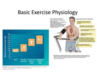
- 19. Blood pressure: • The pressure of the blood against the walls of the arteries during the relaxation (diastolic) and contraction (systolic) of the heart in one cardiac cycle. • The formula for BP is: BP = Q x TPR • Blood pressure is written as systolic (peak value) over diastolic (minimum value); eg – 120/80 mmHg (millimetres mercury). • Total peripheral resistance (TPR) is influenced by the calibre or diameter of the arterioles. Blood pressure increases more in static exercise (weight lifting) due to contracting muscle pushing on blood vessels and increasing TPR.
- 20. Oxygen extraction: • Oxygen extraction occurs at the capillaries, where they are in contact with skeletal muscle fibres. • The amount of oxygen extraction is dependent on the muscle fibre type; slow twitch oxidative fibres will extract the most oxygen. • Blood in the veins and venules (venous blood) has a lower oxygen concentration than blood in the arteries and arterioles (arterial blood). • Not all oxygen is removed from the blood, under any circumstance. • This shows that oxygen supply is not a limiting factor in exercise.
- 21. Oxygen extraction cont.: • Maximal oxygen uptake (VO2max) is the maximum amount of oxygen utilised by the working muscles. • This is the point maximal consumption is measured. At rest 3.5 ml/kg/min of oxygen is needed to sustain life. • Various VO2max protocols exist and are conducted under laboratory conditions – Bruce protocol. • The highest VO2 max recorded was 93 ml/kg/min.
- 23. 23
- 24. The respiratory system: • The lungs are the organs that perform gaseous exchange – removal of CO2 and the re-saturation of O2. • The lungs contain millions of alveoli • As the capillaries are in with skeletal muscle, so to are alveoli in close contact with capillaries in the lungs. • At this level, gasses diffuse from an area of high pressure to low pressure, down their concentration gradients. • CO2 from blood to alveoli and O2 from alveoli to blood.
- 26. Ventilation: • O2 from the atmosphere travels down the airways into the lungs, and CO2 in the lungs is breathed out into the atmosphere. • At rest 250ml of O2 enters the blood and 200ml of CO2 is expired every minute; during exercise it increases as much as 25 times. • Passage of airflow: Nose/mouth> Trachea> Bronchi> Bronchioles> alveoli (then reverse)
- 29. Mechanism of breathing: • The diaphragm and intercostal muscles, two mechanisms control the volume of the chest cavity to increase and decrease, causing air to flow in and our of the lungs. • Inspiration – Increase in the volume of chest cavity, diaphragm moves down and ribs move up, the pressure decreases and is lower than atmospheric pressure, air moves from atmosphere into the lungs. • Expiration – Decrease in the volume of the chest cavity, ribs move down and diaphragm moves up, higher pressure in lungs than atmosphere, air moves from lungs into the atmosphere.
- 30. 30
- 32. Overview: • The neuromuscular system is a combination of the nervous system and muscles working together to allow movement. • Strength, speed and power and flexibility training are all significantly affected by the neuromuscular system. • Fig. 3.9 – neuroderms – nerve pairs per body region.
- 33. Nerve anatomy: • Fig 3.8 – a motor neuron • A neuron is the basic unit of the nervous system. Two types occur – motor and sensory. • An axon is the process that extends from a neuron to connect with another cell. • Dendrites are little tendrils that receive conduction towards the cell body. • Movement can either be voluntary or involuntary.
- 34. Involuntary movement: • The sensory neuron has a sensory ending, in skin, muscle etc. • These endings respond to stimuli and send the information as an electrical impulse. • The impulse travels via the sensory neuron to the central nervous system via the spinal chord.
- 35. Voluntary movement: • The motor neuron receives information from the central nervous system. • The information is in the form of an electrical impulse and it travels down the motor neuron to the muscle fibre. • The impulse travels from the motor unit to the muscle at the neuromuscular junction. • This results in muscle contraction or relaxation.
- 36. Motor Units: • A motor unit is the motor neuron all and the muscle fibres it innervates. • Fast twitch fibres have large motor units and innervates many muscle fibres. • They therefore control gross motor control and produce a great contractile force. • Slow twitch fibres have small motor units and innervate few muscle fibres. • They therefore control fine motor movement and generate a small contractile force.
- 37. Muscle innervation • A muscle will respond to stimulus and if the stimulus is strong enough, the muscle will contract. • It is not possible for some fibres of a motor neuron to contract and others not, its all or none. • Weak muscle contractions are caused by stimuli of small motor units and strong muscle contractions are caused by stimulation of big motor units.
- 38. Structure of skeletal muscle • More than 660 skeletal muscles in the body Levels of Organization • Perimysium covers each bundle (fasiculus) ± 150 fibres • Epimysium covers the entire muscle and tapers to form the tendons that connect muscle to the periosteum of bone • Endomysium covers each fibre • The origin of muscle refers to the relatively stable end, proximal or fixed end of the lever or the end nearest the body’s midline • The insertion end is the distal attachment to the moving bone.
- 40. Muscle fibre type Fast-Twitch Subdivisions Type IIa fibres exhibit fast shortening speed and well-developed energy transfer capacity from aerobic and anaerobic sources, i.e., fast-oxidative-glycolytic (FOG) Type IIb fibres has the greatest anaerobic potential and fastest shortening velocity, i.e., fast-glycolytic (FG) fibres Type IIc fibre is normally rare and undifferentiated , and may contribute to reinnervation and motor unit transformation. Slow twitch fibres: • Low myosin ATPase activity • Slow calcium handling ability and shortening speed • Less well-developed glycolitic capacity than fast twitch fibres • Large and numerous mitochondria Slow twitch fibres generate energy for ATP resynthesis predominantly through the aerobic system of energy transfer.
- 44. Fibre type differences among athletic groups Elite endurance athletes have predominantly ST fibres in the major muscles activated in their specific sport, and vice versa for sprint athletes. Endurance athletes tend toward enlargement of ST fibres. Weightlifters and power athletes show enlargement in both ST and FT fibres, particularly FT fibres, i.e., enlargement of the contractile apparatus, specifically actin and myosin filaments.
- 46. Muscular contraction: • Fig 3.11 – Myosin (Thick) filaments form cross bridges with actin (thin) filaments via tiny projections (myosin heads). • These myosin heads extend from the myosin filament to the actin filament. The myosin head binding site on the actin is distant, during relaxation. • The binding sites are blocked and can only be unblocked when calcium is released by an incoming nerve impulse. • This unblocks the myosin binding sites and allows the myosin heads to extend and form cross bridges, pulling the actin over myosin filaments.
- 48. 48
- 49. 49
- 50. Types of muscular contraction: • Isotonic – dynamic muscle contraction; either concentric (shortening – doing a bicep curl) or eccentric (lengthening – lowering after a bicep curl). • Isometric – tension in the muscle with no movement and change in the length of a muscle; eg. Holding a book in your hand. • Isokinetic – maximal tension of the muscle while moving through the movement range. This can only be obtained on specialized machines - Biodex®.
- 53. Overview: • A series of glands that produce and secrete hormones with regulate body growth, metabolism and sexual development and function. • Hormones are chemical messengers that transfer information from one set of sells to another. This has an effect on functions of that different body part. • E.g. exercise will cause the adrenal cortex to release adrenalin. Adrenalin is a hormone that increases heart rate, breathing rate and prepares the body for the exercise session. • The endocrine system is regulated by a feedback mechanism.
- 54. Glands and functions: • Table 3.6 • The role of hormones is also to maintain homeostasis. • Homeostasis is the balance of biological activities in the body, managed by the hypothalamus. • The pituitary gland is the area that triggers the release or inhibition of hormones.
- 55. Hormones and exercise: • The main hormones that are active during exercise include: - adrenalin – readies body for exercise. - cortisol – protects the body from the stress exercise induces. - glucagon – triggers release of glucose from cells and into the blood. • Others are also active but do not impact as hugely as the list above. • The long term effects of training allows for these hormones to be secreted in lower quantities. • This is due to the body adapting to exercise and improves the efficiency of the body.
- 56. ENERGY SYSTEMS
- 57. Overview: • Processes required to provide the cells of the body with the energy required to sustain life and perform work during activity. • Many of these processes involve different types of cells. • A generalized cell consists of a nucleus, cytoplasm and organelles, all held together by the cell membrane (cytoskeleton), floating in fluid called cytoplasm. • Fig 3.13 – different organelles exist in the cells and all have different functions. For our purposes, take note of the mitochondria (aerobic metabolism).
- 58. Fuels for energy: • 3 sources of energy (Macronutrients) exist and are digested when we eat them, namely: - Carbohydrates - Lipids (Fats) - Proteins • Smaller components of these macronutrients are called micronutrients. • All of these has a different energy value, kcal. • 1 kcal = 1000 calories; amount of heat needed to heat 1 kg(1L) of water by 1 degree Celsius. - Glucose – 4.2 kcal - Fatty acids – 9.4 kcal - Amino acid – 4 kcal
- 59. Lipids: • These big molecules requires lots of oxygen to be mobilized and used as an energy source, more oxygen than carbs or proteins. • The intensity of exercise will determine which energy source is used. • Fats are stored under the skin (subcutaneously), around the vital organs and in muscle as intramuscular triglyceride. • Fat around organs are essential as they protect against mechanical shock • Fat stored subcutaneously is adipose fat and non- essential and they are mobilised for energy.
- 61. • Fat is utilised in its simplest form, free fatty acids, and usually fuel exercise of moderate intensity over a prolonged period of exercise. E.g. – a marathon. • Fig 3.14 – FFA metabolism and storage
- 62. Carbohydrates: • CHO – stored in the liver and skeletal muscle in the form of Glycogen (many glucose molecules). • for CHO to be utilized for energy, it needs to be in its simplest form of glucose in the blood. • Stored glycogen therefore needs to be released from storage before it can be used, this costs 1 ATP. • When blood glucose levels are higher than needed, it gets stored in the form of glycogen. • Glucose is the preferred source of energy to fuel high intensity exercise.
- 64. Proteins: • Not a preferred source of energy for exercise. • Main function is to build, maintain and restore cells. • Complete proteins sources: eggs, milk, meat, fish, and poultry (eggs have the highest quality rating - 100) • Not only do body builders need protein, but endurance athletes as well. • Essential vs non-essential proteins. The Vegetarian Approach • Soy-protein isolates matches in quality some animal proteins. • Nutritional diversity is the key to successful nutrition (supplementation is an option)
- 66. Intracellular energy systems: • Understanding these systems, one needs to understand the fuel sources, chemical structure, where and how they are stored and the amount of energy each one produces. • Fig 3.6 – the metabolic mill – different fuel sources via different pathways, all to produce ATP (energy). • Three energy systems exist – Phosphogen system, Glycolysis and Aerobic metabolism – Table 3.7
- 67. • O2 independent systems (anaerobic) are responsible for immediate energy and uses fast twitch skeletal muscle fibres. • O2 dependent systems (aerobic) are responsible for intermediate and prolonged energy production and predominantly uses slow twitch skeletal muscle fibres. • High energy bonds exist between phosphate groups, when broken down, energy is released for cellular function (muscle contraction). • This is illustrated as: ATP ADP + Pi • ATP stores are limited and can only produce energy for a few seconds, it has to be continually resynthesized.
- 69. 69
- 70. The phosphogen system • Creatine Phosphate (CP) is a high energy compound in skeletal muscle. • The bond between creatine and phosphate contains the energy and when broken, the energy is released for resynthesis of ATP from ADP and Pi. • Small amounts of ATP and CP are stored in muscle and therefore, there is only enough energy stored for about 8 seconds of maximal muscle contraction. • This energy is instantaneous and important for the onset of exercise, for a high intensity and short duration – 100m sprint. • Once this energy is depleted, the body relies on glucose for energy (glycolysis).
- 72. Glycolysis: • Once this energy of the phosphogen system is depleted, the body relies on glucose for energy. • Glycolysis generates anaerobic energy from glucose breakdown to form pyruvate (pyruvic acid) – Fig 3.16. • This process occurs in the cytoplasm and does not need oxygen to produce energy. • Glycolysis only partially breaks down glucose and there is still energy available in the glucose which can be metabolized aerobically. • Two ATP molecules are produced via the pathway of glycolysis along with two lactate molecules.
- 73. • When glycogen is broken down, three ATP molecules are produced, along with two lactate molecules, via glycolysis. • A by-product of lactate formation is H+ ions. Constant exercise intensity will cause a build up of H+ ions in the muscle and blood. This lowers the pH of the blood which would decrease blood pH and eventually causes muscle fatigue, as the enzymes do not function optimally at a lowered pH – Fig 3.18. • Glycolysis will supply energy for approximately 35 – 60sec, depending on fitness. • This pathway will provide a rapid supply of ATP for energy for an intense, short burst of activity. • It also acts as an energy reserve for the middle and long distance athlete to provide a ‘kick’ in the sprint finish, or provide a footballer with instant acceleration to beat an opponent. The end product of glycolysis is pyruvate, which if not used, forms lactic acid, otherwise enters the Krebs cycle for aerobic metabolism.
- 76. Oxygen dependent system (aerobic): • These processes will occur inside the mitochondria – the site for ATP production, the powerhouses of cells. • The greater the amount of mitochondria, the more ATP can be produced – training effect. • The Krebs cycle and electron transport chain are 2 pathways through which energy is produced in aerobic metabolism. • The end product of glycolysis is pyruvate. • Pyruvate will build up and form lactate until the exercise time goes past 60sec, up to 40mins. • Once past 60sec, pyruvate will enter the Krebs cycle as Acetyl-CoA, for aerobic metabolism – Fig. 3.16 • The Krebs cycle alone has 10 conversions, yielding 1 ATP only
- 77. • The complete breakdown of glucose will occur via aerobic metabolism, yielding 36 ATP molecules. • H2O (used in cell) and CO2 (exhaled) are the by- products of these reactions. • When exercise exceeds 40min, lipolysis (the breakdown of adipose tissue/fat) will predominately provide ATP. • One molecule of fat can yield up to 129 ATP, fat has a high caloric density. • Unlike anaerobic metabolism (O2 independent), aerobic metabolism produce large amounts of ATP without fatiguing by-products (blood lactate and H+ ions). • Table 3.7 – Summary.
- 78. The Crossover Concept: • Definition – the exercise intensity at which lipid and CHO utilization for energy is equal. High intensity exercise = CHO Low intensity exercise = Fats • This concept is the changing in fuel sources due to a change in muscle fibre types. • In the trained, crossover = 60 – 70 % VO2max; due decrease in glycogen stores and an increase in adrenaline/noradrenaline concentrations. • These hormones increase lipolysis and therefore changes the fuel source to fuel exercise. • Longer the duration, the more reliant on lipids. • Crossover occurs after 3-5hrs of submaximal work.
- 79. Relationship between O2 independent and O2 dependent systems: • The intensity and duration will main determinants in which energy system is used. - Phosphogen = 8 – 10 sec - Glycolysis = 30 – 60 sec - Aerobic metab. = 60sec – hours • Fig 3.19 – the systems are not mutually exclusive, not like an on/off switch, but rather a dimmer, working at 100% when needed. • These systems can be trained and can become more efficient and the times above will change, depending on training. • When compared, both systems have advantages and disadvantages pg. 102 & 103.
- 80. BODY RESPONSE TO VARIOUS TRAINING
- 81. Aerobic training: • Cardiorespiratory and cardiovascular systems • Short term response – cardiorespiratory system increases HR by the release of noradrenaline (sympathetic) – causes increase in the force of contraction of the heart increased stroke volume increased ejection fraction. • since Q=HRxSV, cardiac output increases from 4.2 L/min to 25 L/min. • Blood vessels will dilate and reduce TPR, increasing blood flow to working muscles. • The body will redirect blood away from the viscera (intestine, pancreas, etc.) and to the working muscles for oxygen delivery, supplying the demand. • Blood pressure will increase due to the cardiac output increasing substantially. • Body core temperature will also increase, resulting in further vasoconstriction and sphlanchic circulation, increasing blood flow to the skin for loss of heat via radiation.
- 82. Aerobic training: • Long term adaptation 1. Heart rate decreases – decrease in sympathetic tone. 2. An increase in capillarisation – more capillaries for gaseous exchange at the muscle. 3. An increase in muscle mitochondria – more mitochondria = more ATP produced. 4. An increase in VO2max – increased O2 extraction ability. 5. An increase in plasma volume – increases Q during exercise. • Table 3.9 + 3.10
- 83. Strength training: • The neuromuscular system will account for the improvements initially. • Short term responses to strength training – pg. 106 – the emphasis being on fast twitch fibres (type IIa). • Short term responses to flexibility training – pg. 106. • The stretch reflex – prevents rapid overstretching of muscle – fig 3.20 – muscle spindles (sensory organs inside muscle fibres) and Golgi tendon organs (sensory organs inside tendons).
- 84. Strength training: • Long term adaptations – hypertrophy – increase in muscle size by addition of myofilaments (actin and myosin). • Hypertrophy of type II muscle fibres are due to an increase in protein formation, while type I fibres are due to a decrease in protein breakdown. • Hypertrophy of type II > hypertrophy of type I. • Hyperplasia, splitting of muscle fibres, occur in animals but are yet to be proven to occur in humans. • Atrophy, decrease in muscle size, is due to lack of strength training, decreased neural and mechanical force on muscle fibres. • Main long term adaptations = pg. 107 & table 3.12 • Table 3.11 – long term adaptations of flexibility training.
- 85. PHYSICAL AND ENVIRONMENTAL FACTORS AFFECTING PERFORMANCE.
- 86. Homeostasis and feedback loop: • Homeostasis – maintenance of the internal environment within a narrow range of temperature, pH and oxygen consumption. • Stress, like exercise, induces changes in the body until that stress is removed/reduced. • Regular exercise will allow the body to adapt and function at a higher physiological level – Fig. 3.21. • Feedback loops, negative (specific to maintain homeostasis) and positive, help maintain homeostasis – Fig 3.22. • If the stressor is applied too intensely, the body cannot adapt and fatigue/exhaustion will occur. • The body needs a controlled stressor to avoid this, which exercise is.
- 87. common stressors Environment influences (Security, working conditions) Changes Emotional and relationships Personality type and assertiveness Values, beliefs and phobias Role conflict incl. gender Time management Earning capacity Work and job clarity Utilization (time)
- 88. www.exerciseacademy.com Stressor Stage 1: Acute • HR & BP • Shallow breathing • Increased sweat • Adrenal secretion No ThreatThreat Intervention and resilience • Relaxation • Life skills • Exercise • Diet • Relationship • Humour • Creativity • Self complexity Stage 2: Chronic • Altered mood • Insomnia • Tension & pain Diffused Stage 3: • Heart disease • CFS Stress response
- 89. A controlled stressor: • Exercise is done to improve health and fitness; fitness defined – pg. 111. • Exercise is a controlled stressor with specific responses that occur in muscle, blood vessels and heart. • The purpose of training is to stress the body, over time, to induce long term adaptation. • If stress is too little, no adaptation will occur. • If stress is too much, over-training/fatigue will occur and lower performance.
- 90. General Adaptation Syndrome (GAS): • The chain of events that occur in response to any stressful exposure, including the stress of exercise. • Hans Seyle – defined ‘adaptation energy’ – an intrinsic property which, when at maximum, coincides with peak performance and when at minimum, coincides with death – Fig. 3.23 • Three stages of GAS: – Alarm stage = initial response to stressor. – Stage of adaptation = improved capacity to deal with stressor. – Stage of exhaustion = body adapts for a long period.
- 91. Acute VS Chronic adaptations: • Acute = temporary nature/short term – E.g. elevated HR, elevated breathing rate. – Revert to pre-exercise state when exercise drops. – Values dependent on fitness of individual. • Chronic = more permanent/long term – E.g. mitochondrial adaptations, fibre changes. – Persist for a sustained period. – Take longer to wear off.
- 93. Exercising in heat: • Metabolic heat during exercise is eliminated via the skin surface, dissipated to the cooler outside environment, via venous blood vessel dilation = vasodilation. • Sweating dissipates this heat as the evaporation of sweat causes cooling, preventing the body temperature to rise 2-3 degrees. • Excessive external heat with increase in body temperature may lead to heat stroke. • Extensive vasodilation may reduce venous return and reduce stroke volume, increase HR to increase Q (double demand on circulation).
- 94. Exercising in cold: • Key concern: the body is wet and sweating; vasodilation is continuing; chilling can occur very rapidly = wear warm clothing immediately after exercise, retains body heat. Exercise performance is affected - blood will be sent to the central core to maintain body temperature. - reduces blood flow to the periphery and working muscles. - reduced blood flow will lead to reduced oxygen delivery and waste removal, reducing performance.
- 95. Exercising in humidity: • Heat combined with humidity can be extremely dangerous. - Sweat cannot evaporate readily - large amount of water vapour in atmosphere - difficult for the body to cool and evaporate sweat - exercise > 30min = reduced intensity
- 96. Exercising at a high altitude: • Moderate to high altitude cause a reduction in the partial pressure of oxygen. - Less air pressure = less pressure to drive O2 into blood, in the lungs - O2 carrying capacity is reduced = reduced O2 carrying capacity to working muscle - can result in respiratory distress & recovery from exercise delayed - headaches may occur = altitude sickness - adaptation to altitude is essential
- 97. Training considerations: • Training threshold – minimum amount of exercise required to produce significant improvements in any physical parameter. • Progression – creating an overload to provide continuing improvements. • Individuality – genetic factors, fibre composition, etc. • Age – some things may be suitable for one age group but not another. • Gender – some anatomical/physiological differences; women respond to exercise the same way men do. • Shape – body composition, structure, influences performance. • Health & medical status – appropriate programming is important for what the client presents with.