Diabetic retinopathy
•Télécharger en tant que PPT, PDF•
32 j'aime•3,541 vues

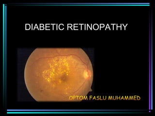
Diabetic retinopathy optom faslu muhammed

Recommandé
Contenu connexe
Tendances
Tendances (20)
En vedette
En vedette (20)
Similaire à Diabetic retinopathy
Similaire à Diabetic retinopathy (20)
Plus de OPTOM FASLU MUHAMMED
Plus de OPTOM FASLU MUHAMMED (20)
Dernier
Dernier (20)
Diabetic retinopathy
- 2. • Retinal changes seen in patients with DM
- 3. pathogenesis • Duration of diabetes • Sex • Poor metabolic control • Heredity • Pregnancy • Hypertension
- 4. Classification of DR Non-proliferative DR (NPDR) • Mild • Moderate • Severe • Very severe II. Proliferative DR (PDR) III. Clinically significant macular oedema (CSME) - May exist by itself or along with NPDR and PDR
- 5. Mild NPDR • At least one microaneurysm - earliest clinically detectable lesion • Retinal hemorrhages • Hard or soft exudates
- 6. Moderate NPDR • Microaneurysms and/or dot and blot hemorrhages in at least 1 quadrant • Soft exudates (Cotton wool spots) • Venous beading or IRMA (intraretinal microvascular abnormalities)
- 7. Mild and Moderate Non- proliferative DR was previously known as Background DR
- 8. Severe NPDR Any one of the following 3 features is present • Microaneurysms and intraretinal hemorrhages in all 4 quadrants • Venous beading in 2 or more quadrants • Moderate IRMA in at least 1 quadrant
- 9. Very severe NPDR • Four quadrants of severe microaneurysms / intra retinal hemorrhages • Two quadrants of venous beading • INMA in one quadrant
- 10. Severe and Very severe Non-proliferative DR was known as the Pre-proliferative DR
- 11. Clinically significant Macular Oedema • Retinal oedema close to fovea • Hard exudates close to fovea • Presents with dimness of vision • By itself or along with NPDR or PDR
- 12. CSME – Hard exudates close to fovea and associated retinal thickening
- 13. Proliferative DR (PDR) Characterized by Proliferation of new vessels from retinal veins • New vessels on the optic disc • New vessels elsewhere on the retina
- 14. Proliferative DR
- 15. COMPLICATIONS OF DIABETIC RETINOPATHY • Vitreous hemorrhage • Tractional retinal detachment • Rubeosis Iridis • Glaucoma • Blindness
- 18. Rubeosis Iridis
- 19. PREVENTION OF COMPLICATIONS • By early institution of appropriate treatment • This requires early detection of DR in its asymptomatic treatable condition • By routine fundus examination of all Diabetics (cost effective screening) • And appropriate referral to ophthalmologist
- 20. Mild and Moderate NPDR No specific treatment for retinopathy - Good metabolic control to delay progression - Control of associated Hypertension, Anemia and Renal failure
- 21. Severe and very severe NPDR • Close follow up by Ophthalmologist
- 22. Clinically significant macular oedema - Laser photocoagulation to minimise risk of visual loss
- 23. Proliferative DR • Retinal laser photocoagulation as per the judgment of ophthalmologist (in high risk eyes) • It converts hypoxic retina (which produces ANGIOGENIC factors) into anoxic retina (which can’t)
- 24. Screening protocol for Diabetic retinopathy 1. Screening once in a 1 year • Diabetics with normal fundus • Mild NPDR 2. Screening once in 6 months • Moderate NPDR
- 25. Referral to Ophthalmologist • Visual Symptoms – Diminished visual acuity – Seeing floaters – Painful eye • Fundus findings - Macular oedema/hard exudates close to fovea - Proliferative DR - Vitreous hemorrhage - Moderate to severe and very severe NPDR - Retinal detachment - Cataract obscuring fundus view
- 26. Simulation of defective vision as experienced by a Diabetic whose vision has been affected by Diabetic retinopathy
- 27. DIRECT OPHTHALMOSCOPY • Examination of the fundus of the eye • To screen for Diabetic Retinopathy • After dilatation of both eyes with 0.5% tropicamide
- 28. View of the retina through an ophthalmoscope
- 29. Normal fundus views of Right and left eye
- 30. Mild NPDR – Microaneurysms, Dot and Blot hemorrhages
- 31. Moderate NPDR
- 32. Moderate NPDR with CSME
- 33. Circinate retinopathy – Hard exudates in a ring around leaking aneurysms
- 34. DRUSEN Age related Macular degeneration: Note the drusen. Not to be confused with Hard exudates. There are no microaneurysms or dot/blot hemorrhages.
- 35. Severe NPDR • Cotton wool patches • Hemorrhages - 4 quadrants With CSME
- 36. Very severe NPDR -Venous beading - scars of laser spots - Absorbing hemorrhages Cotton-wool patches, venous segmentation
- 38. Proliferative DR – New vessels elsewhere on the retina along the supero-temporal vessels
- 39. PDR – New vessels on disc
- 40. PDR – New vessels on disc and new vessels elsewhere on retina
- 41. PDR – with vitreous hemorrhage Vitreous bleed