SlideShare une entreprise Scribd logo
1  sur  93
Télécharger pour lire hors ligne
SMILE DESIGN
DR. FEBEL HUDA, M.D.S,DICOI,FICOI,FAD,DLD.
ORAL MAXILLOFACIAL PROSTHODONTIST AND
IMPLANTOLOGIST
DIPLOMATE FROM THE INTERNATIONAL CONGRESS OF
ORAL IMPLANTOLOGY
FELLOW FROM THE INTERNATIONAL CONGRESS OF
ORAL IMPLANTOLOGY
DIPLOMATE IN LASER DENTISTRY (UNIVERSITY OF
GENOVA - ITALY)
FELLOW IN AESTHETIC DENTISTRY (UNIVERSITY OF
GREIFSWALD - GERMANY)
INTRODUCTION
SMILE DESIGN
ESTHETIC DIAGNOSIS AND
TREATMENT PLANNING
METHOD OF ANALYSIS FOR
DIAGNOSIS AND TREATMENT
PLANNING
• Total Smile Analysis
a.Space Analysis
b.Profile analysis
c. Computer Analysis
TOTAL SMILE ANALYSIS
• FACIAL ANALYSIS (Examination of Lips at rest)
TOTAL SMILE ANALYSIS
Profile analysis
• Profile:
TOTAL SMILE ANALYSIS
• Dentofacial Analysis:
TOTAL SMILE ANALYSIS
• Dentofacial Analysis:
• Incisal edge to lower lip
TOTAL SMILE ANALYSIS
Dentofacial Analysis
• Midline
• Midline skewing to the right
TOTAL SMILE ANALYSIS
• Dentofacial Analysis:
• Tooth lower lip position
• Full smile. No of teeth exposed
TOTAL SMILE ANALYSIS
• Dentofacial Analysis:
• Bilateral Negative Space
• Central incisor proportion
TOTAL SMILE ANALYSIS
Space Analysis
• Dentofacial Analysis:
• Anterior teeth proportion
TOTAL SMILE ANALYSIS
• Computer Analysis
Mc laren’s analysis for Smile Design
• Facial analysis (general facial balance).
• Dento-facial analysis (maxillomandibular
relationships to the face, and the dental midline
relationship to the face).
• Dento-labial analysis (the relationship of the
teeth to the lips).
• Dento-gingival analysis (the relationship of the
teeth to the gingiva, and
• Dental analysis (the intertooth& intratooth
relationships, ie, form & position along with
color).
Anatomy of the Smile: - Ackerman
The normal curvature of the lips Proper exposure of the red zone of the lips
An undistorted philtrum
Undisturbed naso-labial grooves.
Anatomy of the Smile: - Ackerman
Classification of Smile:
According to Solomon E.G.R.
1)Depending on the nature of labial mucous
membrane
a) Papilla smile
b) Gingival smile
c) Mucosa smile
2) Dependant on the lip component
a) Straight smile
b) Convex smile
c) Concave smile
Classification of Smile:
According to Ackerman & Ackerman
There are two basic types of smiles;
i). The social smile
ii). The enjoyment smile.
Each type involves a different anatomic
presentation of the elements of the display
zone.
Classification of Smile:
According to RUBIN L R-
He identified the following neuromuscular smile pattern
• The commissure smile
• The cuspid smile and
• The complex smile.
The commissure smile:
• Most common pattern, 67% of the population.
• Lips contracting to show the upper teeth.
• Lowest incisal edge of max teeth seen is central incissors
• maximum movement of the commissure from 7 to 22 mm.
The cuspid smile:
• Found in 31% of the population.
• Shape of the lips is visualized as a diamond
• Dominance of the levator labii superioris, exposing the cuspid teeth.
• Inferior turn of lips in the maxillary premolars region.
ACCORDING TO EDWARD PHILIPS:
Smile styles
• The commissure smile
• The cuspid smile and
• The complex smile
Stages of a smile
• Stage I- lips closed
• Stage II- resting display
• Stage III- natural smile (three- quarters)
• Stage IV- expanded smile (full)
Types of smile
• Type I- maxillary only
• Type II- maxillary and over 3 mm gingival
• Type III- mandibular only
• Type IV- maxillary and mandibular
• Type V- neither maxillary nor mandibular.
Classification of Smile:
ACCORDING TO ANTHONY H.L.TJAN, GARY D.MILLER AND JOSEPHINE G.P. –
open smiles were divided into 3 types-
• High smile- reveals the total cervicoincisal length of the maxillary anterior teeth and a
contiguous band of gingiva.
• Average smile- reveals 75% to 100% of the maxillary anterior teeth and the interproximal
gingiva only.
• Low smile- displays less than 75% of the anterior teeth.
Classification of Smile:
The complex smile:
• Found in 2% of population.
• The shapes of the lips are typically parallel.
• Simultaneous contraction of the levators of the upper lip & depressors of the lower lip showing
all the upper and lower teeth concurrently.
• The key characteristic of this smile is the strong muscular pull and retraction of the lower lip
downward and back.
• Smile pattern shows maxillary & mandibular incisal flat and parallel.
Requirements of an ideal smile
COMPONENTS OF AN ATTRACTIVE SMILE
AND THEIR ANALYSIS:
• Facial components
• Dental components
• Gingival components
• Physical components
FACIAL COMPONENTS:
• Smile analysis evaluation should begin with the observation of the
facial elements.
• Facial beauty is based on standard esthetic principles of proper
alignment, symmetry, and proportions of the face.
Facial features in smile design include,
– Facial height
– Facial shape
– Facial profile
– Gender, and age.
• FACE HEIGHT is divided into three equal thirds:
– From forehead to brow line
– From brow line to the base of the nose
– From the base of the nose to the base of the chin.
• The width of the face is typically the width of five ‘‘eyes’’.
FACIAL COMPONENTS
• Four Basic FACIAL SHAPES, Trubyte guide are square, tapering, square tapering,
and ovoid.
• LATERAL FACIAL PROFILES -straight, convex, or concave.
• FACIAL FEATURES RELATED TO GENDER AND AGE - soft tissue texture, complexion,
and tissue integrity of the epithelial tissues.
FACIAL COMPONENTS
Geometric Facial analysis:
• Creates horizontal symmetry and act as cohesive forces to unify the facial
composition.
• Cohesive force – to achieve pleasing aesthetics.
• The facial midline is perpendicular to these lines act as segregative
forces.
• Interpupillary line-Incisal Plane - Facial midline.
FACIAL COMPONENTS
A tilt in the incisal plane may be attributed to;
• Dental factors - Attrition, Different Patterns of Eruption, and Periodontal Disease
• Skeletal factors - slanted maxilla.
FROM THE SAGITTAL ASPECT,
SOFT TISSUE ANALYSIS,
• Contour of upper& lower lips
• Lip support
• Lip protrusion
• Prominence of chin
• Recession or prominence of nose and its degree
• Nasolabial angle
• Rickett’s E-plane
Evaluation helps in diagnosis & treatment planning.
 Nasolabial angle – males 90° to 95°; in females 100° to 105°.
 Rickett’s E-plane - Upper lip 4mm behind this imaginary line, while lower lip is 2
mm.
 The protrusion or retrusion of the maxilla can be assessed.
 Prominent maxilla - nasolabial angle < 90° & upper lip < 4 mm to the E-plane -
convex profile.
 Concave profile - nasolabial angle > 90°and upper lip > 4 mm to the E-plane.
 Spear has termed as “facially generated treatment planning”.
 Steiner line - midpoint of the nose to the chin, and touches lips.
 Burstone line - subnasale point to pogonion point.
 The patient’s profile angle extends through the glabella, subnasale, and soft tissue pogonion,
and should be approximately 165° to 175° for Class I occlusion.
LIPS ANALYSIS
Lips analysis
• Boundaries of the smile design.
Lips analysis
• Genetic traits - position of the teeth, alveolar bone, and jaws; and their relationships influence the
shape of the lips.
Width:
• Wide lips make for a wide smile.
• Esthetic smile – smile atleast half the width of the face.
• The upper lip - more arched and wider than the lower lip.
• Lower lip is recessed beneath the upper lip approximately 300
.
• The fullness of the lip, or lip volume, can be categorized as full, average, or thin.
FULLNESS:
Lip symmetry:
• Mirror image appearance of each lip when smiling & on both sides of midline.
• Independent evaluation of the upper and lower lip is essential when analyzing both symmetry and
fullness.
Lip position:
STATIC POSITION
• Static position - Tranquil position and ‘M’.
LARS - Lip Length, Age, Race And Sex
Influence tooth exposure in this tranquil position.
• Length of the upper lip varies from 10-36mm, long maxillary lips show more mandibular rather
than maxillary teeth.
• The amount of tooth exposure at rest is predominantly a muscle-determined position.
Age - Maxillary and Mandibular tooth display.
Multi-factorial phenomenon described by the three Ps:
Programmed ageing
Pathological ageing
Psychological ageing.
RACE & SEX
• Decreasing amount of maxillary, & an increasing amount of mandibular tooth visibility, is seen
from Caucasians to Asians to Blacks.
• Males have longer maxillary lips than females.
• Maxillary tooth display - 1.91 mm for men & 3.40 mm for women.
Inference:
Before the amount of tooth exposure at rest is prescribed for the proposed
prostheses,each patient should be assessed according to the LARS factor.
DYNAMIC POSITION
• Characterized by smile.
The extent of tooth exposure during a smile depends on:
 Skeletal make-up
 Degree of contraction of the facial muscles
 Shape and size of the dental elements
 Shape and size of the lips
Rufenacht’s morphopsychological concepts:
• Thin & taut lips – provide teeth which confer delicacy and fragility.
• Thick or voluptuous lips – provide teeth which display dominance and boldness.
Lip line:
• Relation of the inferior border of the upper lip to the teeth & gingiva.
• Upper lip line - to evaluate the max incisor display & the vertical position of the gingival margins during
smile.
• Lower lip line - to evaluate the buccolingual position of the incisal edge of the maxillary incisors and the
curvature of the incisal plane.
upper lip line:
• Classified as - low, average or high.
• Acceptable if it is within a range of 2 mm apical to the height of the gingiva on the
maxillary centrals.
Extremes:
• Shorter maxillary lips
• Longer maxillary lips
corrective options for gummy smile:
• Vertical maxillary excess - Cephalometric analysis. Skeletal displasias - Orthognathic surgery
(Horizontal le fort I osteotomy) to impact the maxilla.
• Altered passive eruption - diminished tooth size in combination with a high lip line creates a gummy
smile - corrective periodontal procedures are an option.
• Cosmetic crown lengthening – improve normal tooth height and tooth proportions. Creates pleasing
emergence profile, symmetry, positive radicular architecture, and proper zenith points of the gingival
margins.
• Cosmetic dental procedures.
• Exostosies - ledging appearance of the gingival tissues. Re-contouring the alveolar bone is necessary
to recreate & define normal architecture.
Before (top) and after (bottom) crown lengthening, frenectomy and application of 10 maxillary
porcelain veneers.
• Frenum attachment can also affect the upper-lip shape and the amount of tooth exposure. A
frenectomy, can free up the lip for normal lip movement.
• Broad attachment of frenum creates a redundant flap of tissue, termed as a ‘‘lip curtain’’, is
visible hanging beneath the upper lip when smiling.
• These procedures, used in combination with cosmetic dental procedures,can reduce gummy
smiles and produce a more esthetic smile.
Smile zone:
• The exposed portion of teeth & gums within the oral frame formed by lips during a smile.
• Six basic smile-zone shapes:
Negative space:
• The dark space appearing b/w the jaws & the mouth opening either at the corner of the mouth or
around the buccal aspect of the posterior teeth during active smile and laugh.
• Lateral negative space
• Buccal negative spaces.
• Obliteration of these essential spaces by dental elements lead to an unattractive smile.
• Excessive negative space also appear unesthetic.
Smile line:
• The smile line can be defined as an imaginary line running along the incisal edges of the maxillary
anterior teeth and coinciding the curvature of the lower lip / gull-wing course while smiling.
• An attractive smile line is one of the most important features of a pleasing smile.
Smile line:
• Slight to moderate deviations - esthetic recontouring
• Discrepancies to the smile line in situations where there is ideal tooth form and color – orthodontics.
• Reverse smile line - centrals appear shorter than the cuspids along the incisal plane & create an aged
or worn appearance.
• Etiology: Abrasion, erosion or attrition,periodontitis, altered patterns of eruption or poor quality
dentistry.
Intercommissure line and lower lip framing:
• In broad smile position, the imaginary line through the corners of the mouth is known as the
intercommissure line, or ICL.
• The amount of maxillary tooth reveal below the ICL interacts with the viewer’s perception of the
patient’s age. In a youthful smile, approximately 75 percent to 100 percent of the maxillary
teeth would show below this line.
Facial and Dental midline:
• The starting point of the esthetic treatment plan is the facial midline.
• Anatomical landmarks as locating guides - midline of the nose, forehead, interpupillary
plane, philtrum, and chin.
• Variations in genetic structures undermines these landmarks as indicators of the facial
midline.
Two reliable anatomical landmarks to locate facial midline:
• The first is point nasion.
• The second is the base of the philtrum - cupid’s bow in the center of the upper lip, it matches
the papilla between the centrals directly over the dental midline.
• Connecting line locates the position & direction of the facial midline.
• Philtrum of the lip - most accurate. The exceptions are surgical, accident, and cleft-lip cases.
• The dental midline refers to a vertical line formed by the contact of the maxillary central
incisors.
• Ideal – coincidence b/w facial & dental the midline.
• Atleast- the central incisors should be parallel to the facial midline and perpendicular to the
incisal plane.
• Midline discrepancies of up to 4 mm off-centre to facial midline will generally not be perceived
as unesthetic or not noticeable.
• Alignment of the maxillary and mandibular dental midlines is desirable.
• Canted midline – a major design flaw in any natural or restored dentition.
Controversies in the placement of the dental midline:
• One school of thought states that the maxillary dental midline should coincide exactly with the
labial frenum and the facial midline, as it does in 70% of the population.
• The opposing view is that placing the midline exactly in the centre may contribute to a sense of
artificiality.
• Choice is determined after aesthetic appraisal.
• Midline in centre - To detract attention from asymmetries & disharmonies of the face.
• The mandibular midline should not be used as a reference point, because in 75% of the
population this does not coincide with the maxillary midline
• Restorative dentistry - Slight corrections of midlines if maxillary centrals are made relatively
symmetric and correct intertooth relationships are maintained.
• Large midline discrepancy - orthodontics.
Axial inclination of teeth:
• Anterior teeth tends to incline mesially toward the midline & become more pronounced from the central
incisors to the canines.
• Overall esthetic impact - one of a harmonious relationship with the framing of the lower lip.
• Posterior teeth exhibits mesial inclination toward the midline as the cuspid.
• Creates a natural visual gradation - appear to diminish in size as they progress posteriorly.
Contact points / connectors:
• The connector is the zone in which two adjacent teeth appear to touch.
• 50-40-30 connector zone rule – establish esthetic reation.
• Coincidence of incisal edges to curvature of the lower lips enhance the cohesiveness of the
dentofacial composition.
Intratooth Relationships:
• The intratooth relationships refer to the morphology of maxillary anterior teeth.
• Shape & Size.
Shape:
• The morphology of maxillary anterior teeth is a fusion of the three basic shapes circle,
square and triangle.
• Teeth have inimitable shapes based on only three variables.
• Studies by Williams' and Frush and Fisher.
• Williams - shape of the central incisor is inverted frontal view of the face – invalidated.
• Frush and Fisher suggested that sex, age and personality related to the contour of the
anterior dental segment.
• Chronological age and dental age.
• Genetically determined factor – relatives records as guides.
• Morphopsychology - The concept of transposing the gender variations onto the shape of the
teeth.
• Females - curvaceous features, Males - ruggedness and sharp line angles.
Size:
• Tooth size - mesio-distal width divided by the inciso-gingival length - yields the width/length (w/l) ratio.
• Sizing with 1 to 16 theory, postulated by House and Loop.
• Height of C.I - 1/16th of the distance from the inferior border of the chin to the inter pupillary line.
• Width is 1/16th of the distance measured from either zygomatic prominence.
TWO SCHOOLS OF THOUGHT ON SIZE OF
CENTRAL INCISORS
• Rufenacht suggested that the width and length of the central incisor should be constant
throughout life based on morpho-psychological determination of an ideal proportion.
• The second theory states that our bodies are in perpetual change throughout life. The dentition is
no exception to this transformation. Creating teeth with a youthful appearance in an older person
and creates a sense of artificiality.
Inter-tooth relationships
Tooth proportions:
Golden proportion:
• Greek’s mathematical concept of beauty.
• Pythagoras - Golden Proportion - (1/1.618 = 0.618)
• Plato - Beautiful Proportion (1/1.733 = 0.577).
• Kepler called it the “DIVINE PROPORTION”.
• The application of golden no. to dentistry was first mentioned by LOMBARDI(1973) and
developed by LEVIN(1978).
• Levinused the golden proportion to relate the successive widths of the anterior teeth as
viewed from the frontal.
• Width of the central incisor 0.618, or approximately 62% width of the lateral incisor.
Repeated Ratio:
• Lombardi described the use of a "continuous proportion or repeated ratio which
has been established between the width of central and lateral incisor and is
continued in the ratio of the placement of the remaining teeth and spaces."
• Eg. A lateral incisor should be 80% the width of central incisor and a canine should
be 80% width of the lateral incisors.
Recurring esthetic dental (RED) proportion:
• The 62% golden proportion is used, the lateral incisor appears too narrow, and the resulting
canine is not prevalent enough.
• Preston reported the golden proportion only 17%.
• Using continuous proportion or repeated ratio is not convincing.
• By combining elements of concepts Daniel.H.Ward has defined as the recurring esthetic
dental (RED) proportion.
• The RED proportion states that the proportion of the successive widths of teeth as viewed
from the frontal should remain constant as one move distally.
Physical attributes of the elements of the dento-facial composition :
• Fundamental principles of esthetics - artistic parameters.
• Co-relating them to the dento-facial complex to scale esthetics of the dento-facial composition.
Various physical attributes of elements of a composition impart the esthetic value:
• Contrast
• Unity
• Cohesive & segregate force
• Symmetry
• Proportion
o Dominance
o Balance
o Visual tension
o Progressive abating in dental composition
o Shade progression & selection
GINGIVAL COMPONENT:
• A feature of smile design that is often overlooked yet very significant is the health, symmetry, and
architecture of the gingival tissues. These tissues frame the teeth and add to the symmetry of the smile.
HEALTH, COLOR AND TEXTURE :
• Paramount for long-term success & the esthetic value of the treatment.
• Pale pink, vary in degree of vascularity, epithelial kertinization, and pigmentation, in the thickness of the
epithelium.
• Pointed papillary contour and should fill the interdental spaces to the contact point.
• Black triangles - makes a smile less attractive.
• Managing the soft tissues in this area improves the smile.
• Firm in consistency and the attached part should be firmly anchored.
• A normal, healthy gingival sulcus should not exceed 3 mm in depth.
• The texture - stippled (orange-peel–like appearance) in most cases. Fine or coarse. Younger
females has more finely textured & finer stippling when compared with that of males.
ARCHITECTURE & SYMMETRY:
• Positive radicular shape forming a scalloped appearance should be symmetric on both
sides of the midline.
• The marginal contour of the gingival should be sloped coronally to the end in a thin edge.
• Marginal gingival line extending from cuspid to cuspid. Ideally, the laterals reach slightly
short of that line.
Gingival zenith:
• The most apical point of the free gingival margin along the long axis of the tooth. Their position are
dictated by:
• Root form anatomy.
• CEJ.
• Osseous crest, where gingiva is scalloped the most.
• Located distal to the long axis on the maxillary centrals and cuspids.
• Zenith point of max.lat & mand.incisors is coincident with the long axis of these teeth.
• The compromised zenith can be reestablished by replacing the defective restoration.
Importance of zenith point
o When closing diastemas or changing the mesial or distal tilt position of the tooth i.e., moving
zenith points horizontally.
o In cases where teeth needs to be shown longer or more taper at the gingival 1/3rd
Zenith point
can be moved apically.
By horizontal & vertical alterations, zenith point can enhance:
1. Perception of tooth axis.
2. Length.
3. Gingival shapes.
Gingival Biotype :
• The gingival biotype is classified as thin or thick.
• Prevalence - thick biotype is 85% & thin biotype is 15%.
Thin periodontal biotypes:
• Gingiva – delicate, almost translucent, appears friable with a minimal zone of attached gingiva.
• Escalates the risk of recession following crown preparation, periodontal or implant surgery.
Thick periodontal biotype
• Dense fibrotic and resilient, making it resistant to surgical procedures
• Reacts with pocket formation & during disease.
• Favorable esthetic outcomes with restorations.
• Relatively large amount of attached gingiva.
• Thick underlying osseous form.
• Relatively resistant to acute trauma.
TISSUE BIOTYPE IN PROSTHODONTIC TREATMENT
Tooth preparation:
• Thin biotypes – supra-gingival margins.
• Subgingival for PFM - grayish discoloration - esthetic failure.
• Thick biotypes - subgingivally in esthetic zones.
• Over contoured restorations will lead to gingival recession in thin biotypes. Pocket formation is
seen in thick biotype.
Gingival retraction:
• Thin biotypes - Thin retraction cord to avoid injury
• > 15 minutes - chances of recession are more.
• Thick biotype – comfortable retraction.
Gingival aesthetic line (GAL):
• The GAL
• The GAL.
• GAL is considered for creating pleasing gingival level transition between the maxillary anterior teeth.
Four classes of GAL can be described:
• Class I — The GAL angle is between 45° and 90° and the lateral incisor is touching or below (1 mm to 2
mm) the GAL .
• Class II — observed in Class II angles or pseudo-Class II conditions.
• Class III — The GAL angle equals 90°, and the canine, lateral, and central all lie below the
GAL.
• Class IV —The GAL angle can be acute or obtuse. Gingival asymmetries due to recession,
altered patterns of eruption, loss of interdental papillae, clefts, and high frenal insertions.
• GAL should be relatively horizontal to the horizon and relatively symmetric on both sides
of the midline. It may radiate up slightly as it goes posterior.
Mclaren, inside dentistry—july/august 2009
PHYSIOLOGICAL COMPONENT:
Perceptual aspects – the art of illusion:
• Illusion is a figment of imagination where a perception of an object is created.
Fundamentals and Principles:
• The art of creating illusions consists of changing perception, to cause an object to
appear different from what it actually is.
• When an object is subjected to light it exhibits two dimensions, that is, length and
width. True natural light is multi-directional and on striking the surface of the
object, also reveals texture and shadows, this adds the third life like dimension of
depth.
Illusion works on two basic principles;
• the principle of illumination
• the principle of line.
• ‘Principle of Illumination” is based on the perception that light approaches and dark recedes. The
artistic predilection exhibited in the principle of illumination can be maintained to change the size,
shape and the overall form of the tooth through illusions.
• The second artistic predilection of great importance in dentistry is the use of horizontal and vertical
lines and ridges. Horizontal lines make the object appear wider and vertical lines make the object
appear longer. This is termed as the “Principle of Line”.
Esthetic treatment planning and sequencing :
An ideal esthetic treatment plan should be;
• Minimally invasive
• Preserve as much of the natural structures as possible.
• Realign the ideal form and function of the teeth and tissues.
• Enhance the esthetics.
• Never compromise the patient’s oral health or the stability of his or her teeth.
• Attempts to achieve perfection in every way.
• Patient interview – treatment options mock-ups.
• Treatment sequencing - phase wise distribution of treatment procedures, execution.
Esthetic treatment options:
• Cosmetic contouring – the reshaphing of anterior teeth for esthetic purposes involving
mesial, distal, labial and lingual surfaces.
• Bleaching discoloured teeth
• Composite resin bonding – to change the shape of the teeth as well as their colour
• Etched porcelain restorations- inlays, onlays, veneers – discoloured teeth, attrition,
erosion, abfraction, tooth chipping, endodontically treated teeth.
• Crown restoration
• Periodontal theraphy – in altered passive eruptions, crown lengthening, esthethic
gingival recontouring, frenectomies, flap surgeries, osseous recontouring.
• Orthodontic and surgical orthodontical interventions – in diastema, crowded
teeth, surgical orthodontic corrections of dento-facial deformities such as vertical
maxillary insufficiency, vertical maxillary excess etc.,
Role of Technology in Changing Smile:
Extra oral video camera:
• Record of the patient's face while moving and talking.
• It can record the patient in various moods and gestures
• Computer imaging .
Intraoral video camera:
• Reliability, performance and image quality.
• Smallest details of tooth can be viewed as close as 2mm.
• Pt education & treatment options
• Application wit computer imaging system.
T- Scan and Digital Radiography
• Buccolingual width of the jaws as well as the
• Location of anatomic features for placing an implant in the esthetic position.
CAD/CAM
• To design veneers and crowns to enhance smile while the patient waits.
• “Mock- up" of a planned cosmetic teatment
• Solve potential problems before providing treatment to the patient.
Lasers
• Delicate gum surgery and crown lengthening/ gingivectomy and gingivoplasty.
• Controlled bleeding - dry operating field - excellent visibility - Reduced operating time - reduced post
operative swelling, pain and scarring.
Abrasive Technology
• Jet air stream with micro abrasive particles.
• Stain removal, decay removal.
• Painless & faster.
•
Repairing existing composite, ceramics.
Conclusion
“A well designed smile is a product of
consolidated efforts accomplished by accurate
diagnosis, methodical treatment planning, use of
advanced materials and contemporary
techniques rendered by the skilled dentist”.
References
• Goldstein, RE: Change your Smile, ed 3 Chicago, Quintessence, 1997.
• Nicholas C. Davis, Smile Design, Dent Clin N Am 51 (2007) 299–318
• Ahmed I. anterior dental esthetics: dentofacial perspective. BDJ 2005 198:81-88
• Levin El. Dental esthetics and the golden proportion. J Prosthet Dent 1978; 40:
244–252.
• Ackermann JL et al. smile analysis and design in digital era, Am J Orthod
Dentofacial Orthop 2007;131:305-10
• Kamal Shigli, Swaraj Bharati: Role of technology in designing a confident smile. J.
IPS Dec. 2001, vol.1, no.4.6)
• Vig RG Brundo GC. The kinetics of anterior tooth display. J ProsthetDent
1978;39:502-4.
• Jeff.morley et al., macroesthetic elements of smile design, JADA, Vol. 132, January
2001.
• T.G.Mathews, anatomy of the smile, J ProsthetDent 1978;39:2 128-135.

Contenu connexe

Tendances

Skeletal maturity indicators
Skeletal maturity indicators Skeletal maturity indicators
Skeletal maturity indicators Moosa Ahmed
 
Esthetics in FPD
Esthetics in FPDEsthetics in FPD
Esthetics in FPDHemal Patel
 
Principles of smile design
Principles of smile designPrinciples of smile design
Principles of smile designMohammed Rhael
 
Head gear in orthodontics
Head gear in orthodonticsHead gear in orthodontics
Head gear in orthodonticsIshtiaq Hasan
 
lateral cephalometric analysis in orthodontic
 lateral cephalometric analysis in orthodontic lateral cephalometric analysis in orthodontic
lateral cephalometric analysis in orthodonticbilal falahi
 
Canted occlusal plane ; etiology and evaluation part 1
Canted occlusal plane ; etiology and evaluation part 1Canted occlusal plane ; etiology and evaluation part 1
Canted occlusal plane ; etiology and evaluation part 1Maher Fouda
 
Smile analysis and digital smile design
Smile analysis and digital smile designSmile analysis and digital smile design
Smile analysis and digital smile designSherif Sultan
 
Orthodontic diagnosis
Orthodontic diagnosisOrthodontic diagnosis
Orthodontic diagnosisManish Kumar
 
orientation jaw relation in complete denture fabrication
orientation jaw relation in complete denture fabricationorientation jaw relation in complete denture fabrication
orientation jaw relation in complete denture fabricationDr. PRAGATI AGRAWAL
 
Cephalometrics in orthodontics
Cephalometrics in orthodonticsCephalometrics in orthodontics
Cephalometrics in orthodonticsDinesh Raj
 

Tendances (20)

Centric relation
Centric relationCentric relation
Centric relation
 
Downs analysis
Downs analysisDowns analysis
Downs analysis
 
Space analysis
Space analysisSpace analysis
Space analysis
 
Soft tissue morphology
Soft tissue morphologySoft tissue morphology
Soft tissue morphology
 
Skeletal maturity indicators
Skeletal maturity indicators Skeletal maturity indicators
Skeletal maturity indicators
 
Ricketts analysis
Ricketts analysisRicketts analysis
Ricketts analysis
 
Smile analysis
Smile analysisSmile analysis
Smile analysis
 
Esthetics in FPD
Esthetics in FPDEsthetics in FPD
Esthetics in FPD
 
Principles of smile design
Principles of smile designPrinciples of smile design
Principles of smile design
 
Head gear in orthodontics
Head gear in orthodonticsHead gear in orthodontics
Head gear in orthodontics
 
lateral cephalometric analysis in orthodontic
 lateral cephalometric analysis in orthodontic lateral cephalometric analysis in orthodontic
lateral cephalometric analysis in orthodontic
 
Digital Smile Designing
Digital Smile DesigningDigital Smile Designing
Digital Smile Designing
 
Canted occlusal plane ; etiology and evaluation part 1
Canted occlusal plane ; etiology and evaluation part 1Canted occlusal plane ; etiology and evaluation part 1
Canted occlusal plane ; etiology and evaluation part 1
 
Smile analysis and digital smile design
Smile analysis and digital smile designSmile analysis and digital smile design
Smile analysis and digital smile design
 
Reference points for facebow
Reference points for facebowReference points for facebow
Reference points for facebow
 
Cephalometrics
CephalometricsCephalometrics
Cephalometrics
 
Orthodontic diagnosis
Orthodontic diagnosisOrthodontic diagnosis
Orthodontic diagnosis
 
orientation jaw relation in complete denture fabrication
orientation jaw relation in complete denture fabricationorientation jaw relation in complete denture fabrication
orientation jaw relation in complete denture fabrication
 
Servo system in orthodontics
Servo system in orthodonticsServo system in orthodontics
Servo system in orthodontics
 
Cephalometrics in orthodontics
Cephalometrics in orthodonticsCephalometrics in orthodontics
Cephalometrics in orthodontics
 

Similaire à Smile Design Guide by Dr. Febel Huda

Diagnosis and treatment of gummy smile
Diagnosis and treatment of gummy smileDiagnosis and treatment of gummy smile
Diagnosis and treatment of gummy smileMarwan Mouakeh
 
The smile line and its importance in treatment planning
The smile line and its importance in treatment planningThe smile line and its importance in treatment planning
The smile line and its importance in treatment planningIndian dental academy
 
smile and orthodontic implication.docx
smile and orthodontic implication.docxsmile and orthodontic implication.docx
smile and orthodontic implication.docxDr.Mohammed Alruby
 
Dynamic smile analysis in young childern jc
Dynamic smile analysis in young childern jcDynamic smile analysis in young childern jc
Dynamic smile analysis in young childern jcIndian dental academy
 
Smile in orthodontics
Smile in orthodonticsSmile in orthodontics
Smile in orthodonticsJicky Rajan
 
Smile analysis in vertical dimention:- factors to be considered when observed...
Smile analysis in vertical dimention:- factors to be considered when observed...Smile analysis in vertical dimention:- factors to be considered when observed...
Smile analysis in vertical dimention:- factors to be considered when observed...Dr.Maulik patel
 
Softtissue based diagnosis and treatment planning /certified fixed orthodonti...
Softtissue based diagnosis and treatment planning /certified fixed orthodonti...Softtissue based diagnosis and treatment planning /certified fixed orthodonti...
Softtissue based diagnosis and treatment planning /certified fixed orthodonti...Indian dental academy
 
Soft tissue based diagnosis and treatment planning /certified fixed orthodont...
Soft tissue based diagnosis and treatment planning /certified fixed orthodont...Soft tissue based diagnosis and treatment planning /certified fixed orthodont...
Soft tissue based diagnosis and treatment planning /certified fixed orthodont...Indian dental academy
 
Soft tissue based diagnosis and treatment planning
Soft tissue based diagnosis and treatment planningSoft tissue based diagnosis and treatment planning
Soft tissue based diagnosis and treatment planningIndian dental academy
 
Assessment of smile
Assessment of smileAssessment of smile
Assessment of smilekashmira483
 

Similaire à Smile Design Guide by Dr. Febel Huda (20)

Management of Gummy smile
Management of Gummy smileManagement of Gummy smile
Management of Gummy smile
 
Smile Design
Smile DesignSmile Design
Smile Design
 
Diagnosis and treatment of gummy smile
Diagnosis and treatment of gummy smileDiagnosis and treatment of gummy smile
Diagnosis and treatment of gummy smile
 
The smile line and its importance in treatment planning
The smile line and its importance in treatment planningThe smile line and its importance in treatment planning
The smile line and its importance in treatment planning
 
Principles of smile design
Principles of smile designPrinciples of smile design
Principles of smile design
 
smile and orthodontic implication.docx
smile and orthodontic implication.docxsmile and orthodontic implication.docx
smile and orthodontic implication.docx
 
Dynamic smile analysis in young childern jc
Dynamic smile analysis in young childern jcDynamic smile analysis in young childern jc
Dynamic smile analysis in young childern jc
 
Smile in orthodontics
Smile in orthodonticsSmile in orthodontics
Smile in orthodontics
 
Smile Design
Smile DesignSmile Design
Smile Design
 
Smile analysis in vertical dimention:- factors to be considered when observed...
Smile analysis in vertical dimention:- factors to be considered when observed...Smile analysis in vertical dimention:- factors to be considered when observed...
Smile analysis in vertical dimention:- factors to be considered when observed...
 
jc.pptx
jc.pptxjc.pptx
jc.pptx
 
jc.pptx
jc.pptxjc.pptx
jc.pptx
 
Softtissue based diagnosis and treatment planning /certified fixed orthodonti...
Softtissue based diagnosis and treatment planning /certified fixed orthodonti...Softtissue based diagnosis and treatment planning /certified fixed orthodonti...
Softtissue based diagnosis and treatment planning /certified fixed orthodonti...
 
Soft tissue based diagnosis and treatment planning /certified fixed orthodont...
Soft tissue based diagnosis and treatment planning /certified fixed orthodont...Soft tissue based diagnosis and treatment planning /certified fixed orthodont...
Soft tissue based diagnosis and treatment planning /certified fixed orthodont...
 
Soft tissue based diagnosis and treatment planning
Soft tissue based diagnosis and treatment planningSoft tissue based diagnosis and treatment planning
Soft tissue based diagnosis and treatment planning
 
Understanding soft tissues
Understanding soft tissuesUnderstanding soft tissues
Understanding soft tissues
 
Assessment of smile
Assessment of smileAssessment of smile
Assessment of smile
 
Dynamic evaluation of soft tissues
Dynamic evaluation of soft tissuesDynamic evaluation of soft tissues
Dynamic evaluation of soft tissues
 
Dynamic evaluation of soft tissues
Dynamic evaluation of soft tissuesDynamic evaluation of soft tissues
Dynamic evaluation of soft tissues
 
Dynamic evaluation of soft tissues
Dynamic evaluation of soft tissuesDynamic evaluation of soft tissues
Dynamic evaluation of soft tissues
 

Plus de Febel Huda

DIODE LASER MECHANISM OF ACTION
DIODE LASER MECHANISM OF ACTION DIODE LASER MECHANISM OF ACTION
DIODE LASER MECHANISM OF ACTION Febel Huda
 
LASER DENTISTRY PHOTO ALBUM
LASER DENTISTRY PHOTO ALBUMLASER DENTISTRY PHOTO ALBUM
LASER DENTISTRY PHOTO ALBUMFebel Huda
 
Full mouth rehabilitation using pankey mann schulyer technique
Full mouth rehabilitation using pankey mann schulyer techniqueFull mouth rehabilitation using pankey mann schulyer technique
Full mouth rehabilitation using pankey mann schulyer techniqueFebel Huda
 
Model and die materials
Model and die materialsModel and die materials
Model and die materialsFebel Huda
 
Extra oral defects,
Extra oral defects,Extra oral defects,
Extra oral defects,Febel Huda
 
Biocompatibility of dental materials
Biocompatibility of dental materialsBiocompatibility of dental materials
Biocompatibility of dental materialsFebel Huda
 
Anterior teeth selection
Anterior teeth selectionAnterior teeth selection
Anterior teeth selectionFebel Huda
 

Plus de Febel Huda (8)

DIODE LASER MECHANISM OF ACTION
DIODE LASER MECHANISM OF ACTION DIODE LASER MECHANISM OF ACTION
DIODE LASER MECHANISM OF ACTION
 
LASER DENTISTRY PHOTO ALBUM
LASER DENTISTRY PHOTO ALBUMLASER DENTISTRY PHOTO ALBUM
LASER DENTISTRY PHOTO ALBUM
 
Dental Waxes
Dental WaxesDental Waxes
Dental Waxes
 
Full mouth rehabilitation using pankey mann schulyer technique
Full mouth rehabilitation using pankey mann schulyer techniqueFull mouth rehabilitation using pankey mann schulyer technique
Full mouth rehabilitation using pankey mann schulyer technique
 
Model and die materials
Model and die materialsModel and die materials
Model and die materials
 
Extra oral defects,
Extra oral defects,Extra oral defects,
Extra oral defects,
 
Biocompatibility of dental materials
Biocompatibility of dental materialsBiocompatibility of dental materials
Biocompatibility of dental materials
 
Anterior teeth selection
Anterior teeth selectionAnterior teeth selection
Anterior teeth selection
 

Dernier

Wessex Health Partners Wessex Integrated Care, Population Health, Research & ...
Wessex Health Partners Wessex Integrated Care, Population Health, Research & ...Wessex Health Partners Wessex Integrated Care, Population Health, Research & ...
Wessex Health Partners Wessex Integrated Care, Population Health, Research & ...Wessex Health Partners
 
Introduction to Sports Injuries by- Dr. Anjali Rai
Introduction to Sports Injuries by- Dr. Anjali RaiIntroduction to Sports Injuries by- Dr. Anjali Rai
Introduction to Sports Injuries by- Dr. Anjali RaiGoogle
 
Apiculture Chapter 1. Introduction 2.ppt
Apiculture Chapter 1. Introduction 2.pptApiculture Chapter 1. Introduction 2.ppt
Apiculture Chapter 1. Introduction 2.pptkedirjemalharun
 
World-Health-Day-2024-My-Health-My-Right.pptx
World-Health-Day-2024-My-Health-My-Right.pptxWorld-Health-Day-2024-My-Health-My-Right.pptx
World-Health-Day-2024-My-Health-My-Right.pptxEx WHO/USAID
 
Role of medicinal and aromatic plants in national economy PDF.pdf
Role of medicinal and aromatic plants in national economy PDF.pdfRole of medicinal and aromatic plants in national economy PDF.pdf
Role of medicinal and aromatic plants in national economy PDF.pdfDivya Kanojiya
 
Lippincott Microcards_ Microbiology Flash Cards-LWW (2015).pdf
Lippincott Microcards_ Microbiology Flash Cards-LWW (2015).pdfLippincott Microcards_ Microbiology Flash Cards-LWW (2015).pdf
Lippincott Microcards_ Microbiology Flash Cards-LWW (2015).pdfSreeja Cherukuru
 
Culture and Health Disorders Social change.pptx
Culture and Health Disorders Social change.pptxCulture and Health Disorders Social change.pptx
Culture and Health Disorders Social change.pptxDr. Dheeraj Kumar
 
Screening for colorectal cancer AAU.pptx
Screening for colorectal cancer AAU.pptxScreening for colorectal cancer AAU.pptx
Screening for colorectal cancer AAU.pptxtadehabte
 
SHOCK (Medical SURGICAL BASED EDITION)).pptx
SHOCK (Medical SURGICAL BASED EDITION)).pptxSHOCK (Medical SURGICAL BASED EDITION)).pptx
SHOCK (Medical SURGICAL BASED EDITION)).pptxAbhishek943418
 
Presentation on Parasympathetic Nervous System
Presentation on Parasympathetic Nervous SystemPresentation on Parasympathetic Nervous System
Presentation on Parasympathetic Nervous SystemPrerana Jadhav
 
Informed Consent Empowering Healthcare Decision-Making.pptx
Informed Consent Empowering Healthcare Decision-Making.pptxInformed Consent Empowering Healthcare Decision-Making.pptx
Informed Consent Empowering Healthcare Decision-Making.pptxSasikiranMarri
 
VarSeq 2.6.0: Advancing Pharmacogenomics and Genomic Analysis
VarSeq 2.6.0: Advancing Pharmacogenomics and Genomic AnalysisVarSeq 2.6.0: Advancing Pharmacogenomics and Genomic Analysis
VarSeq 2.6.0: Advancing Pharmacogenomics and Genomic AnalysisGolden Helix
 
ANEMIA IN PREGNANCY by Dr. Akebom Kidanemariam
ANEMIA IN PREGNANCY by Dr. Akebom KidanemariamANEMIA IN PREGNANCY by Dr. Akebom Kidanemariam
ANEMIA IN PREGNANCY by Dr. Akebom KidanemariamAkebom Gebremichael
 
History and Development of Pharmacovigilence.pdf
History and Development of Pharmacovigilence.pdfHistory and Development of Pharmacovigilence.pdf
History and Development of Pharmacovigilence.pdfSasikiranMarri
 
Phytochemical Investigation of Drugs PDF.pdf
Phytochemical Investigation of Drugs PDF.pdfPhytochemical Investigation of Drugs PDF.pdf
Phytochemical Investigation of Drugs PDF.pdfDivya Kanojiya
 
Basic principles involved in the traditional systems of medicine PDF.pdf
Basic principles involved in the traditional systems of medicine PDF.pdfBasic principles involved in the traditional systems of medicine PDF.pdf
Basic principles involved in the traditional systems of medicine PDF.pdfDivya Kanojiya
 
Nutrition of OCD for my Nutritional Neuroscience Class
Nutrition of OCD for my Nutritional Neuroscience ClassNutrition of OCD for my Nutritional Neuroscience Class
Nutrition of OCD for my Nutritional Neuroscience Classmanuelazg2001
 
Presentation for Bella Mahl 2024-03-28-24-MW-Overview-Bella.pptx
Presentation for Bella Mahl 2024-03-28-24-MW-Overview-Bella.pptxPresentation for Bella Mahl 2024-03-28-24-MW-Overview-Bella.pptx
Presentation for Bella Mahl 2024-03-28-24-MW-Overview-Bella.pptxpdamico1
 
Presentation on General Anesthetics pdf.
Presentation on General Anesthetics pdf.Presentation on General Anesthetics pdf.
Presentation on General Anesthetics pdf.Prerana Jadhav
 

Dernier (20)

Wessex Health Partners Wessex Integrated Care, Population Health, Research & ...
Wessex Health Partners Wessex Integrated Care, Population Health, Research & ...Wessex Health Partners Wessex Integrated Care, Population Health, Research & ...
Wessex Health Partners Wessex Integrated Care, Population Health, Research & ...
 
Introduction to Sports Injuries by- Dr. Anjali Rai
Introduction to Sports Injuries by- Dr. Anjali RaiIntroduction to Sports Injuries by- Dr. Anjali Rai
Introduction to Sports Injuries by- Dr. Anjali Rai
 
Apiculture Chapter 1. Introduction 2.ppt
Apiculture Chapter 1. Introduction 2.pptApiculture Chapter 1. Introduction 2.ppt
Apiculture Chapter 1. Introduction 2.ppt
 
World-Health-Day-2024-My-Health-My-Right.pptx
World-Health-Day-2024-My-Health-My-Right.pptxWorld-Health-Day-2024-My-Health-My-Right.pptx
World-Health-Day-2024-My-Health-My-Right.pptx
 
Role of medicinal and aromatic plants in national economy PDF.pdf
Role of medicinal and aromatic plants in national economy PDF.pdfRole of medicinal and aromatic plants in national economy PDF.pdf
Role of medicinal and aromatic plants in national economy PDF.pdf
 
Lippincott Microcards_ Microbiology Flash Cards-LWW (2015).pdf
Lippincott Microcards_ Microbiology Flash Cards-LWW (2015).pdfLippincott Microcards_ Microbiology Flash Cards-LWW (2015).pdf
Lippincott Microcards_ Microbiology Flash Cards-LWW (2015).pdf
 
Culture and Health Disorders Social change.pptx
Culture and Health Disorders Social change.pptxCulture and Health Disorders Social change.pptx
Culture and Health Disorders Social change.pptx
 
Screening for colorectal cancer AAU.pptx
Screening for colorectal cancer AAU.pptxScreening for colorectal cancer AAU.pptx
Screening for colorectal cancer AAU.pptx
 
SHOCK (Medical SURGICAL BASED EDITION)).pptx
SHOCK (Medical SURGICAL BASED EDITION)).pptxSHOCK (Medical SURGICAL BASED EDITION)).pptx
SHOCK (Medical SURGICAL BASED EDITION)).pptx
 
Presentation on Parasympathetic Nervous System
Presentation on Parasympathetic Nervous SystemPresentation on Parasympathetic Nervous System
Presentation on Parasympathetic Nervous System
 
JANGAMA VISHA .pptx-
JANGAMA VISHA .pptx-JANGAMA VISHA .pptx-
JANGAMA VISHA .pptx-
 
Informed Consent Empowering Healthcare Decision-Making.pptx
Informed Consent Empowering Healthcare Decision-Making.pptxInformed Consent Empowering Healthcare Decision-Making.pptx
Informed Consent Empowering Healthcare Decision-Making.pptx
 
VarSeq 2.6.0: Advancing Pharmacogenomics and Genomic Analysis
VarSeq 2.6.0: Advancing Pharmacogenomics and Genomic AnalysisVarSeq 2.6.0: Advancing Pharmacogenomics and Genomic Analysis
VarSeq 2.6.0: Advancing Pharmacogenomics and Genomic Analysis
 
ANEMIA IN PREGNANCY by Dr. Akebom Kidanemariam
ANEMIA IN PREGNANCY by Dr. Akebom KidanemariamANEMIA IN PREGNANCY by Dr. Akebom Kidanemariam
ANEMIA IN PREGNANCY by Dr. Akebom Kidanemariam
 
History and Development of Pharmacovigilence.pdf
History and Development of Pharmacovigilence.pdfHistory and Development of Pharmacovigilence.pdf
History and Development of Pharmacovigilence.pdf
 
Phytochemical Investigation of Drugs PDF.pdf
Phytochemical Investigation of Drugs PDF.pdfPhytochemical Investigation of Drugs PDF.pdf
Phytochemical Investigation of Drugs PDF.pdf
 
Basic principles involved in the traditional systems of medicine PDF.pdf
Basic principles involved in the traditional systems of medicine PDF.pdfBasic principles involved in the traditional systems of medicine PDF.pdf
Basic principles involved in the traditional systems of medicine PDF.pdf
 
Nutrition of OCD for my Nutritional Neuroscience Class
Nutrition of OCD for my Nutritional Neuroscience ClassNutrition of OCD for my Nutritional Neuroscience Class
Nutrition of OCD for my Nutritional Neuroscience Class
 
Presentation for Bella Mahl 2024-03-28-24-MW-Overview-Bella.pptx
Presentation for Bella Mahl 2024-03-28-24-MW-Overview-Bella.pptxPresentation for Bella Mahl 2024-03-28-24-MW-Overview-Bella.pptx
Presentation for Bella Mahl 2024-03-28-24-MW-Overview-Bella.pptx
 
Presentation on General Anesthetics pdf.
Presentation on General Anesthetics pdf.Presentation on General Anesthetics pdf.
Presentation on General Anesthetics pdf.
 

Smile Design Guide by Dr. Febel Huda

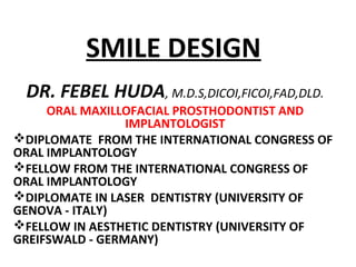
  • 1. SMILE DESIGN DR. FEBEL HUDA, M.D.S,DICOI,FICOI,FAD,DLD. ORAL MAXILLOFACIAL PROSTHODONTIST AND IMPLANTOLOGIST DIPLOMATE FROM THE INTERNATIONAL CONGRESS OF ORAL IMPLANTOLOGY FELLOW FROM THE INTERNATIONAL CONGRESS OF ORAL IMPLANTOLOGY DIPLOMATE IN LASER DENTISTRY (UNIVERSITY OF GENOVA - ITALY) FELLOW IN AESTHETIC DENTISTRY (UNIVERSITY OF GREIFSWALD - GERMANY)
  • 5. METHOD OF ANALYSIS FOR DIAGNOSIS AND TREATMENT PLANNING • Total Smile Analysis a.Space Analysis b.Profile analysis c. Computer Analysis
  • 6. TOTAL SMILE ANALYSIS • FACIAL ANALYSIS (Examination of Lips at rest)
  • 7. TOTAL SMILE ANALYSIS Profile analysis • Profile:
  • 8. TOTAL SMILE ANALYSIS • Dentofacial Analysis:
  • 9. TOTAL SMILE ANALYSIS • Dentofacial Analysis: • Incisal edge to lower lip
  • 10. TOTAL SMILE ANALYSIS Dentofacial Analysis • Midline • Midline skewing to the right
  • 11. TOTAL SMILE ANALYSIS • Dentofacial Analysis: • Tooth lower lip position • Full smile. No of teeth exposed
  • 12. TOTAL SMILE ANALYSIS • Dentofacial Analysis: • Bilateral Negative Space • Central incisor proportion
  • 13. TOTAL SMILE ANALYSIS Space Analysis • Dentofacial Analysis: • Anterior teeth proportion
  • 14. TOTAL SMILE ANALYSIS • Computer Analysis
  • 15. Mc laren’s analysis for Smile Design • Facial analysis (general facial balance). • Dento-facial analysis (maxillomandibular relationships to the face, and the dental midline relationship to the face). • Dento-labial analysis (the relationship of the teeth to the lips). • Dento-gingival analysis (the relationship of the teeth to the gingiva, and • Dental analysis (the intertooth& intratooth relationships, ie, form & position along with color).
  • 16. Anatomy of the Smile: - Ackerman The normal curvature of the lips Proper exposure of the red zone of the lips An undistorted philtrum Undisturbed naso-labial grooves.
  • 17. Anatomy of the Smile: - Ackerman
  • 18. Classification of Smile: According to Solomon E.G.R. 1)Depending on the nature of labial mucous membrane a) Papilla smile b) Gingival smile c) Mucosa smile 2) Dependant on the lip component a) Straight smile b) Convex smile c) Concave smile
  • 19. Classification of Smile: According to Ackerman & Ackerman There are two basic types of smiles; i). The social smile ii). The enjoyment smile. Each type involves a different anatomic presentation of the elements of the display zone.
  • 20. Classification of Smile: According to RUBIN L R- He identified the following neuromuscular smile pattern • The commissure smile • The cuspid smile and • The complex smile. The commissure smile: • Most common pattern, 67% of the population. • Lips contracting to show the upper teeth. • Lowest incisal edge of max teeth seen is central incissors • maximum movement of the commissure from 7 to 22 mm. The cuspid smile: • Found in 31% of the population. • Shape of the lips is visualized as a diamond • Dominance of the levator labii superioris, exposing the cuspid teeth. • Inferior turn of lips in the maxillary premolars region.
  • 21. ACCORDING TO EDWARD PHILIPS: Smile styles • The commissure smile • The cuspid smile and • The complex smile Stages of a smile • Stage I- lips closed • Stage II- resting display • Stage III- natural smile (three- quarters) • Stage IV- expanded smile (full) Types of smile • Type I- maxillary only • Type II- maxillary and over 3 mm gingival • Type III- mandibular only • Type IV- maxillary and mandibular • Type V- neither maxillary nor mandibular. Classification of Smile:
  • 22. ACCORDING TO ANTHONY H.L.TJAN, GARY D.MILLER AND JOSEPHINE G.P. – open smiles were divided into 3 types- • High smile- reveals the total cervicoincisal length of the maxillary anterior teeth and a contiguous band of gingiva. • Average smile- reveals 75% to 100% of the maxillary anterior teeth and the interproximal gingiva only. • Low smile- displays less than 75% of the anterior teeth. Classification of Smile:
  • 23. The complex smile: • Found in 2% of population. • The shapes of the lips are typically parallel. • Simultaneous contraction of the levators of the upper lip & depressors of the lower lip showing all the upper and lower teeth concurrently. • The key characteristic of this smile is the strong muscular pull and retraction of the lower lip downward and back. • Smile pattern shows maxillary & mandibular incisal flat and parallel.
  • 24. Requirements of an ideal smile
  • 25. COMPONENTS OF AN ATTRACTIVE SMILE AND THEIR ANALYSIS: • Facial components • Dental components • Gingival components • Physical components
  • 26. FACIAL COMPONENTS: • Smile analysis evaluation should begin with the observation of the facial elements. • Facial beauty is based on standard esthetic principles of proper alignment, symmetry, and proportions of the face. Facial features in smile design include, – Facial height – Facial shape – Facial profile – Gender, and age.
  • 27. • FACE HEIGHT is divided into three equal thirds: – From forehead to brow line – From brow line to the base of the nose – From the base of the nose to the base of the chin. • The width of the face is typically the width of five ‘‘eyes’’. FACIAL COMPONENTS
  • 28. • Four Basic FACIAL SHAPES, Trubyte guide are square, tapering, square tapering, and ovoid. • LATERAL FACIAL PROFILES -straight, convex, or concave. • FACIAL FEATURES RELATED TO GENDER AND AGE - soft tissue texture, complexion, and tissue integrity of the epithelial tissues. FACIAL COMPONENTS
  • 29. Geometric Facial analysis: • Creates horizontal symmetry and act as cohesive forces to unify the facial composition. • Cohesive force – to achieve pleasing aesthetics. • The facial midline is perpendicular to these lines act as segregative forces. • Interpupillary line-Incisal Plane - Facial midline. FACIAL COMPONENTS
  • 30. A tilt in the incisal plane may be attributed to; • Dental factors - Attrition, Different Patterns of Eruption, and Periodontal Disease • Skeletal factors - slanted maxilla.
  • 31. FROM THE SAGITTAL ASPECT, SOFT TISSUE ANALYSIS, • Contour of upper& lower lips • Lip support • Lip protrusion • Prominence of chin • Recession or prominence of nose and its degree • Nasolabial angle • Rickett’s E-plane Evaluation helps in diagnosis & treatment planning.
  • 32.  Nasolabial angle – males 90° to 95°; in females 100° to 105°.  Rickett’s E-plane - Upper lip 4mm behind this imaginary line, while lower lip is 2 mm.  The protrusion or retrusion of the maxilla can be assessed.  Prominent maxilla - nasolabial angle < 90° & upper lip < 4 mm to the E-plane - convex profile.
  • 33.  Concave profile - nasolabial angle > 90°and upper lip > 4 mm to the E-plane.  Spear has termed as “facially generated treatment planning”.  Steiner line - midpoint of the nose to the chin, and touches lips.  Burstone line - subnasale point to pogonion point.  The patient’s profile angle extends through the glabella, subnasale, and soft tissue pogonion, and should be approximately 165° to 175° for Class I occlusion.
  • 35. Lips analysis • Boundaries of the smile design.
  • 36. Lips analysis • Genetic traits - position of the teeth, alveolar bone, and jaws; and their relationships influence the shape of the lips.
  • 37. Width: • Wide lips make for a wide smile. • Esthetic smile – smile atleast half the width of the face. • The upper lip - more arched and wider than the lower lip. • Lower lip is recessed beneath the upper lip approximately 300 . • The fullness of the lip, or lip volume, can be categorized as full, average, or thin. FULLNESS:
  • 38. Lip symmetry: • Mirror image appearance of each lip when smiling & on both sides of midline. • Independent evaluation of the upper and lower lip is essential when analyzing both symmetry and fullness.
  • 40. STATIC POSITION • Static position - Tranquil position and ‘M’. LARS - Lip Length, Age, Race And Sex Influence tooth exposure in this tranquil position.
  • 41. • Length of the upper lip varies from 10-36mm, long maxillary lips show more mandibular rather than maxillary teeth. • The amount of tooth exposure at rest is predominantly a muscle-determined position.
  • 42. Age - Maxillary and Mandibular tooth display. Multi-factorial phenomenon described by the three Ps: Programmed ageing Pathological ageing Psychological ageing.
  • 43. RACE & SEX • Decreasing amount of maxillary, & an increasing amount of mandibular tooth visibility, is seen from Caucasians to Asians to Blacks. • Males have longer maxillary lips than females. • Maxillary tooth display - 1.91 mm for men & 3.40 mm for women. Inference: Before the amount of tooth exposure at rest is prescribed for the proposed prostheses,each patient should be assessed according to the LARS factor.
  • 44. DYNAMIC POSITION • Characterized by smile. The extent of tooth exposure during a smile depends on:  Skeletal make-up  Degree of contraction of the facial muscles  Shape and size of the dental elements  Shape and size of the lips
  • 45. Rufenacht’s morphopsychological concepts: • Thin & taut lips – provide teeth which confer delicacy and fragility. • Thick or voluptuous lips – provide teeth which display dominance and boldness.
  • 46. Lip line: • Relation of the inferior border of the upper lip to the teeth & gingiva. • Upper lip line - to evaluate the max incisor display & the vertical position of the gingival margins during smile. • Lower lip line - to evaluate the buccolingual position of the incisal edge of the maxillary incisors and the curvature of the incisal plane.
  • 47. upper lip line: • Classified as - low, average or high. • Acceptable if it is within a range of 2 mm apical to the height of the gingiva on the maxillary centrals.
  • 48. Extremes: • Shorter maxillary lips • Longer maxillary lips
  • 49. corrective options for gummy smile: • Vertical maxillary excess - Cephalometric analysis. Skeletal displasias - Orthognathic surgery (Horizontal le fort I osteotomy) to impact the maxilla. • Altered passive eruption - diminished tooth size in combination with a high lip line creates a gummy smile - corrective periodontal procedures are an option. • Cosmetic crown lengthening – improve normal tooth height and tooth proportions. Creates pleasing emergence profile, symmetry, positive radicular architecture, and proper zenith points of the gingival margins. • Cosmetic dental procedures. • Exostosies - ledging appearance of the gingival tissues. Re-contouring the alveolar bone is necessary to recreate & define normal architecture.
  • 50. Before (top) and after (bottom) crown lengthening, frenectomy and application of 10 maxillary porcelain veneers.
  • 51. • Frenum attachment can also affect the upper-lip shape and the amount of tooth exposure. A frenectomy, can free up the lip for normal lip movement. • Broad attachment of frenum creates a redundant flap of tissue, termed as a ‘‘lip curtain’’, is visible hanging beneath the upper lip when smiling. • These procedures, used in combination with cosmetic dental procedures,can reduce gummy smiles and produce a more esthetic smile.
  • 52. Smile zone: • The exposed portion of teeth & gums within the oral frame formed by lips during a smile. • Six basic smile-zone shapes:
  • 53. Negative space: • The dark space appearing b/w the jaws & the mouth opening either at the corner of the mouth or around the buccal aspect of the posterior teeth during active smile and laugh. • Lateral negative space • Buccal negative spaces. • Obliteration of these essential spaces by dental elements lead to an unattractive smile. • Excessive negative space also appear unesthetic.
  • 54. Smile line: • The smile line can be defined as an imaginary line running along the incisal edges of the maxillary anterior teeth and coinciding the curvature of the lower lip / gull-wing course while smiling. • An attractive smile line is one of the most important features of a pleasing smile.
  • 55. Smile line: • Slight to moderate deviations - esthetic recontouring • Discrepancies to the smile line in situations where there is ideal tooth form and color – orthodontics. • Reverse smile line - centrals appear shorter than the cuspids along the incisal plane & create an aged or worn appearance. • Etiology: Abrasion, erosion or attrition,periodontitis, altered patterns of eruption or poor quality dentistry.
  • 56. Intercommissure line and lower lip framing: • In broad smile position, the imaginary line through the corners of the mouth is known as the intercommissure line, or ICL. • The amount of maxillary tooth reveal below the ICL interacts with the viewer’s perception of the patient’s age. In a youthful smile, approximately 75 percent to 100 percent of the maxillary teeth would show below this line.
  • 57. Facial and Dental midline: • The starting point of the esthetic treatment plan is the facial midline. • Anatomical landmarks as locating guides - midline of the nose, forehead, interpupillary plane, philtrum, and chin. • Variations in genetic structures undermines these landmarks as indicators of the facial midline.
  • 58. Two reliable anatomical landmarks to locate facial midline: • The first is point nasion. • The second is the base of the philtrum - cupid’s bow in the center of the upper lip, it matches the papilla between the centrals directly over the dental midline. • Connecting line locates the position & direction of the facial midline. • Philtrum of the lip - most accurate. The exceptions are surgical, accident, and cleft-lip cases.
  • 59. • The dental midline refers to a vertical line formed by the contact of the maxillary central incisors. • Ideal – coincidence b/w facial & dental the midline. • Atleast- the central incisors should be parallel to the facial midline and perpendicular to the incisal plane.
  • 60. • Midline discrepancies of up to 4 mm off-centre to facial midline will generally not be perceived as unesthetic or not noticeable. • Alignment of the maxillary and mandibular dental midlines is desirable. • Canted midline – a major design flaw in any natural or restored dentition.
  • 61. Controversies in the placement of the dental midline: • One school of thought states that the maxillary dental midline should coincide exactly with the labial frenum and the facial midline, as it does in 70% of the population. • The opposing view is that placing the midline exactly in the centre may contribute to a sense of artificiality. • Choice is determined after aesthetic appraisal. • Midline in centre - To detract attention from asymmetries & disharmonies of the face. • The mandibular midline should not be used as a reference point, because in 75% of the population this does not coincide with the maxillary midline • Restorative dentistry - Slight corrections of midlines if maxillary centrals are made relatively symmetric and correct intertooth relationships are maintained. • Large midline discrepancy - orthodontics.
  • 62. Axial inclination of teeth: • Anterior teeth tends to incline mesially toward the midline & become more pronounced from the central incisors to the canines. • Overall esthetic impact - one of a harmonious relationship with the framing of the lower lip. • Posterior teeth exhibits mesial inclination toward the midline as the cuspid. • Creates a natural visual gradation - appear to diminish in size as they progress posteriorly.
  • 63. Contact points / connectors: • The connector is the zone in which two adjacent teeth appear to touch. • 50-40-30 connector zone rule – establish esthetic reation. • Coincidence of incisal edges to curvature of the lower lips enhance the cohesiveness of the dentofacial composition.
  • 64. Intratooth Relationships: • The intratooth relationships refer to the morphology of maxillary anterior teeth. • Shape & Size. Shape: • The morphology of maxillary anterior teeth is a fusion of the three basic shapes circle, square and triangle. • Teeth have inimitable shapes based on only three variables.
  • 65. • Studies by Williams' and Frush and Fisher. • Williams - shape of the central incisor is inverted frontal view of the face – invalidated. • Frush and Fisher suggested that sex, age and personality related to the contour of the anterior dental segment. • Chronological age and dental age. • Genetically determined factor – relatives records as guides.
  • 66. • Morphopsychology - The concept of transposing the gender variations onto the shape of the teeth. • Females - curvaceous features, Males - ruggedness and sharp line angles.
  • 67. Size: • Tooth size - mesio-distal width divided by the inciso-gingival length - yields the width/length (w/l) ratio. • Sizing with 1 to 16 theory, postulated by House and Loop. • Height of C.I - 1/16th of the distance from the inferior border of the chin to the inter pupillary line. • Width is 1/16th of the distance measured from either zygomatic prominence.
  • 68. TWO SCHOOLS OF THOUGHT ON SIZE OF CENTRAL INCISORS • Rufenacht suggested that the width and length of the central incisor should be constant throughout life based on morpho-psychological determination of an ideal proportion. • The second theory states that our bodies are in perpetual change throughout life. The dentition is no exception to this transformation. Creating teeth with a youthful appearance in an older person and creates a sense of artificiality.
  • 69. Inter-tooth relationships Tooth proportions: Golden proportion: • Greek’s mathematical concept of beauty. • Pythagoras - Golden Proportion - (1/1.618 = 0.618) • Plato - Beautiful Proportion (1/1.733 = 0.577).
  • 70. • Kepler called it the “DIVINE PROPORTION”. • The application of golden no. to dentistry was first mentioned by LOMBARDI(1973) and developed by LEVIN(1978). • Levinused the golden proportion to relate the successive widths of the anterior teeth as viewed from the frontal. • Width of the central incisor 0.618, or approximately 62% width of the lateral incisor.
  • 71.
  • 72. Repeated Ratio: • Lombardi described the use of a "continuous proportion or repeated ratio which has been established between the width of central and lateral incisor and is continued in the ratio of the placement of the remaining teeth and spaces." • Eg. A lateral incisor should be 80% the width of central incisor and a canine should be 80% width of the lateral incisors.
  • 73. Recurring esthetic dental (RED) proportion: • The 62% golden proportion is used, the lateral incisor appears too narrow, and the resulting canine is not prevalent enough. • Preston reported the golden proportion only 17%. • Using continuous proportion or repeated ratio is not convincing. • By combining elements of concepts Daniel.H.Ward has defined as the recurring esthetic dental (RED) proportion. • The RED proportion states that the proportion of the successive widths of teeth as viewed from the frontal should remain constant as one move distally.
  • 74. Physical attributes of the elements of the dento-facial composition : • Fundamental principles of esthetics - artistic parameters. • Co-relating them to the dento-facial complex to scale esthetics of the dento-facial composition. Various physical attributes of elements of a composition impart the esthetic value: • Contrast • Unity • Cohesive & segregate force • Symmetry • Proportion o Dominance o Balance o Visual tension o Progressive abating in dental composition o Shade progression & selection
  • 75. GINGIVAL COMPONENT: • A feature of smile design that is often overlooked yet very significant is the health, symmetry, and architecture of the gingival tissues. These tissues frame the teeth and add to the symmetry of the smile. HEALTH, COLOR AND TEXTURE : • Paramount for long-term success & the esthetic value of the treatment. • Pale pink, vary in degree of vascularity, epithelial kertinization, and pigmentation, in the thickness of the epithelium. • Pointed papillary contour and should fill the interdental spaces to the contact point. • Black triangles - makes a smile less attractive. • Managing the soft tissues in this area improves the smile.
  • 76. • Firm in consistency and the attached part should be firmly anchored. • A normal, healthy gingival sulcus should not exceed 3 mm in depth. • The texture - stippled (orange-peel–like appearance) in most cases. Fine or coarse. Younger females has more finely textured & finer stippling when compared with that of males.
  • 77. ARCHITECTURE & SYMMETRY: • Positive radicular shape forming a scalloped appearance should be symmetric on both sides of the midline. • The marginal contour of the gingival should be sloped coronally to the end in a thin edge. • Marginal gingival line extending from cuspid to cuspid. Ideally, the laterals reach slightly short of that line.
  • 78. Gingival zenith: • The most apical point of the free gingival margin along the long axis of the tooth. Their position are dictated by: • Root form anatomy. • CEJ. • Osseous crest, where gingiva is scalloped the most. • Located distal to the long axis on the maxillary centrals and cuspids. • Zenith point of max.lat & mand.incisors is coincident with the long axis of these teeth. • The compromised zenith can be reestablished by replacing the defective restoration.
  • 79. Importance of zenith point o When closing diastemas or changing the mesial or distal tilt position of the tooth i.e., moving zenith points horizontally. o In cases where teeth needs to be shown longer or more taper at the gingival 1/3rd Zenith point can be moved apically. By horizontal & vertical alterations, zenith point can enhance: 1. Perception of tooth axis. 2. Length. 3. Gingival shapes.
  • 80. Gingival Biotype : • The gingival biotype is classified as thin or thick. • Prevalence - thick biotype is 85% & thin biotype is 15%. Thin periodontal biotypes: • Gingiva – delicate, almost translucent, appears friable with a minimal zone of attached gingiva. • Escalates the risk of recession following crown preparation, periodontal or implant surgery.
  • 81. Thick periodontal biotype • Dense fibrotic and resilient, making it resistant to surgical procedures • Reacts with pocket formation & during disease. • Favorable esthetic outcomes with restorations. • Relatively large amount of attached gingiva. • Thick underlying osseous form. • Relatively resistant to acute trauma.
  • 82. TISSUE BIOTYPE IN PROSTHODONTIC TREATMENT Tooth preparation: • Thin biotypes – supra-gingival margins. • Subgingival for PFM - grayish discoloration - esthetic failure. • Thick biotypes - subgingivally in esthetic zones. • Over contoured restorations will lead to gingival recession in thin biotypes. Pocket formation is seen in thick biotype. Gingival retraction: • Thin biotypes - Thin retraction cord to avoid injury • > 15 minutes - chances of recession are more. • Thick biotype – comfortable retraction.
  • 83. Gingival aesthetic line (GAL): • The GAL • The GAL. • GAL is considered for creating pleasing gingival level transition between the maxillary anterior teeth. Four classes of GAL can be described: • Class I — The GAL angle is between 45° and 90° and the lateral incisor is touching or below (1 mm to 2 mm) the GAL .
  • 84. • Class II — observed in Class II angles or pseudo-Class II conditions. • Class III — The GAL angle equals 90°, and the canine, lateral, and central all lie below the GAL. • Class IV —The GAL angle can be acute or obtuse. Gingival asymmetries due to recession, altered patterns of eruption, loss of interdental papillae, clefts, and high frenal insertions. • GAL should be relatively horizontal to the horizon and relatively symmetric on both sides of the midline. It may radiate up slightly as it goes posterior. Mclaren, inside dentistry—july/august 2009
  • 85. PHYSIOLOGICAL COMPONENT: Perceptual aspects – the art of illusion: • Illusion is a figment of imagination where a perception of an object is created. Fundamentals and Principles: • The art of creating illusions consists of changing perception, to cause an object to appear different from what it actually is. • When an object is subjected to light it exhibits two dimensions, that is, length and width. True natural light is multi-directional and on striking the surface of the object, also reveals texture and shadows, this adds the third life like dimension of depth.
  • 86. Illusion works on two basic principles; • the principle of illumination • the principle of line. • ‘Principle of Illumination” is based on the perception that light approaches and dark recedes. The artistic predilection exhibited in the principle of illumination can be maintained to change the size, shape and the overall form of the tooth through illusions. • The second artistic predilection of great importance in dentistry is the use of horizontal and vertical lines and ridges. Horizontal lines make the object appear wider and vertical lines make the object appear longer. This is termed as the “Principle of Line”.
  • 87. Esthetic treatment planning and sequencing : An ideal esthetic treatment plan should be; • Minimally invasive • Preserve as much of the natural structures as possible. • Realign the ideal form and function of the teeth and tissues. • Enhance the esthetics. • Never compromise the patient’s oral health or the stability of his or her teeth. • Attempts to achieve perfection in every way. • Patient interview – treatment options mock-ups. • Treatment sequencing - phase wise distribution of treatment procedures, execution.
  • 88. Esthetic treatment options: • Cosmetic contouring – the reshaphing of anterior teeth for esthetic purposes involving mesial, distal, labial and lingual surfaces. • Bleaching discoloured teeth • Composite resin bonding – to change the shape of the teeth as well as their colour • Etched porcelain restorations- inlays, onlays, veneers – discoloured teeth, attrition, erosion, abfraction, tooth chipping, endodontically treated teeth.
  • 89. • Crown restoration • Periodontal theraphy – in altered passive eruptions, crown lengthening, esthethic gingival recontouring, frenectomies, flap surgeries, osseous recontouring. • Orthodontic and surgical orthodontical interventions – in diastema, crowded teeth, surgical orthodontic corrections of dento-facial deformities such as vertical maxillary insufficiency, vertical maxillary excess etc.,
  • 90. Role of Technology in Changing Smile: Extra oral video camera: • Record of the patient's face while moving and talking. • It can record the patient in various moods and gestures • Computer imaging . Intraoral video camera: • Reliability, performance and image quality. • Smallest details of tooth can be viewed as close as 2mm. • Pt education & treatment options • Application wit computer imaging system. T- Scan and Digital Radiography • Buccolingual width of the jaws as well as the • Location of anatomic features for placing an implant in the esthetic position.
  • 91. CAD/CAM • To design veneers and crowns to enhance smile while the patient waits. • “Mock- up" of a planned cosmetic teatment • Solve potential problems before providing treatment to the patient. Lasers • Delicate gum surgery and crown lengthening/ gingivectomy and gingivoplasty. • Controlled bleeding - dry operating field - excellent visibility - Reduced operating time - reduced post operative swelling, pain and scarring. Abrasive Technology • Jet air stream with micro abrasive particles. • Stain removal, decay removal. • Painless & faster. • Repairing existing composite, ceramics.
  • 92. Conclusion “A well designed smile is a product of consolidated efforts accomplished by accurate diagnosis, methodical treatment planning, use of advanced materials and contemporary techniques rendered by the skilled dentist”.
  • 93. References • Goldstein, RE: Change your Smile, ed 3 Chicago, Quintessence, 1997. • Nicholas C. Davis, Smile Design, Dent Clin N Am 51 (2007) 299–318 • Ahmed I. anterior dental esthetics: dentofacial perspective. BDJ 2005 198:81-88 • Levin El. Dental esthetics and the golden proportion. J Prosthet Dent 1978; 40: 244–252. • Ackermann JL et al. smile analysis and design in digital era, Am J Orthod Dentofacial Orthop 2007;131:305-10 • Kamal Shigli, Swaraj Bharati: Role of technology in designing a confident smile. J. IPS Dec. 2001, vol.1, no.4.6) • Vig RG Brundo GC. The kinetics of anterior tooth display. J ProsthetDent 1978;39:502-4. • Jeff.morley et al., macroesthetic elements of smile design, JADA, Vol. 132, January 2001. • T.G.Mathews, anatomy of the smile, J ProsthetDent 1978;39:2 128-135.