6. e270 Circulation April 6, 2010
Appendix 1. Author Relationships With Industry and industry during his/her tenure, he/she is required to notify
Other Entities . . . . . . . . . . . . . . . . . . . . . . .e365 guideline staff in writing. These statements are reviewed by
Appendix 2. Reviewer Relationships With Industry and the Task Force on Practice Guidelines and all members during
Other Entities . . . . . . . . . . . . . . . . . . . . . .e367 each conference call and/or meeting of the writing committee,
Appendix 3. Abbreviation List . . . . . . . . . . . . . . . . . . .e369 updated as changes occur, and ultimately published as an
References . . . . . . . . . . . . . . . . . . . . . . . . . . . . . . . . . . .e348 appendix to the document. For detailed information regarding
guideline policies and procedures, please refer to the methodol-
Preamble ogy manual for ACCF/AHA Guideline Writing Committees.1
It is essential that the medical profession play a central role in RWI and other entities pertinent to this guideline for authors and
critically evaluating the evidence related to drugs, devices, peer reviewers are disclosed in Appendixes 1 and 2, respec-
and procedures for the detection, management, or prevention tively. Disclosure information for the ACCF/AHA Task Force
of disease. Properly applied, rigorous, expert analysis of the on Practice Guidelines is also available online at (http://www.acc.
available data documenting absolute and relative benefits and org/about/overview/ ClinicalDocumentsTaskForces.cfm).
risks of these therapies and procedures can improve These practice guidelines are intended to assist healthcare
outcomes and reduce costs of care by focusing resources providers in clinical decision making by describing a range of
on the most effective strategies. One important use of such generally acceptable approaches for diagnosis, management,
data is the production of clinical practice guidelines that, in and prevention of specific diseases or conditions. Clinicians
turn, can provide a foundation for a variety of other should consider the quality and availability of expertise in the
applications such as performance measures, appropriate area where care is provided. These guidelines attempt to
use criteria, clinical decision support tools, and quality define practices that meet the needs of most patients in most
improvement tools. circumstances. The recommendations reflect a consensus
The American College of Cardiology Foundation (ACCF) after a thorough review of the available current scientific
and the American Heart Association (AHA) have jointly en-
evidence and are intended to improve patient care. The Task
gaged in the production of guidelines in the area of cardiovas-
Force recognizes that situations arise where additional data
cular disease since 1980. The ACCF/AHA Task Force on
are needed to better inform patient care; these areas will be
Practice Guidelines is charged with developing, updating, and
identified within each respective guideline when appropriate.
revising practice guidelines for cardiovascular diseases and
Patient adherence to prescribed and agreed upon medical
procedures, and the Task Force directs and oversees this effort.
regimens and lifestyles is an important aspect of treatment.
Writing committees are charged with assessing the evidence as
Prescribed courses of treatment in accordance with these recom-
an independent group of authors to develop, update, or revise
mendations are effective only if they are followed. Because lack
recommendations for clinical practice.
of patient understanding and adherence may adversely affect
Experts in the subject under consideration have been selected
outcomes, physicians and other healthcare providers should
from both organizations to examine subject-specific data and
make every effort to engage the patient’s active participation in
write guidelines in partnership with representatives from other
medical practitioner and specialty groups. Writing committees prescribed medical regimens and lifestyles.
are specifically charged to perform a formal literature review, If these guidelines are used as the basis for regulatory or
weigh the strength of evidence for or against particular treat- payer decisions, the goal should be improvement in quality of
ments or procedures, and include estimates of expected health care and aligned with the patient’s best interest. The ultimate
outcomes where data exist. Patient-specific modifiers, comor- judgment regarding care of a particular patient must be made
bidities, and issues of patient preference that may influence the by the healthcare provider and the patient in light of all of the
choice of tests or therapies are considered. When available, circumstances presented by that patient. Consequently, there
information from studies on cost is considered, but data on are circumstances in which deviations from these guidelines
efficacy and clinical outcomes constitute the primary basis for are appropriate.
recommendations in these guidelines. The guidelines will be reviewed annually by the ACCF/
The ACCF/AHA Task Force on Practice Guidelines makes AHA Task Force on Practice Guidelines and considered
every effort to avoid actual, potential, or perceived conflicts current unless they are updated, revised, or withdrawn
of interest that may arise as a result of industry relationships from distribution.
or personal interests among the writing committee. Specifi- Alice K. Jacobs, MD, FACC, FAHA
cally, all members of the writing committee, as well as peer Chair, ACCF/AHA Task Force on Practice Guidelines
reviewers of the document, are asked to disclose all current
relationships and those 24 months prior to initiation of the Sidney C. Smith, Jr, MD, FACC, FAHA
writing effort that may be perceived as relevant. All guideline Immediate Past Chair, ACCF/AHA Task Force on
recommendations require a confidential vote by the writing Practice Guidelines
committee and must be approved by a consensus of the
members voting. Members who were recused from voting are
1. Introduction
noted on the title page of this document. Members must
recuse themselves from voting on any recommendation 1.1. Methodology and Evidence Review
where their relationships with industry (RWI) apply. If a The writing committee conducted a comprehensive search of
writing committee member develops a new relationship with the medical and scientific literature through the use of
Downloaded from circ.ahajournals.org by on June 10, 2010
7. Hiratzka et al 2010 Guidelines on Thoracic Aortic Disease e271
PubMed/MEDLINE. Searches were limited to publications odds ratio (OR), relative risk (RR), or hazard ratio (HR)
written in the English language. Compiled reports were depending on the format in the original publication. Along
reviewed and additional articles were provided by committee with all other point statistics, confidence intervals (CIs) for
members. Specifically targeted searches were conducted on those statistics are added when available.
the following subtopics: acute aortic dissection, ankylosing The writing committee recognized that the evidence base
spondylitis, aortic dissection and litigation, aortic neoplasm, for this guideline is less robust in terms of randomized
aortic tumors, Behcet disease, bicuspid aortic valve, calcified
¸ clinical trials than prior ACCF/AHA guidelines, particularly
aorta, chronic dissection, coarctation of the aorta, D-dimer, those focused on coronary artery disease (CAD) and heart
dissecting aneurysm, Ehlers-Danlos syndrome, endovascular failure. As the reader will discern, much of the evidence base
and aortic aneurysms, medial degeneration, porcelain aorta, for this topic consists of cohort studies and retrospective
giant cell arteritis, imaging and thoracic aortic disease, reviews, which largely emanate from centers with a special-
inflammatory disease, intramural hematoma, Loeys-Dietz ized interest in specific types of thoracic aortic disease. The
syndrome, Marfan syndrome, Noonan syndrome, penetrating writing committee attempted to focus on providing the
aortic ulcer, polycystic kidney disease, thoracic and aortic practitioner with recommendations for evaluation and treat-
aneurysms, thoracic aortic disease and patient care, thoracic ment wherever possible and where controversy exists, iden-
aortic disease and surgery, thoracic aorta and Kawasaki tified as such in the text.
disease, Takayasu arteritis, thoracoabdominal and aorta or The writing committee acknowledges the expertise of the
aortic disease, and Turner syndrome. More than 850 refer- highly experienced and effective practice guidelines staff of
ences were reviewed, with 830 used as the primary evidence the ACCF and AHA. The writing committee chair also
base for the final guideline. The ACCF/AHA Task Force on acknowledges the commitment and dedication of the diverse
Practice Guidelines methodology processes were followed to writing committee members who were able to put aside issues
write the text and recommendations. In general, published of specialty “turf” and focus on providing the medical
manuscripts appearing in journals listed in Index Medicus community with a guideline aimed at optimal patient care.
were used as the evidence base. Published abstracts were used
only for emerging information but were not used in the 1.2. Organization of the Writing Committee
formulation of recommendations. The guideline was written by a committee comprised of
The committee reviewed and ranked evidence supporting experts in cardiovascular medicine, surgery, radiology, and
current recommendations with the weight of evidence ranked nursing. For many of the previous ACCF/AHA practice
as Level A if the data were derived from multiple randomized guidelines, writing expertise has been available within these 2
clinical trials or meta-analyses. The committee ranked avail- organizations. Because of the broad scope and diversity of
able evidence as Level B when data were derived from a thoracic aortic diseases, as well as the specialists who treat
single randomized trial or nonrandomized studies. Evidence such patients, the ACCF and AHA sought greater involve-
was ranked as Level C when the primary source of the ment from many specialty organizations. Most, but not all,
recommendation was consensus opinion, case studies, or specialty organizations that represent the major stakeholders
standard of care. In the narrative portions of these guidelines, caring for patients with thoracic aortic diseases provided
evidence is generally presented in chronological order of writing committee members and financial support of the
development. Studies are identified as observational, retro- project, and they are recognized as marquee level partners
spective, prospective, or randomized. For certain conditions with the ACCF and AHA. These organizations included the
for which inadequate data are available, recommendations are American Association for Thoracic Surgery (AATS), Amer-
based on expert consensus and clinical experience and are ican College of Radiology (ACR), American Stroke Associ-
ranked as Level C. An analogous example is the use of ation (ASA), Society of Cardiovascular Anesthesiologists
penicillin for pneumococcal pneumonia, where there are no (SCA), Society for Cardiovascular Angiography and Inter-
randomized trials and treatment is based on clinical experi- ventions (SCAI), Society of Interventional Radiology (SIR),
ence. When recommendations at Level C are supported by Society of Thoracic Surgeons (STS), and Society for Vascu-
historical clinical data, appropriate references (including lar Medicine (SVM). The American College of Emergency
clinical reviews) are cited if available. For issues where Physicians (ACEP) and the American College of Physicians
sparse data are available, a survey of current practice among (ACP) were also represented on the writing committee.
the clinicians on the writing committee formed the basis for Where additional expertise was needed, the scientific coun-
Level C recommendations and no references are cited. The cils of the AHA were contacted for writing committee
schema for classification of recommendations and level of representatives. Representation was provided or facilitated by
evidence is summarized in Table 1, which also illustrates how the Councils on Cardiovascular Nursing, Cardiovascular
the grading system provides an estimate of the size of the Surgery and Anesthesia, Cardiovascular Radiology and Inter-
treatment effect and an estimate of the certainty of the vention, and Clinical Cardiology, Council for High Blood
treatment effect. Pressure Research, and Stroke Council.
To provide clinicians with a comprehensive set of data,
whenever possible, the exact event rates in various treatment 1.3. Document Review and Approval
arms of clinical trials are presented to permit calculation of This document was reviewed by 3 outside reviewers nomi-
the absolute risk difference (ARD), number needed to harm nated by the ACCF and 2 outside reviewers nominated by the
(NNH); the relative treatment effects are described either as AHA, as well as 1 or 2 reviewers from each of the following
Downloaded from circ.ahajournals.org by on June 10, 2010
8. e272 Circulation April 6, 2010
Table 1. Applying Classification of Recommendations and Level of Evidence
*Data available from clinical trials or registries about the usefulness/efficacy in different subpopulations, such as sex, age, history of diabetes, history of prior
myocardial infarction, history of heart failure, and prior aspirin use. A recommendation with Level of Evidence B or C does not imply that the recommendation is weak.
Many important clinical questions addressed in the guidelines do not lend themselves to clinical trials. Even though randomized trials are not available, there may
be a very clear clinical consensus that a particular test or therapy is useful or effective.
†In 2003, the ACCF/AHA Task Force on Practice Guidelines developed a list of suggested phrases to use when writing recommendations. All guideline
recommendations have been written in full sentences that express a complete thought, such that a recommendation, even if separated and presented apart from
the rest of the document (including headings above sets of recommendations), would still convey the full intent of the recommendation. It is hoped that this will
increase readers’ comprehension of the guidelines and will allow queries at the individual recommendation level.
organizations: the AATS, ACP, ACEP, ACR, ASA, SCA, matic disease states and presentations. According to the
SCAI, SIR, STS, and the SVM. It was also reviewed by 6 Centers for Disease Control and Prevention death certificate
individual content reviewers—2 content reviewers from the data, diseases of the aorta and its branches account for 43 000
ACCF Catherization Committee and 1 content reviewer from to 47 000 deaths annually in the United States.2 The precise
the ACCF Interventional Council. All reviewer RWI infor- number of deaths attributable to thoracic aortic diseases is
mation was collected and distributed to the writing committee unclear. However, autopsy studies suggest that the presenta-
and is published in this document (see Appendix 2). tion of thoracic aortic disease is often death due to aortic
This document was approved for publication by the gov- dissection (AoD) and rupture, and these deaths account for
erning bodies of the ACCF and the AHA and the AATS, twice as many deaths as attributed to ruptured abdominal
ACEP, ACR, ASA, SCA, SCAI, SIR, STS, and SVM and aortic aneurysms (AAAs).3 The diagnosis of acute thoracic
was endorsed by the North American Society for Cardiovas- AoD or rupture is often difficult and delayed, and errors in
cular Imaging. diagnosis may account for deaths otherwise attributed to
cardiac arrhythmia, myocardial infarction (MI), pulmonary
1.4. Scope of the Guideline embolism, or mesenteric ischemia.
The term “thoracic aortic disease” encompasses a broad range The University HealthSystem Consortium (UHC) is an
of degenerative, structural, acquired, genetic-based, and trau- alliance of more than 100 academic medical centers and
Downloaded from circ.ahajournals.org by on June 10, 2010
9. Hiratzka et al 2010 Guidelines on Thoracic Aortic Disease e273
Table 2A. ICD-9 Procedure Codes For Aortic Aneurysms 2007. Subcategories include those with dissection, those with
Aortic Aneurysm No Mention of
rupture, and those with neither. In the most recent 1-year
Category Dissection Ruptured Rupture period assessed (fourth quarter 2006 through third quarter
Thoracic 441.01 441.1 441.2
2007), there were nearly 9000 cases representing all patients
with thoracic aortic disease discharged from UHC hospitals.
Abdominal 441.02 441.3 441.4
Although these data are unavailable for the entire United
Thoracoabdominal 441.03 441.6 441.7
States, they provide important estimates of the magnitude of
Aortic (unspecified) 441.00 441.5 441.9
the prevalence of thoracic aortic disease. Additional informa-
Table courtesy of UHC Clinical DataBase/Resource Manager. tion regarding the patient admission source, particularly those
with acute presentations, is pertinent to the discussion regard-
affiliate hospitals. UHC’s Clinical DataBase/Resource Man- ing interinstitutional transfer (see Section 17.2).
ager allows comparison of patient-level risk-adjusted out- Most patients with significant thoracic aortic disease will
comes for performance improvement. The UHC provided the be directed to specialized practitioners and institutions. How-
writing committee with a summary of recent information ever, the importance of early recognition and prompt treatment
based on ICD-9 codes for thoracic aortic disease–related and/or referral for various thoracic aortic diseases by all health-
hospitalizations from the Clinical DataBase/Resource Man- care professionals provides the rationale for this document. This
ager (Tables 2A and 2B). This data table demonstrates a high guideline will attempt to provide the practitioner with a suffi-
number of hospital discharges (more than 135 000) for cient description of background information, diagnostic modal-
thoracic, abdominal, thoracoabdominal, and “unspecified” ities, and treatment strategies so that appropriate care of these
aortic aneurysms in the 5-year period between 2002 and patients can be facilitated and better understood. The goal of this
Table 2B. Number of Discharges by Year by Category of Aortic Aneurysm Among Academic Medical Centers Reporting Data to the
UHC Clinical Database*
Aortic Aneurysm 5-Year Category % % of All
Category 2002q4 –2003q3 2003q4 –2004q3 2004q4 –2005q3 2005q4 –2006q3 2006q4 –2007q3 Total Distribution Categories
Thoracic 25.9
Dissection 1607 1683 2028 2321 2355 9994 28.5
Ruptured 187 225 219 234 251 1116 3.2
No mention of 3086 4026 4953 5730 6156 23 954 68.3
rupture
Subtotal 4880 5934 7200 8285 8762 35 064
Abdominal 62.7
Dissection 553 630 747 867 867 3664 4.3
Ruptured 651 657 720 734 702 3464 4.1
No mention of 12 075 13 280 15 882 17 818 18 683 77 738 91.6
rupture
Subtotal 13 279 14 567 17 349 19 419 20 252 84 866
Thoracoabdominal 8.3
Dissection 583 587 674 755 834 3433 30.5
Ruptured 150 129 110 157 127 673 6.0
No mention of 1091 1183 1472 1621 1773 7140 63.5
rupture
Subtotal 1824 1899 2256 2533 2734 11 246
Aortic (unspecified) 3.0
Dissection 223 310 326 343 339 1541 37.6
Ruptured 9 3 15 9 9 45 1.1
No mention of 310 385 505 612 701 2513 61.3
rupture
Subtotal 542 698 846 964 1049 4099
Total No. of cases 20 525 23 098 27 651 31 201 32 797 135 275
Total No. of 2 679 334 2 777 880 3 018 141 3 222 542 3 297 834 14 995 731
inpatient
discharges
UHC indicates University HealthSystem Consortium.
*Note: Year-to-year increases are due in part to changes in number of reporting hospitals.
Table courtesy of UHC Clinical DataBase/Resource Manager.
Downloaded from circ.ahajournals.org by on June 10, 2010
10. e274 Circulation April 6, 2010
guideline is to improve the health outcomes and quality of life ● For many thoracic aortic diseases, results of treatment for
for all patients with thoracic aortic disease. stable, often asymptomatic, but high-risk conditions are far
Other practice guidelines developed by ACCF and AHA better than the results of treatment required for acute and
address the management of patients with cardiac and often catastrophic disease presentations. Thus, the identifi-
vascular diseases. The ACCF/AHA guidelines on periph- cation and treatment of patients at risk for acute and
eral arterial disease4 include recommendations for lower catastrophic disease presentations (eg, thoracic AoD, tho-
extremity, renal, mesenteric, and abdominal aortic dis- racic aneurysm rupture) prior to such an occurrence are
eases. Data standards on this topic are currently in devel- paramount to eliminating the high morbidity and mortality
opment, as is a practice guideline on extracranial carotid associated with acute presentations (see Section 8.1).
and vertebral artery diseases. The ACCF/AHA guidelines ● A subset of patients with acute AoD are subject to missed
are published in the Journal of the American College of or delayed detection of this catastrophic disease state.
Cardiology and Circulation and are available on both the Many present with atypical symptoms and findings, mak-
ACC (www.acc.org) and AHA (my.americanheart.org) ing diagnosis even more difficult (see Sections 8.1.4 and
Web sites. 8.6). This issue has come under greater medical-legal
This guideline includes diseases involving any or all parts scrutiny, and specific cases have been widely discussed in
of the thoracic aorta with the exception of aortic valve
the public domain. Widespread awareness of the varied and
diseases5 and includes the abdominal aorta when contiguous
complex nature of thoracic aortic disease presentations has
thoracic aortic diseases are present. Specific disease states are
been lacking, especially for acute AoD. Risk factors and
described in the following sections, and the reader is referred
clinical presentation clues are noted in Section 8.1.4. The
to the glossary of terminology in Section 1.5 and Appendix 3
collaboration and cosponsorship of multiple medical special-
for abbreviations used throughout the guideline.
ties in the writing of this guideline will provide unique
The reader will note that several topics or referenced areas
may appear more than once throughout the guideline. This opportunities for widespread dissemination of knowledge to
will appear to be redundant to those who choose to read the raise the level of awareness among all medical specialties.
● There is rapidly accumulating evidence that genetic alter-
entire document, but the writing committee believed that
because of the multidisciplinary nature of and organizational ations or mutations predispose some individuals to aortic
involvement in this project, individuals representing specific diseases (see Section 5). Therefore, identification of the
disciplines may choose to read and extract portions of the genetic alterations leading to these aortic diseases has the
document for their own use. Inclusion of the narrative text potential for early identification of individuals at risk. In
and references was thought to be appropriate to facilitate a addition, biochemical abnormalities involved in the pro-
more complete understanding for these disciplines and indi- gression of aortic disease are being identified through
viduals. Accordingly, the organization of the guideline is studies of patients’ aortic samples and animal models of the
meant to be less of a textbook presentation of the various disease.6,7 The biochemical alterations identified in the
topics but rather a more clinically oriented document appli- aortic tissue have the potential to serve as biomarkers for
cable to a variety of disciplines. aortic disease. Understanding the molecular pathogenesis
may lead to targeted therapy to prevent aortic disease.
1.4.1. Critical Issues Medical and gene-based treatments are beginning to show
As the writing committee developed this guideline, several
promise for reducing or delaying catastrophic complica-
critical issues emerged:
tions of thoracic aortic diseases (see Section 9.2).
● Thoracic aortic diseases are usually asymptomatic and not ● As noted in Section 18, there are several areas where
easily detectable until an acute and often catastrophic greater resources for research and both short- and long-
complication occurs. Imaging of the thoracic aorta with term outcomes registries are needed.
computed tomographic imaging (CT), magnetic resonance
imaging (MR), or in some cases, echocardiographic exam- 1.5. Glossary of Terms and Abbreviations Used
ination is the only method to detect thoracic aortic diseases Throughout Guideline
and determine risk for future complications (see Section 4).
● Radiologic imaging technologies have improved in terms Aneurysm (or true aneurysm): a permanent localized dila-
of accuracy of detection of thoracic aortic disease. How- tation of an artery, having at least a 50% increase in
ever, as the use of these technologies has increased, so also diameter compared with the expected normal diameter of
has the potential risk associated with repeated radiation the artery in question. Although all 3 layers (intima, media,
and adventitia) may be present, the intima and media in
exposure, as well as contrast medium–related toxicity.
large aneurysms may be so attenuated that in some sections
Whether these technologies should be used repeatedly as a
of the wall they are undetectable.
widespread screening tool is discussed in Section 4. In Pseudoaneurysm (or false aneurysm): contains blood re-
addition, the writing committee formulated recommenda- sulting from disruption of the arterial wall with extravasa-
tions on a standard reporting format for thoracic aortic tion of blood contained by periarterial connective tissue
findings as discussed in Section 4.5. and not by the arterial wall layers (see Section 8.4). Such
● Imaging for asymptomatic patients at high risk based on an extravascular hematoma that freely communicates with
history or associated diseases is expensive and not always the intravascular space is also known as a pulsating
covered by payers. hematoma.8 –10
Downloaded from circ.ahajournals.org by on June 10, 2010
11. Hiratzka et al 2010 Guidelines on Thoracic Aortic Disease e275
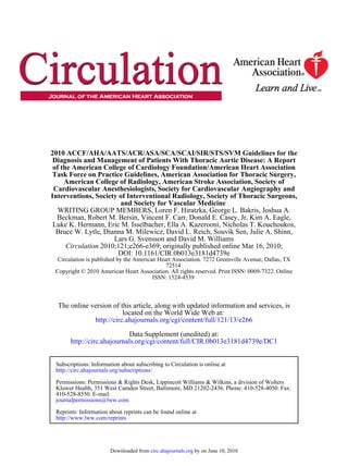
Figure 1. Aortic pathology associated with tho-
racic aortic aneurysm involving the ascending
aorta. All panels are identically oriented with the
adventitia at the top and the intima at the bottom.
H&E staining of aortic sections from a control (a)
and a patient (b) with a TAA demonstrates medial
degeneration with the fragmentation of elastic
fibers, accumulation of proteoglycans, and regions
of smooth muscle cell loss. Movat staining of aor-
tic sections from control (c) and patient with an
aneurysm (d) shows fragmentation of elastic fibers
(stained black), loss of smooth muscle cells (cells
stained red and nuclei stained violet), and accu-
mulation of proteoglycans (stained blue) in the
medial layer. 40 magnification; scale bars repre-
sent 500 mcg. H&E indicates hematoxylin and
eosin; and TAA, thoracic aortic aneurysm. Modi-
fied from Milewicz et al.11
Ectasia: arterial dilatation less than 150% of normal arterial bounded by an external elastic lamina, another fenestrated
diameter. sheet of elastic fibers.
Arteriomegaly: diffuse arterial dilatation involving several Adventitia: resilient layer of collagen containing the vasa
arterial segments with an increase in diameter greater than vasorum and nerves. Some of the vasa vasorum can
50% by comparison to the expected normal arterial penetrate into the outer third of the media.
diameter.
Thoracoabdominal aneurysm (TAA): aneurysm involving 2.2. Normal Thoracic Aortic Diameter
the thoracic and abdominal aorta (see Section 9.2.2.3). In 1991, the Society for Vascular Surgery created a table
Abdominal aortic aneurysm (AAA): aneurysm involving (Table 3) describing the normal diameter of the adult thoracic
the infradiaphragmatic abdominal aorta. aorta based on CT and chest x-ray.12
Aortic dissection (AoD): disruption of the media layer of the Since then, it has been recognized that the “normal aortic
aorta with bleeding within and along the wall of the aorta. diameter” is influenced by a number of factors, including
Dissection may, and often does, occur without an aneu- patient age, sex, and body size; location of aortic measure-
rysm being present. An aneurysm may, and often does,
ment; method of measurement; and the robustness and type of
occur without dissection. The term “dissecting aortic an-
eurysm” is often used incorrectly and should be reserved imaging methods used. Hannuksela et al13 noted that diameter
only for those cases where a dissection occurs in an increased by 0.12 to 0.29 mm/y at each level measured by CT
aneurysmal aorta (see Section 8.1). for 41 men and 36 women aged 18 to 82 years (Figure 2).
Aortic diameter for men was larger than that for women, but
2. The Thoracic Aorta the difference decreased with age. Body mass index also
affected aortic diameter by 0.27 mm (0.14 to 0.44 mm) per
2.1. The Normal Aorta unit of body mass index.13
The thoracic aorta is divided into 4 parts: the aortic root Aortic diameter gradually tapers downstream from the
(which includes the aortic valve annulus, the aortic valve sinuses of Valsalva. Hager et al14 examined 46 men and 24
cusps, and the sinuses of Valsalva); the ascending aorta
(which includes the tubular portion of the ascending aorta Table 3. Normal Adult Thoracic Aortic Diameters
beginning at the sinotubular junction and extending to the
Range of Reported Reported Assessment
brachiocephalic artery origin); the aortic arch (which begins
Thoracic Aorta Mean (cm) SD (cm) Method
at the origin of the brachiocephalic artery and is the origin of
Root (female) 3.50 to 3.72 0.38 CT
the head and neck arteries, coursing in front of the trachea and
to the left of the esophagus and the trachea); and the Root (male) 3.63 to 3.91 0.38 CT
descending aorta (which begins at the isthmus between the Ascending 2.86 NA CXR
origin of the left subclavian artery and the ligamentum (female, male)
arteriosum and courses anterior to the vertebral column, and Mid-descending 2.45 to 2.64 0.31 CT
then through the diaphragm into the abdomen). (female)
The normal human adult aortic wall is composed of 3 Mid-descending 2.39 to 2.98 0.31 CT
(male)
layers, listed from the blood flow surface outward (Figure 1):
Diaphragmatic 2.40 to 2.44 0.32 CT
Intima: endothelial layer on a basement membrane with (female)
minimal ground substance and connective tissue. Diaphragmatic 2.43 to 2.69 0.27 to 0.40 CT, arteriography
Media: bounded by an internal elastic lamina, a fenestrated (male)
sheet of elastic fibers; layers of elastic fibers arranged CT indicates computed tomographic imaging; CXR, chest x-ray; and NA, not
concentrically with interposed smooth muscle cells; applicable. Reprinted with permission from Johnston et al.12
Downloaded from circ.ahajournals.org by on June 10, 2010
12. e276 Circulation April 6, 2010
ascending aorta exceeds the diameter of the aorta at the level of
the sinuses Valsalva, even if both are within normal range, then
the ascending aorta is considered to be enlarged. To adjust for
body habitus variation, the use of aortic diameter indexed to
height has been reported to better indicate surgical timing than
might be recommended from aortic diameter alone for an
otherwise asymptomatic patient with Marfan syndrome or bi-
cuspid aortic valve.16 Whenever possible, the writing committee
has inserted aortic diameter thresholds for further action,
whether the action is for continued surveillance or for endovas-
cular or surgical intervention.
3. Thoracic Aortic Histopathology
3.1. Atherosclerosis
Atherosclerosis is characterized by intimal lesions called
atheromata, or atheromatous or fibrofatty plaques, which
protrude into the arterial lumen and weaken the underlying
Figure 2. Normal diameter and upper limit of ascending and
descending aorta related to age. Reprinted with permission from media often associated with calcification. With aging, pres-
Hannuksela et al.13 ence of risk factors, and genetic predisposition, this
progresses to complicated lesions with surface defects, hem-
women without cardiovascular disease (age range 1 to 89 orrhage, and/or thrombosis. A 1995 consensus document
years; mean age 50.2 years) using helical CT (Figure 3). For from the AHA defines the types and histologic classes of
these patients, there was no correlation with weight, height, or atherosclerosis17 (Figure 5).
body surface area, but aortic diameter increased with age and Thoracic aortic atherosclerosis is less common than abdominal
was larger for men than for women.14 aortic atherosclerosis, but the clinical importance is great. Clinical
Two-dimensional echocardiography has been used to de- presentations and problems associated with aortic atherosclerosis
fine the “normal” range for aortic diameter at the sinuses of and atheroma are discussed extensively in Section 11.
Valsalva in different age categories (and stratified by body
surface area).15 Adjusting for 2 of the key determinants of 3.2. Aneurysms and Dissections
aortic diameter allows a more precise characterization of The pathology associated with thoracic aortic aneurysms and
aortic size in otherwise healthy individuals15 (Figure 4). dissections was initially termed cystic medial necrosis but
Again, age and sex affected aortic root diameter, but the this term is a misnomer; the disease is not associated with
influence of sex was neutralized when diameter was indexed necrosis of the aorta or with cyst formation. Aortic aneurysm
to body surface area (Table 4). histopathology, more accurately termed medial degeneration,
These tables and definitions help define the presence or is characterized by disruption and loss of elastic fibers and
absence of a thoracic aortic aneurysm and help define the increased deposition of proteoglycans (Figure 1). Typically,
threshold for considering further treatment for such patients. there are areas of loss of smooth muscle cells in the aortic
However, patients with certain genetic syndromes and abnormal media, but whether there is a total loss of smooth muscle cells
tissue morphology may in fact have a normal aortic diameter at in the aortic wall is not clear. There can be atherosclerosis
the time of acute AoD rupture (see Section 5.1.2). Another lesions present, but again, these changes are typically super-
challenge relates to abnormal morphology of one aortic segment imposed on medial degenerative disease. Although medial
compared with another. For example, if the diameter of the degeneration was initially described as a noninflammatory
Figure 3. Mean aortic diameters (in cm) at various
levels measured by helical CT in 70 adults. Thin
lines represent 2 SDs, representing 95% refer-
ence area. CT indicates computed tomographic
imaging; and SD, standard deviation. Reprinted
with permission from Hager et al.14
Downloaded from circ.ahajournals.org by on June 10, 2010
13. Hiratzka et al 2010 Guidelines on Thoracic Aortic Disease e277
Figure 4. Sinus of Valsalva diameter, by
body surface area. Left, The 95% normal
confidence limits for aortic root diameter
at the sinuses of Valsalva in relation to
body surface area in adults 40 years of
age. Right, The 95% normal confidence
limits for the proximal ascending aortic
diameter in relation to body surface area
in adults 40 years of age. SEE indicates
standard error of the estimate. Reprinted
with permission from Roman et al.15
disease, recent literature supports the presence of inflamma- smooth muscle cell loss, morphometric analysis of aortic tissue
tory cell infiltration in this disease.18,19 has suggested that hyperplastic cellular remodeling of the media
The biochemical pathways and proteins involved with medial in ascending thoracic aortic aneurysms may be an initial adap-
degeneration have not been clearly delineated. However, multi- tive response to minimize increased wall stress resulting from
ple studies have found increased immunostaining for a subset of vascular dilatation.25 More recent studies of the aortic pathology
matrix metalloproteinases (MMPs) in the media of thoracic associated with myosin heavy chain 11, smooth muscle
aortic aneurysms, particularly MMP-2 and MMP-9.20 –23 Immu- (MYH11), and actin, alpha 2, smooth muscle aorta (ACTA2)
nostaining of aortic media from patients with Marfan syndrome mutations leading to ascending aortic aneurysms demonstrate a
has demonstrated increases in MMP-2 and MMP-9, which were hyperplastic response by smooth muscle cells in the aortic
associated with smooth muscle cells at the borders of areas of
media. The aortic media in aneurysm tissue taken from patients
medial degeneration and on the surface of disrupted elastic
harboring mutations in these genes demonstrated focal hyper-
fibers. Elevated MMP-2 and MMP-9 immunostaining has been
plasia associated with smooth muscle cells that were remarkable
demonstrated in ascending aneurysms from patients with either
tricuspid or bicuspid aortic valves21,23 and inconsistently in for a lack of structured orientation parallel to the lumen of the
ascending aortic tissue from patients with tricuspid aortic aorta, but instead, the smooth muscle cells were oriented
valves.22 These 2 MMPs are known to have elastolytic activity. randomly with respect to one another.26,27
Variable expression of MMPs and tissue inhibitors of MMPs has
also been demonstrated in aortic tissue of patients with Marfan 3.3. Vasculitis and Inflammatory Diseases
syndrome versus patients without Marfan syndrome.24 Although A variety of inflammatory vasculitides may also result in
accumulation of proteoglycans in the aortic media is another thoracic aortic disease. These include giant cell arteritis
consistent finding in thoracic aortic aneurysms, no studies have (GCA), Takayasu arteritis, and Behcet disease (see Section
¸
determined why this accumulation occurs or whether these are 7). The pathophysiology of GCA shares important features
causative in nature. with Takayasu arteritis.28 T-cell clonal expansion suggests a
Medial degeneration is also associated with focal loss of specific antigenic response, which currently remains unelu-
vascular smooth muscle cells. Although there are regions of cidated. The inflammatory response, which begins in the
Table 4. Sex Differences in Aortic Root Dimensions in Adults
Absolute Indexed
Aortic Root Values (cm) Men P Value Women Values (cm/m2) Men P Value Women
Annulus 2.6 0.3 0.001 2.3 0.2 1.3 0.1 NS 1.3 0.1
Sinuses of Valsalva 3.4 0.3 0.001 3.0 0.3 1.7 0.2 NS 1.8 0.2
Sinotubular junction 2.9 0.3 0.001 2.6 0.3 1.5 0.2 NS 1.5 0.2
Proximal ascending aorta 3.0 0.4 0.001 2.7 0.4 1.5 0.2 NS 1.6 0.3
NS indicates not significant.
Adapted from Roman et al.15
Downloaded from circ.ahajournals.org by on June 10, 2010
14. e278 Circulation April 6, 2010
Figure 5. Atherosclerotic lesions. Flow
diagram in center column indicates path-
ways in evolution and progression of
human atherosclerotic lesions. Roman
numerals indicate histologically character-
istic types of lesions defined at the left of
the flow diagram. The direction of the
arrows indicates the sequence in which
characteristic morphologies may change.
From Type I to Type IV, changes in lesion
morphology occur primarily because of
increasing accumulation of lipid. The loop
between Types V and VI illustrates how
lesions increase in thickness when throm-
botic deposits form on their surfaces.
Thrombotic deposits may form repeatedly
over varied time spans in the same loca-
tion and may be the principal mechanism
for gradual occlusion of medium-sized
arteries. Adapted from Stary et al.17
adventitial layer, is marked by augmented cytokine and MMP 6. Techniques to minimize episodic and cumulative radi-
production causing granuloma formation. Granuloma forma- ation exposure should be utilized whenever possi-
tion both shields the vessel from the inciting antigen and ble.30,31 (Level of Evidence: B)
causes vessel destruction.29 Behcet disease is a vasculitis
¸
affecting both arteries and veins, of all sizes. Class IIa
4. Imaging Modalities 1. If clinical information is available, it can be useful to
relate aortic diameter to the patient’s age and body
4.1. Recommendations for Aortic Imaging size. (Level of Evidence: C)
Techniques to Determine the Presence and
Progression of Thoracic Aortic Disease Definitive identification or exclusion of thoracic aortic dis-
Class I ease or one of its anatomic variants requires dedicated aortic
1. Measurements of aortic diameter should be taken at Table 5. Essential Elements of Aortic Imaging Reports
reproducible anatomic landmarks, perpendicular to
the axis of blood flow, and reported in a clear and 1. The location at which the aorta is abnormal (see Section 2).
consistent format (see Table 5). (Level of Evidence: C) 2. The maximum diameter of any dilatation, measured from the external
2. For measurements taken by computed tomographic wall of the aorta, perpendicular to the axis of flow, and the length of the
imaging or magnetic resonance imaging, the exter- aorta that is abnormal.
nal diameter should be measured perpendicular to 3. For patients with presumed or documented genetic syndromes at risk for
the axis of blood flow. For aortic root measurements, aortic root disease measurements of aortic valve, sinuses of Valsalva,
the widest diameter, typically at the mid-sinus level, sinotubular junction, and ascending aorta.
should be used. (Level of Evidence: C) 4. The presence of internal filling defects consistent with thrombus or
3. For measurements taken by echocardiography, the atheroma.
internal diameter should be measured perpendicular 5. The presence of IMH, PAU, and calcification.
to the axis of blood flow. For aortic root measure-
6. Extension of aortic abnormality into branch vessels, including dissection
ments, the widest diameter, typically at the mid-
and aneurysm, and secondary evidence of end-organ injury (eg, renal or
sinus level, should be used. (Level of Evidence: C) bowel hypoperfusion).
4. Abnormalities of aortic morphology should be rec-
ognized and reported separately even when aortic 7. Evidence of aortic rupture, including periaortic and mediastinal
hematoma, pericardial and pleural fluid, and contrast extravasation from
diameters are within normal limits. (Level of Evi-
the aortic lumen.
dence: C)
5. The finding of aortic dissection, aneurysm, trau- 8. When a prior examination is available, direct image to image comparison
matic injury and/or aortic rupture should be imme- to determine if there has been any increase in diameter.
diately communicated to the referring physician. IMH indicates intramural hematoma; and PAU, penetrating atherosclerotic
(Level of Evidence: C) ulcer.
Downloaded from circ.ahajournals.org by on June 10, 2010