SlideShare a Scribd company logo
1 of 35
Enamel
1-enamel is ectodermal in origins
2-it is derived from inner enamel
  epithelium of enamel organ.
3-mature enamel is completely
non cellular
Ameloblasts are derived from outer enamel epithelium)
Mature enamel is completely acellular (True or false )
Physical characters
   1-Thickness
   2-Color
   3- Hardness
   4- Tensile strength
   5-Permeability
   6-Solubility
thickness
 It is thick at the incisal edge and cusp tip of molars and
 premolars (2-2.5 mm) and ends cervically as knife edge


The thickest part of the enamel is present in---------, and………….
color
Thick enamel---------------------- bluish white
 Decrease in thickness-----------------------yellow due
             to the underlying dentin

Healthy thick enamel is -------- in color, while thin enamel is …………in color
hardness
Enamel is the hardest calcified tissue in the body
Because of its high content of minerals.
Enamel can withstand load of mastication and
resist wear process.
------- is the hardest calcified tissue in the body.
Enamel is the hardest calcified tissue in the body (give a reason)
Tensile strength
Although it is hard , enamel is extremely
brittle and depends on the strength of
the underlying dentin.

Tensile strength of enamel is greater than that of dentin ( true or false)
permeability
Enamel is selectively permeable,
permitting partial or complete passage
of ions due to the presence of
microscopic pores.
            Enamel is completely impermeable to ions
                                      (False or true)
solubility
Enamel dissolves in acid media. Its
surface is less soluble than deep
enamel.
Chemical composition
Mature enamel consists of:
         Mainly(96%)
      inorganic substance
          Small (3%)
 amount of organic substance
            & 1%)
             water
Inorganic component
 Mainly calcium phosphate crystals
     (hydroxyapatite crystals)
organic component
Mainly non collagenous proteins:
        A- amelogenins
        B- non-amelogenins
Microscopic structure
1-Rods (prism).
2- Rod sheath.
3-Inter rodal cement substance
Ground section        Decalcified section
   the organic           the inorganic
substance is burnt   substance is dissolved
and the inorganic       and the organic
substance remain       substance remain
Enamel rods
1- Course.
2- Number.
3- Transverse striation.
4- Direction.
5- Gnarled enamel.
Course: the enamel rod starts straight
at dentino-enamel junction (D.E.J.)
for about 30 µ then has a wavy
course till near the outer surface of
enamel where it become straight
once more.
The number
 of the enamel rods varies from 5 millions in
 lower lateral incisor to 12 millions in the
 upper first perm.The number of the rods
 equals the number of the ameloblasts.
 anent molar.
 At the tooth surface there are about 20000-
 30000 enamel rods in 1 mm2
 The density of the rods is at the DEJ about 10%
 higher than at the enamel surface.

The total number of enamel rods varies from------- to ---------
transmission electron microscope
Scanning electron microscope
Transverse striation of rods
Direction of rods


permanent tooth   Deciduous tooth
Gnarled enamel
                                  Twisted
             Gnarled              course of
             enamel               enamel rods
                         D


Gnarled enamel is twisted and intertwined rode
structure associated with increase strength of the
enamel ,Present mainly in the incisal edges and tip
of the cusps. (enamel rods develop in planes of
tension).
Give short account on gnarled enamel
Gnarled enamel
Rod sheath

L.M. distinct thin layer peripheral to the rods
       Different refractive index, darker and more
       acid resistant, and less calcified and more
       organic.

E.M. not distinct layer, but organically rich inter-
       rod space devoid of crystals.
Inter rod substance
L.M. distinct cement substance with higher
       refractive index.

E.M.tail of the adjacent rod with different
      direction of the crystals
INCREMENTAL LINES OF
      RETZIUS
NEONATAL LINE

            Prenatal enamel

    Neonatal line
                      Postnatal enamel
PERIKYMATA
The external manifestation of the
incremental lines of Retzius represented
as transverse wave like grooves on the
surface of the enamel are known as
perikymata.
HUNTER SCHREGER
           BANDS
The dark bands (Diazones) absorb the •
light where the light bands (Parazones)
reflect the light.
ENAMEL TUFTAND
ENAMEL LAMELLAE
ENAMEL SPINDLE


It makes the area hypersensitive to pain
Enamel spindles are :
A- odontoblastic processes cross the dentinoenamel junction.
B- remnants of ameloblasts.
C- enamel rods develop in planes of tension.
Surface structure
It is relatively structure-less layer covers the cervical
 region of the teeth. 30um in thickness in 70% of
People, and it is hyper-mineralized.
ENAMEL CUTICLE


Primary cuticle                       secondary cuticle
It is delicate membrane covers   It is non cellular keratinized layer
 the crown of the newly           formed by reduced enamel
Erupted tooth. It has            epithelium after tooth eruption,
the same Structure as            After removal of the primary one
 basement membrane               by wear and brushing
ENAMEL pellicle
It is a glycoprotein of saliva that covers the tooth
 immediately after eruption. It reforms within hours
 after removal.

             ENAMEL plaque
Within a day or two after the pellicle has formed it
colonized With microorganisms to form bacterial
Plaque.

More Related Content

What's hot

What's hot (20)

Enamel
EnamelEnamel
Enamel
 
Maxillary second and third molars
Maxillary second and third molarsMaxillary second and third molars
Maxillary second and third molars
 
Permanent Maxillary Central Incisor
Permanent Maxillary Central IncisorPermanent Maxillary Central Incisor
Permanent Maxillary Central Incisor
 
Enamel
EnamelEnamel
Enamel
 
mandibular second molars
mandibular second molarsmandibular second molars
mandibular second molars
 
Maxillary central incisor (ORAL ANATOMY)
Maxillary central incisor (ORAL ANATOMY)Maxillary central incisor (ORAL ANATOMY)
Maxillary central incisor (ORAL ANATOMY)
 
Mandibular central & lateral incisors
Mandibular central & lateral incisorsMandibular central & lateral incisors
Mandibular central & lateral incisors
 
Mandibular central incisor
Mandibular central incisorMandibular central incisor
Mandibular central incisor
 
Mandibular Premolars
Mandibular PremolarsMandibular Premolars
Mandibular Premolars
 
Dentinogenesis & histology of dentin
Dentinogenesis & histology of dentinDentinogenesis & histology of dentin
Dentinogenesis & histology of dentin
 
Enamel - structure and development
Enamel - structure and developmentEnamel - structure and development
Enamel - structure and development
 
Permanent maxillary molars
Permanent maxillary molarsPermanent maxillary molars
Permanent maxillary molars
 
Dentin
DentinDentin
Dentin
 
Amelogenesis Slideshare.pptx
Amelogenesis Slideshare.pptxAmelogenesis Slideshare.pptx
Amelogenesis Slideshare.pptx
 
Enamel
EnamelEnamel
Enamel
 
Enamel
EnamelEnamel
Enamel
 
ENAMEL
ENAMELENAMEL
ENAMEL
 
Mandibular Canine
Mandibular CanineMandibular Canine
Mandibular Canine
 
Permanent Mandibular Lateral Incisor
Permanent Mandibular Lateral IncisorPermanent Mandibular Lateral Incisor
Permanent Mandibular Lateral Incisor
 
Mandibular 2nd premolar
Mandibular 2nd premolar Mandibular 2nd premolar
Mandibular 2nd premolar
 

Viewers also liked

Amelogeneis Imperfecta
Amelogeneis ImperfectaAmelogeneis Imperfecta
Amelogeneis Imperfecta
shabeel pn
 
Dental Cements by Silas M. Toka
Dental Cements by Silas M. TokaDental Cements by Silas M. Toka
Dental Cements by Silas M. Toka
Silas Toka
 

Viewers also liked (12)

Pulpal & periapical diseases
Pulpal & periapical diseases Pulpal & periapical diseases
Pulpal & periapical diseases
 
Enamel Erosion
Enamel ErosionEnamel Erosion
Enamel Erosion
 
Developmental disturbances in structure of teeth Amelogenesis imperfecta /en...
Developmental disturbances in structure of teeth Amelogenesis imperfecta /en...Developmental disturbances in structure of teeth Amelogenesis imperfecta /en...
Developmental disturbances in structure of teeth Amelogenesis imperfecta /en...
 
Tooth development
Tooth developmentTooth development
Tooth development
 
Amelogenesis imperfecta
Amelogenesis imperfecta Amelogenesis imperfecta
Amelogenesis imperfecta
 
Amelogeneis Imperfecta
Amelogeneis ImperfectaAmelogeneis Imperfecta
Amelogeneis Imperfecta
 
Dental Cements by Silas M. Toka
Dental Cements by Silas M. TokaDental Cements by Silas M. Toka
Dental Cements by Silas M. Toka
 
Amelogenesis imperfecta
Amelogenesis imperfectaAmelogenesis imperfecta
Amelogenesis imperfecta
 
dental cements (3) dental material
dental cements (3) dental materialdental cements (3) dental material
dental cements (3) dental material
 
Dental Anatomy: Enamel
Dental Anatomy: Enamel Dental Anatomy: Enamel
Dental Anatomy: Enamel
 
Histology of Enamel
Histology of EnamelHistology of Enamel
Histology of Enamel
 
Dental Cement
Dental CementDental Cement
Dental Cement
 

Similar to Enamel

Enamel presentation. prepared by mohammed yahia
Enamel presentation. prepared by mohammed yahiaEnamel presentation. prepared by mohammed yahia
Enamel presentation. prepared by mohammed yahia
Maher Aziz
 
Enamle
EnamleEnamle

Similar to Enamel (20)

Enamel done
Enamel   doneEnamel   done
Enamel done
 
Enamel.pptx
Enamel.pptxEnamel.pptx
Enamel.pptx
 
ENAMEL
 ENAMEL ENAMEL
ENAMEL
 
Enamel
Enamel Enamel
Enamel
 
Enamel seminar.pptx
Enamel seminar.pptxEnamel seminar.pptx
Enamel seminar.pptx
 
Enamel
EnamelEnamel
Enamel
 
Enamel
EnamelEnamel
Enamel
 
Enamel presentation. prepared by mohammed yahia
Enamel presentation. prepared by mohammed yahiaEnamel presentation. prepared by mohammed yahia
Enamel presentation. prepared by mohammed yahia
 
enamel.ppt
enamel.pptenamel.ppt
enamel.ppt
 
TOOTH ENAMEL - HISTOPATHOLOGY FOR STUDENTS
TOOTH ENAMEL - HISTOPATHOLOGY FOR STUDENTSTOOTH ENAMEL - HISTOPATHOLOGY FOR STUDENTS
TOOTH ENAMEL - HISTOPATHOLOGY FOR STUDENTS
 
Enamel.pptx
Enamel.pptxEnamel.pptx
Enamel.pptx
 
Enamel
EnamelEnamel
Enamel
 
Enamel
EnamelEnamel
Enamel
 
Enamel
EnamelEnamel
Enamel
 
Enamel
EnamelEnamel
Enamel
 
enamel and dentin biology.ppt
enamel and dentin biology.pptenamel and dentin biology.ppt
enamel and dentin biology.ppt
 
Enamel significance in operative dentistry /certified fixed orthodontic cour...
Enamel significance in operative dentistry  /certified fixed orthodontic cour...Enamel significance in operative dentistry  /certified fixed orthodontic cour...
Enamel significance in operative dentistry /certified fixed orthodontic cour...
 
HYPOCALCIFIED STRUTURES OF ENAMEL.pptx
HYPOCALCIFIED STRUTURES OF ENAMEL.pptxHYPOCALCIFIED STRUTURES OF ENAMEL.pptx
HYPOCALCIFIED STRUTURES OF ENAMEL.pptx
 
Enamle
EnamleEnamle
Enamle
 
Dento enamel junction
Dento enamel junction Dento enamel junction
Dento enamel junction
 

Enamel

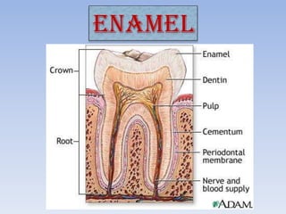
  • 2. 1-enamel is ectodermal in origins 2-it is derived from inner enamel epithelium of enamel organ. 3-mature enamel is completely non cellular Ameloblasts are derived from outer enamel epithelium) Mature enamel is completely acellular (True or false )
  • 3. Physical characters 1-Thickness 2-Color 3- Hardness 4- Tensile strength 5-Permeability 6-Solubility
  • 4. thickness It is thick at the incisal edge and cusp tip of molars and premolars (2-2.5 mm) and ends cervically as knife edge The thickest part of the enamel is present in---------, and………….
  • 5. color Thick enamel---------------------- bluish white Decrease in thickness-----------------------yellow due to the underlying dentin Healthy thick enamel is -------- in color, while thin enamel is …………in color
  • 6. hardness Enamel is the hardest calcified tissue in the body Because of its high content of minerals. Enamel can withstand load of mastication and resist wear process. ------- is the hardest calcified tissue in the body. Enamel is the hardest calcified tissue in the body (give a reason)
  • 7. Tensile strength Although it is hard , enamel is extremely brittle and depends on the strength of the underlying dentin. Tensile strength of enamel is greater than that of dentin ( true or false)
  • 8. permeability Enamel is selectively permeable, permitting partial or complete passage of ions due to the presence of microscopic pores. Enamel is completely impermeable to ions (False or true)
  • 9. solubility Enamel dissolves in acid media. Its surface is less soluble than deep enamel.
  • 10. Chemical composition Mature enamel consists of: Mainly(96%) inorganic substance Small (3%) amount of organic substance & 1%) water
  • 11. Inorganic component Mainly calcium phosphate crystals (hydroxyapatite crystals)
  • 12. organic component Mainly non collagenous proteins: A- amelogenins B- non-amelogenins
  • 13. Microscopic structure 1-Rods (prism). 2- Rod sheath. 3-Inter rodal cement substance
  • 14. Ground section Decalcified section the organic the inorganic substance is burnt substance is dissolved and the inorganic and the organic substance remain substance remain
  • 15. Enamel rods 1- Course. 2- Number. 3- Transverse striation. 4- Direction. 5- Gnarled enamel.
  • 16. Course: the enamel rod starts straight at dentino-enamel junction (D.E.J.) for about 30 µ then has a wavy course till near the outer surface of enamel where it become straight once more.
  • 17. The number of the enamel rods varies from 5 millions in lower lateral incisor to 12 millions in the upper first perm.The number of the rods equals the number of the ameloblasts. anent molar. At the tooth surface there are about 20000- 30000 enamel rods in 1 mm2 The density of the rods is at the DEJ about 10% higher than at the enamel surface. The total number of enamel rods varies from------- to ---------
  • 19.
  • 22. Direction of rods permanent tooth Deciduous tooth
  • 23. Gnarled enamel Twisted Gnarled course of enamel enamel rods D Gnarled enamel is twisted and intertwined rode structure associated with increase strength of the enamel ,Present mainly in the incisal edges and tip of the cusps. (enamel rods develop in planes of tension). Give short account on gnarled enamel
  • 25. Rod sheath L.M. distinct thin layer peripheral to the rods Different refractive index, darker and more acid resistant, and less calcified and more organic. E.M. not distinct layer, but organically rich inter- rod space devoid of crystals.
  • 26. Inter rod substance L.M. distinct cement substance with higher refractive index. E.M.tail of the adjacent rod with different direction of the crystals
  • 28. NEONATAL LINE Prenatal enamel Neonatal line Postnatal enamel
  • 29. PERIKYMATA The external manifestation of the incremental lines of Retzius represented as transverse wave like grooves on the surface of the enamel are known as perikymata.
  • 30. HUNTER SCHREGER BANDS The dark bands (Diazones) absorb the • light where the light bands (Parazones) reflect the light.
  • 32. ENAMEL SPINDLE It makes the area hypersensitive to pain Enamel spindles are : A- odontoblastic processes cross the dentinoenamel junction. B- remnants of ameloblasts. C- enamel rods develop in planes of tension.
  • 33. Surface structure It is relatively structure-less layer covers the cervical region of the teeth. 30um in thickness in 70% of People, and it is hyper-mineralized.
  • 34. ENAMEL CUTICLE Primary cuticle secondary cuticle It is delicate membrane covers It is non cellular keratinized layer the crown of the newly formed by reduced enamel Erupted tooth. It has epithelium after tooth eruption, the same Structure as After removal of the primary one basement membrane by wear and brushing
  • 35. ENAMEL pellicle It is a glycoprotein of saliva that covers the tooth immediately after eruption. It reforms within hours after removal. ENAMEL plaque Within a day or two after the pellicle has formed it colonized With microorganisms to form bacterial Plaque.