Glaucoma Detection and Treatment Options
•Télécharger en tant que PPS, PDF•
495 j'aime•157,468 vues


Recommandé
Contenu connexe
Tendances
En vedette
En vedette (20)
Similaire à Glaucoma Detection and Treatment Options
Similaire à Glaucoma Detection and Treatment Options (20)
Plus de Hossein Mirzaie
Plus de Hossein Mirzaie (20)
Dernier
Dernier (20)
Glaucoma Detection and Treatment Options
- 1. Glaucoma The silent thief of sight
- 2. Glaucoma Glaucoma Types, diagnosis, and Treatment
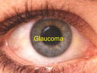
- 3. Glaucoma In general, Glaucoma occurs as a result of increased intraocular pressure (IOP) caused by a malformation or malfunction of the eyes drainage system. Normal IOP is 19 – 21 inches of mercury. The increased pressure causes compresssion of the retina and the optic nerve, and causes progressive , PERMANENT loss of eyesight if left untreated.
- 5. Glaucoma Primary Open Angle Glaucoma
- 6. Glaucoma Primary Open Angle Glaucoma is caused when the normal drainage system of the eye becomes partially blocked, causing pressure to build within the eye. Normal Eye
- 7. Glaucoma Primary Open Angle Glaucoma is caused when the normal drainage system of the eye becomes partially blocked, causing pressure to build within the eye. Glaucoma usually affects the perimitery vision first, with sight gradually being lost towards the center of the eye. Vision loss with Glaucoma
- 8. Glaucoma Primary Open Angle Glaucoma Normal Eye Pressure builds when the drainage system is blocked. This increasing pressure presses against the Optic Nerve and causes a gradual loss of sight. Total loss of vision
- 10. Glaucoma Angle – closure Glaucoma (Acute Glaucoma)
- 11. Angle Closure Glaucoma (Sometimes referred to as Narrow Angle Glaucoma) is caused when the normal drainage system of the eye becomes suddenly blocked, causing pressure to build within the eye at a very rapid rate. Complete blindness can occur in as little as 3 to 5 days! Normal Eye Sudden blockage causes pressure to build rapidly. Glaucoma
- 14. Glaucoma Congenital Glaucoma results as a condition from birth. Children are born with conditions such as an abnormal development of the Anterior Chamber angles which prohibit the normal drainage of fluid from the eyes, which then causes an increase in the pressure within the eye, and subsequent Retinal and Optic Disc damage.
- 15. Glaucoma Parents normally are the first to recognize the symptoms of Congenital Glaucoma: Cloudiness of the cornea due to Edema Distension of the eye Photophobia (sensitive to light)
- 16. Glaucoma In most cases, numerous surgeries are required to correct Congenital Glaucoma. Lasers are sometimes used, as well as Filtration Surgery and insertion of Tube shunts:
- 18. Glaucoma Secondary Glaucoma is usually the result of a trauma to the eye, although it can develop due to several causes: Abnormal deposits in the eye fluid Uveitis Lens Changes Drugs Haemorrhage
- 20. Glaucoma Pigmentary Glaucoma can develop as a result of small pieces of the Iris breaking off. These small particles can lodge themselves in the normal drainage canals and subsequently interfere with the normal drainage of fluids from the eye.
- 21. Glaucoma Normal Tension Glaucoma
- 22. Glaucoma Normal Tension Glaucoma occurs when there is damage to the Optic nerve detected in patients who have completely normal Inter – Occular pressure. It has the same characteristics as Primary Open – Angle Glaucoma.
- 23. Glaucoma Diagnosis and Treatment
- 24. Diagnosis Tonometry is often used as a diagnostic tool. The Tonometer is gently pressed against the eyeball, and the resistance (internal pressure) is measured. This requires that the eye be numbed prior to the test. Gonioscopy can be used to determine if the angle where the iris meets the cornea is open or closed. Glaucoma
- 25. Diagnosis Perimetry is an essential method used to determine if there is any loss of the visual field. Glaucoma
- 26. Primary Open Angle Glaucoma Diagnosis Slit Lamp Examination is another method of diagnosis of patients with suspected Glaucoma. Glaucoma
- 28. Measurement of Retinal Nerve Fiber Layer thickness with the StratusOCT is the most recent advancement in technology that aids in the diagnosis of Glaucoma. Glaucoma