Management of deviated midline
•
33 likes•10,541 views

This document discusses the management of deviated midlines. It notes that differential diagnosis is important to determine the cause of the midline deviation and appropriate treatment. Treatment may involve correcting dental asymmetries through orthodontics or expanding a narrow maxillary arch first if caused by a posterior crossbite. Functional appliances can be used to shift the mandible into the proper position if a skeletal deviation is present. Surgical correction may be needed for true skeletal asymmetries. The goal is to adapt the occlusion and correct dental or facial asymmetries causing the deviated midline.

Recommended
More Related Content
What's hot
What's hot (20)
Viewers also liked
Viewers also liked (20)
Similar to Management of deviated midline
Similar to Management of deviated midline (20)
More from Indian dental academy
More from Indian dental academy (20)
Recently uploaded
Recently uploaded (20)
Management of deviated midline
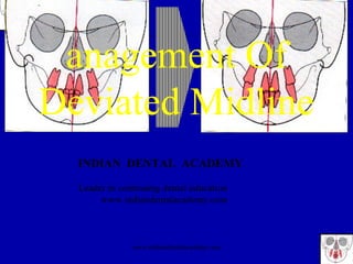
- 1. anagement Of Deviated Midline INDIAN DENTAL ACADEMY Leader in continuing dental education www.indiandentalacademy.com www.indiandentalacademy.com
- 2. Contents Introduction Definition Classification Etiological factors causing the midline deviation Diagnosis of the midline deviation Management of the deviated midline www.indiandentalacademy.com
- 3. Introduction Perfect bilateral symmetry is largely a theoretical concept that seldom exists in the living organisms. Asymmetry of the face and dentition is a naturally occurring phenomenon. Relative symmetry and midline coordination are basic to an appreciation of facial harmony and balance.www.indiandentalacademy.com
- 4. Definition Symmetry is defined as equality or correspondence in forms of parts distributed around a centre or an axis at the two extreme pole or on the two opposite sides of the body. Asymmetry in craniofacial areas can be recognized as difference in size or relationship of the two sides of the face. This may be a result of discrepancies in the form of individual bones or malposition of one or more bones in the craniofacial complex, or the asymmetry may be confined to the soft tissue. www.indiandentalacademy.com
- 5. Classification Sarver evaluates the facial symmetry under the following reference planes Nasal tip to the mid sagittal plane Maxillary dental midline to the mid sagittal plane Maxillary dental midline to mandibular dental midline Mandibular dental midline to mid symphysis Mid symphysis to the mid sagittal plane www.indiandentalacademy.com
- 6. Midlines can also be classified as Dental midline – related to the symmetry of the dentition of the maxilla and mandible Functional midline - related to the functions of the stomatognathic system Skeletal midline – related to the symmetry of the osseous structures of the craniofacial region Soft tissue midline – related to the symmetry of the soft tissue of the craniofacial regionwww.indiandentalacademy.com
- 7. Functional midline Centric relation is the relationship of the mandible to the cranium when the condyles are in an orthopaedically stable position. It is the most retruded position of the condyle.{superoanterior position} Centric occlusion is the maximum intercuspation of the teeth Postural rest position - the synergistic and the antagonistic muscular components are in dynamic equilibrium. The balance is maintained with minimal basic muscle tonus.www.indiandentalacademy.com
- 8. Postural rest position Postural rest position Myostatic anti stretch reflex Permanent exogenous factors Gravity Dependent and altered with the head positionwww.indiandentalacademy.com
- 9. Movement of the mandible During the closing maneuver from the rest position to the habitual occlusion two phases of the movement can be observed The free phase - from the postural rest position to the point of initial contact or occlusal prematurity The articular phase - from the initial contact position to the centric occlusion or habitual occlusal position www.indiandentalacademy.com
- 10. Laterocclusion When the occlusal prematurity is present Midline shift observed only during the centric occlusion or intercuspation. During the postural rest position the midlines are coincident and well centered . The mandible slides laterally from the rest position into a cross bite and is caused by the tooth guidance,after the initial contact of the free phase LATEROCCLUSION also called the pseudo cross bitewww.indiandentalacademy.com
- 11. Mandibular dental midline coincident with the facial midline in postural rest position Mandibular dental midline shifted to the right in habitual occlusion www.indiandentalacademy.com
- 12. Postural rest position Initial contact Habitual occlusion www.indiandentalacademy.com
- 14. Laterognathy Midline shift is present in both centric occlusion and in the postural rest position This condition is generally seen in the true asymmetry of the mandible Functional appliances have poor prognosis Surgical correction required LATEROGNATHY www.indiandentalacademy.com
- 15. Mandibular dental midline not coincident with the facial midline in postural rest position Mandibular dental midline not coincident with the facial midline in habitual occlusion www.indiandentalacademy.com
- 16. Etiology One of the common etiologic factor for the deviation of midline, irrespective of the type of the midline shift is GENETICS – due to the genetic imperfections in the mechanism which was meant to create symmetry and environmental factors producing decided right and left differences Examples – multiple neurofibromatosis- familial incidence associated with dominant gene Hemifacial microsomia Cleft lip and palatewww.indiandentalacademy.com
- 17. Lundstorm classified the etiological factors as Genetic Non genetic combination Another classification by the same author Qualitative – Size of the teeth Location in the arches Position of the arches Quantitative Differences in the number of teeth on each side or the presence of cleft lip or palatewww.indiandentalacademy.com
- 18. Midline diastema Mesiodens Generalized spacing Frenal attachments Congenital absence of a tooth Morphological variation of a tooth eg. Microdontia or macrodontia Asymmetrical exfoliation Retained deciduous teeth Early loss of primary teeth Crowding Trauma Habits such as thumb sucking and tongue thrusting Traumatic occlusion leading to pathological migration Pathological condition such as cysts EtiologyDental midline shift www.indiandentalacademy.com
- 20. The upper right central incisor has shifted to the right due to the congenital absence of lateral incisor The contact point upper central incisor do not coincide with the center of the philtrum www.indiandentalacademy.com
- 21. Functional midline shift Neurological disturbances Disturbances in the tooth – tooth inter relationship Anterior cross bite Posterior cross bite Contracted maxillary arch Any other occlusal prematurity preventing the smooth closure from the free phase to the articular phase Compensation of a skeletal discrepancy Etiology www.indiandentalacademy.com
- 23. Skeletal midline shift Genetics Local Trauma Ankylosis Damage to nerve – loss of muscle function and tone Pathological state in the form of cysts and tumors Unilateral posterior edentulous area Post operative sequale of orthognathic surgery Etiology Temperomandibular joint Systemic Intrauterine pressure during pregnancy and significant pressure in the birth canal Condylar resorption Rheumatoid arthritis Systemic Lupus Erythamatosis Sjogren’s syndrome Marfan’s syndromewww.indiandentalacademy.com
- 24. Soft tissue midline shift Neurological disturbances such as cerebral palsy and Hemifacial microsomia Massetric hypertrophy Trauma Scars including surgical scars Dermatomyositis Neoplasm Adaptation to the existing skeletal asymmetries Etiology www.indiandentalacademy.com
- 25. Diagnosis Asymmetry of the face is one of the more difficult problems with which the orthodontists have to contend and which presents serious diagnostic problems. The recognition of actual site of asymmetry is essential for correct treatment planning. The point at which normal symmetry becomes abnormal cannot be easily identified and is often identified by the clinician’s sense of balance and patient’s perception of their imbalance. Vig and Hewitt studywww.indiandentalacademy.com
- 26. Diagnostic aids History Clinical examination Photographic evaluation Radiographic examination Orientation of the cast Occlusograms Computed tomography Video cepalometry Multi view fluoroscopy www.indiandentalacademy.com
- 27. History Patient history is important for the diagnosis, as it aids in the knowledge of Exfoliation of the primary teeth Extractions undergone if any Trauma Familial tendencies Congenital problems Surgical procedurewww.indiandentalacademy.com
- 28. Clinical examination Frontal view evaluation Nasal tip to mid sagittal plane Maxillary dental midline to midsagittal plane Mandibular dental midline to midsagittal plane Mandibular dental midline to midsymphysis Midsymphysis to midsagittal plane Evaluation of dental midline in the Mouth open Centric relation At initial contact Centric occlusion www.indiandentalacademy.com
- 29. Other features to be noted during clinical examination are Missing and supernumerary teeth Tooth shape and size Arch form symmetry Frenal attachments www.indiandentalacademy.com
- 32. Advantages Inexpensive No exposure to potentially harmful radiation Better evaluation of the harmony relationship among the craniofacial structure including the contribution of muscle and adipose tissue Readily used to posture the head and face and to compare these with the relationship existing among the different craniofacial structureswww.indiandentalacademy.com
- 34. Occlusograms 1992, JCO, Occlusograms in Orthodontic Treatment Planning - RICHARD D. FABER, Lower occlusal tracing placed over arch symmetry chart to establish midline and perpendicular reference crosshairs. www.indiandentalacademy.com
- 35. Radiographs OPG Posteroanterior view (PA view) Submento vertex Lateral cephalometric radiograph IOPA www.indiandentalacademy.com
- 36. Orthopantamogram Temperomandibular joint can be viewed Asymmetry of the body or ramus of the mandible Missing or supernumerary teeth Pathological condition like cysts and neoplasmwww.indiandentalacademy.com
- 37. Rickett’s analysis Svanholt and Solow Grummons analysis Grayson analysis Hewitt analysis Posteroanterior view www.indiandentalacademy.com
- 38. Landmarks of Grummons analysis JCO, 1987, A Frontal Asymmetry Analysis – Duane et alwww.indiandentalacademy.com
- 41. JCO 1982: Orthognathic and Craniofacial Surgical Diagnosis and Treatment Planning: A Visual Approach. Farhad et al Horizontal and vertical lines are drawn to indicate areas of asymmetry. www.indiandentalacademy.com
- 42. Posteroanterior view Disadvantages Midline assessment is difficult Difficulty in reproducing head posture Difficulty in identifying landmarks because of superimposition of structures Exposure to radiation www.indiandentalacademy.com
- 43. Submentovertex (SV) Berger was the first to suggest the use of SV (problems and promises of basilar view cephalogram) in orthodontics. Gibert associated the film cassette parallel to the FH plane Pearson found exceptional degree of symmetry in the sphenoid bone Keith and Campion used sphenoid bone as a fixed reference in comparing the development of growth of skull Marmary and associates showed that perpendicular bisector of a line joining the foramina spinosa was a reliable and accurate midline Ritucci and burstone developed the ceph system for the assessment of craniofacial regionwww.indiandentalacademy.com
- 46. Diagnosis and treatment planning of skeletal asymmetry with submento vertex radiograph, 1984 AJO, Forsberg et al www.indiandentalacademy.com
- 48. Basilar multi plane cephalometric analysis Put forward by Grayson et al 1985, AJO Developed to facilitate the measurement of the craniofacial complex from the submento vertex view. Basilar view is a two dimensional representation of a three dimensional object The cranium can be reconstructed in 3 dimensions from the basilar view cephalograph by separately tracing each of the three suggested horizontal plane www.indiandentalacademy.com
- 49. Multi View Fluoroscopy Permits three dimensional analysis of the oropharyngeal components in motion Combines the lateral, frontal and basal projection Contrast medium is used to define the soft tissue landmarks and to determine their function during the variety of functions of the stomatognathic system. www.indiandentalacademy.com
- 50. Management Differential diagnosis and appropriate inter arch and intra arch mechanotherapy is necessary to determine and correct the midline problem. Review of literature Breakspear advocates adapting the occlusion by ‘stoning’- occlusal equilibration. This mode of treatment allows the settling of occlusion to function better but not to correct the dental or facial asymmetry. www.indiandentalacademy.com
- 51. Paul Lewis – dental asymmetries are more commonly seen with class II malocclusion The correction of mid line caused by the shift of the mandible or rotation of the mandible is attempted only after teeth in both arches are put into quite ideal occlusion. In midline deviation that occurs with posterior cross bite,the narrow maxillary arch must be expanded first. A class II elastic worn from a hook or sliding yoke on the side towards which the mandible has shifted. A second elastic is worn across the anterior teeth to swing or pivot the mandible until midline correction is achieved. www.indiandentalacademy.com
- 52. Angle’s treatment modality . From Angle EH. Malocclusion of the teeth, 1907. Angle suggests Class III elastic with tandem anterior diagonal elastic in conjunction with area expansion for correction of midline discrepancies. www.indiandentalacademy.com
- 53. Begg mechanotherapy . From Begg PR, Kesling P. Begg orthodontic theory and technique, 3rd ed. Space-closing elastics and Class II intermaxillary elastics applied at start of second stage of treatment. Anterior diagonal elastics,class II elastics and class III elastics and Uprighting springs (Mollenhaeur) www.indiandentalacademy.com
- 54. Wick Alexander Midline is corrected during the finishing stages ¼ inch, 6 ounce elastic ,one end attached to maxillary lateral bracket and adjacent central bracket. The other end attached to mandibular lateral incisor bracket on the opposite side. In case of class II tendency, a class II elastic is worn and in class III tendency,class III elastics are worn. The class II elastic is attached to the maxillary lateral incisor and mandibular second molar. The two elastics impart roughly parallel force vectors. www.indiandentalacademy.com
- 56. In cases of midline discrepancy in class I buccal relationship, only the midline elastic is worn, extra class II or class III elastics are not worn. Midline elastics are worn during the finishing stages of active treatment with one exception. In an extraction case during space closure the midline may be shifted significantly, during space closure the elastic can be attached to the closing loops. This will help to control the direction of space closure, thus improving the midline. Over correction Ideal occlusion and midline www.indiandentalacademy.com
- 57. Correction of midline in class II subdivision Suppose class II on right and class I on left, the midline is generally shifted to the left To re establish the midline the extraction pattern would be Upper arch – left side – second premolar right side – first premolar Lower arch - left side – first premolar right side – second premolar 4 5 5 4 www.indiandentalacademy.com
- 58. Gianelly Introduced a biomechanical system with second order bends to move the teeth distally and create space for the midline correction Class II and class III elastics are used to enhance the couple force systems Gianelly AA, Paul IA. A procedure for midline correction. AJO 1970www.indiandentalacademy.com
- 59. Strang Double vertical spring loop auxiliary adjusted for the mass movement of the four incisor teeth to the left. From strang R, Thompson W. A textbook of orthodontia, 1958. www.indiandentalacademy.com
- 60. Source: AJO-DO , 1990 Jun : The midline – diagnosis and treatment, Jerrold www.indiandentalacademy.com
- 61. Profitt Minor midline discrepancy can be corrected during the finishing stages Large discrepancy correction becomes difficult after the closure of extraction spaces A correct maxillary midline is more important for good facial aesthetics and mild mandibular midline creates no esthetic difficulty Use of class II or class III elastics bilaterally with a heavier force on one side Combination class II and class III elastics www.indiandentalacademy.com
- 63. Parallel cross elastics used to correct mild tranverse discrepancy leading to the lateral mandibular shift late in the treatment Anterior diagonal elastics with rectangular arch wire in the lower arch and a round wire in the upper to shift the maxillary arch www.indiandentalacademy.com
- 64. When midlines are deviated to the opposite side,correction accomplished with Uprighting springs Functions are normal Not healthy from periodontal point of view Esthetic results are poor www.indiandentalacademy.com
- 78. Careful attention to midline coordination and attendant facial symmetry helps to achieve Maximum intercuspation Normal function with anterior disocclusion and without any loading of the anteriors Stability in the finished result Promotion of anterior dental and facial esthetics Decreased potential for Temperomandibular joint dysfunction www.indiandentalacademy.com
- 79. Functional midline deviation The functional midline shift can be corrected by unlocking the mandible Removal of the occlusal prematurities Expansion of the upper arch Functional appliances Inter arch elasticswww.indiandentalacademy.com
- 80. Surgical options Nasal tip to midsagittal plane Rhinoplasty Camouflaging grafting of the tip of and /or the dorsum Maxillary dental midline Subapical procedure to rotate midlines Mandibular dental midline to symphysis Subapical procedures to rotate the mandible Mandibular asymmetry{functional mandibular shift} Two or three piece maxillary expansion via Le fort I osteotomy Surgically assisted maxillary expansionwww.indiandentalacademy.com
- 81. True mandibular asymmetry Distraction osteogenesis Bilateral ramal osteotomies Camouflage through bone grafting or alloplastic augmentation Transverse cant of the maxilla Maxillo mandibular surgery Chin asymmetry Rotational genioplasty Lateral or vertical movement of chin via inferior border osteotomy Camouflage via bone graft,ostectomy or alloplastic augmentation www.indiandentalacademy.com
- 82. Soft tissue asymmetry Augmentation with bone grafts, alloplastic material and silicone implants to re contour the desired areas of the face Muscular stripping www.indiandentalacademy.com
- 86. Source: Angle Orthodontist on CD-ROM (Copyright © 1998 Angle Orthodontist, Inc.), 1995 No. 3, 233 - 239: Figures. Figure 4 Schematic picture of a face including reference plane A which is positioned between the bilateral landmarks Exocanthion, while it is defined perpendicular in all directions to the line through these landmarks. www.indiandentalacademy.com
- 87. Ajo 1991 Assessment of structural and displacement mandibular asymmetries - Schmid, Mongini, and Felisio The following conclusions may be drawn: 1. In the growing patient, craniomandibular asymmetry with transverse deviation of the mandible and the chin, with no genetic or congenital origin and without a history of trauma, infection, or tumor, is possibly the result of mandibular displacement consequent to occlusal alterations. 2. If the mandibular displacement is not detected and treated in a timely manner, adaptive mandibular asymmetry may develop. 3. Depending on the elapsed time between the onset of mandibular displacement and the examination, the patient can show displacement asymmetry, structural asymmetry, or a combination of both. The last possibility may be the most frequent in a population of growing patients. 4. The different patterns of asymmetry can be identified and to some extent quantified in each patient. 5. Successful treatment during the growing period is possible in some patients. If the subject remains untreated, asymmetry can become a permanent feature in the adult. 6. However, mandibular displacement may not be all or even part of the cause of a craniomandibular dysfunction. In such cases any kind of orthopedic treatment may be completely or partially ineffective. 7. Because the symmetry in one of the control subjects improved in the absence of intervention, other factors besides treatment may be responsible for the different www.indiandentalacademy.com
- 89. Correction of arch asymmetries as suggested by Lewis www.indiandentalacademy.com Thank you For more details please visit www.indiandentalacademy.com