General anatomy of urinary system ppt
•Download as PPT, PDF•
256 likes•186,895 views

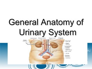
The urinary system, also known as the renal system or urinary tract, consists of the kidneys, ureters, bladder, and the urethra. The purpose of the urinary system is to eliminate waste from the body, regulate blood volume and blood pressure, control levels of electrolytes and metabolites, and regulate blood pH.

Recommended
More Related Content
What's hot
What's hot (20)
Similar to General anatomy of urinary system ppt
Similar to General anatomy of urinary system ppt (20)
More from jagan _jaggi
More from jagan _jaggi (20)
Recently uploaded
Recently uploaded (20)
General anatomy of urinary system ppt
- 1. General Anatomy ofGeneral Anatomy of Urinary SystemUrinary System
- 2. IntroductionIntroduction TheThe urinary systemurinary system, also known as, also known as thethe renal systemrenal system The urinary system refers to the structuresThe urinary system refers to the structures that produce and conductthat produce and conduct urineurine to theto the point of excretion.point of excretion.
- 3. URINARY SYSTEM ORGANSURINARY SYSTEM ORGANS Kidneys (2)Kidneys (2) Ureters (2)Ureters (2) Urinary bladderUrinary bladder UrethraUrethra
- 5. KidneyKidney The human body normally has two pairedThe human body normally has two paired kidneyskidneys, one on the left and one on the, one on the left and one on the right.right. The functional unit of the kidney isThe functional unit of the kidney is nephron.nephron. Urine is formed byUrine is formed by nephronsnephrons
- 6. Location and External Anatomy ofLocation and External Anatomy of KidneysKidneys LocatedLocated retroperitoneallyretroperitoneally Lateral to TLateral to T1212–L–L33 vertebraevertebrae Average kidneyAverage kidney 12 cm tall, 6 cm wide,12 cm tall, 6 cm wide, 3 cm thick3 cm thick
- 7. Protected by three connective tissueProtected by three connective tissue layerslayers Renal fasciaRenal fascia Attaches to abdominal wallAttaches to abdominal wall Adipose capsuleAdipose capsule Fat cushioning kidneyFat cushioning kidney Renal capsuleRenal capsule Fibrous sacFibrous sac Protects from trauma and infectionProtects from trauma and infection
- 9. KIDNEYKIDNEY Gross anatomyGross anatomy Renal parenchymaRenal parenchyma Renal sinusRenal sinus
- 10. KIDNEY ANATOMYKIDNEY ANATOMY Renal parenchymaRenal parenchyma Two zonesTwo zones Outer cortexOuter cortex Inner medullaInner medulla
- 12. Anatomy of the kidneysAnatomy of the kidneys Superficial outer cortex and inner medullaSuperficial outer cortex and inner medulla The medulla consists of 6-18 renalThe medulla consists of 6-18 renal pyramidspyramids The cortex is composed of roughly 1.25The cortex is composed of roughly 1.25 million nephronsmillion nephrons
- 13. KIDNEY ANATOMYKIDNEY ANATOMY Renal parenchymaRenal parenchyma Renal pyramidsRenal pyramids Extensions of cortex (renal columns)Extensions of cortex (renal columns) divide medulla into 6 – 10divide medulla into 6 – 10 renalrenal pyramidspyramids Pyramid + overlying cortex = LobePyramid + overlying cortex = Lobe Point of pyramid = PapillaPoint of pyramid = Papilla Papilla nested in cup (minor calyx)Papilla nested in cup (minor calyx) 2 – 3 minor calices2 – 3 minor calices Major calyxMajor calyx 2 – 3 major calices2 – 3 major calices Renal pelvisRenal pelvis Renal pelvisRenal pelvis UreterUreter
- 14. KIDNEY ANATOMYKIDNEY ANATOMY Renal sinusRenal sinus Surrounded by renal parenchymaSurrounded by renal parenchyma Contains blood & lymph vessels, nerves,Contains blood & lymph vessels, nerves, urine-collecting structuresurine-collecting structures HilusHilus On concave surfaceOn concave surface Vessels and nerves enter and exitVessels and nerves enter and exit
- 15. Major and minor calyces along with theMajor and minor calyces along with the pelvis drain urine to the ureterspelvis drain urine to the ureters
- 16. Human Anatomy, 3rd editionHuman Anatomy, 3rd edition Prentice Hall, © 2001Prentice Hall, © 2001 Structure of the KidneyStructure of the Kidney
- 18. NEPHRONSNEPHRONS NephronsNephrons Functional units of kidneyFunctional units of kidney ~1.2 million per kidney~1.2 million per kidney Three main partsThree main parts Blood vesselsBlood vessels Renal corpuscleRenal corpuscle Renal tubuleRenal tubule
- 19. Blood vessels servicing kidneyBlood vessels servicing kidney GlomerulusGlomerulus Fenestrated capillariesFenestrated capillaries Capillary filtration in glomerulus initiates urineCapillary filtration in glomerulus initiates urine productionproduction Filtrate lacks cells & proteinsFiltrate lacks cells & proteins Drained by efferent arterioleDrained by efferent arteriole Peritubular capillariesPeritubular capillaries Renal veinRenal vein
- 21. Renal corpuscleRenal corpuscle Composed of aComposed of a glomerulusglomerulus and theand the Bowman's capsuleBowman's capsule,, TheThe renal corpusclerenal corpuscle is the beginning ofis the beginning of the nephron.the nephron. It is the nephron's initial filteringIt is the nephron's initial filtering component.component.
- 22. GlomerulusGlomerulus TheThe glomerulusglomerulus is ais a capillarycapillary tuft thattuft that receives its blood supply from an afferentreceives its blood supply from an afferent arteriolearteriole of theof the renal circulationrenal circulation.. The glomerular blood pressure providesThe glomerular blood pressure provides the driving force for water and solutes tothe driving force for water and solutes to be filtered out of the blood and into thebe filtered out of the blood and into the space made byspace made byBowman's capsuleBowman's capsule
- 23. The remainder of the blood passes intoThe remainder of the blood passes into the efferent arteriole.the efferent arteriole. The diameter of efferent arterioles isThe diameter of efferent arterioles is smaller than that of afferent arterioles,smaller than that of afferent arterioles, increasing the hydrostatic pressure in theincreasing the hydrostatic pressure in the glomerulus.glomerulus.
- 24. Bowman's capsuleBowman's capsule The Bowman's capsule, also called theThe Bowman's capsule, also called the glomerular capsule.glomerular capsule. surrounds the glomerulus.surrounds the glomerulus. It is composed of a visceral inner layerIt is composed of a visceral inner layer formed by specialized cellsformed by specialized cells called podocytes.called podocytes. Parietal outer layer composed of simpleParietal outer layer composed of simple squamous epithelium.squamous epithelium.
- 25. Fluids from blood in the glomerulus areFluids from blood in the glomerulus are filtered through the visceral layer offiltered through the visceral layer of podocytes, resulting in the glomerularpodocytes, resulting in the glomerular filtrate.filtrate.
- 26. NOTENOTE Renal corpuscleRenal corpuscle Glomerulus plus capsuleGlomerulus plus capsule Glomerulus enclosed in two-layered glomerularGlomerulus enclosed in two-layered glomerular capsulecapsule ““Bowman’s capsule”Bowman’s capsule” Fluid filters from glomerular capillariesFluid filters from glomerular capillaries ““Glomerular filtrate”Glomerular filtrate” Fluid collects in capsular spaceFluid collects in capsular space Fluid flows into renal tubuleFluid flows into renal tubule
- 28. Renal tubuleRenal tubule Leads from glomerular capsuleLeads from glomerular capsule Ends at tip of medullary pyramidEnds at tip of medullary pyramid ~3 cm long~3 cm long Four major regionsFour major regions Proximal convoluted tubuleProximal convoluted tubule Nephron loopNephron loop Distal convoluted tubuleDistal convoluted tubule Collecting ductCollecting duct
- 30. Proximal convoluted tubuleProximal convoluted tubule (PCT)(PCT) Arises from glomerular capsuleArises from glomerular capsule Longest, most coiled regionLongest, most coiled region lies in cortexlies in cortex lined by simple cuboidal epithelium with brushlined by simple cuboidal epithelium with brush borders which help to increase the area ofborders which help to increase the area of absorption greatly.)absorption greatly.) Prominent microvilliProminent microvilli Function in absorptionFunction in absorption
- 32. Nephron loop (“Loop of Henle”)Nephron loop (“Loop of Henle”) ““U” – shaped, distal to PCTU” – shaped, distal to PCT lies in medullalies in medulla 2 parts2 parts Descending limb of loop of HenleDescending limb of loop of Henle Ascending limb of loop of HenleAscending limb of loop of Henle
- 33. Ascending limb of loop of HenleAscending limb of loop of Henle The ascending limb of loop of Henle is dividedThe ascending limb of loop of Henle is divided into 2 segments:into 2 segments: Lower end of ascending limbLower end of ascending limb is very thin andis very thin and is lined by simple squamous epithelium.is lined by simple squamous epithelium. The distal portion of ascending limbThe distal portion of ascending limb is thickis thick and is lined by simple cuboidal epithelium.and is lined by simple cuboidal epithelium. Thin ascending limb of loop of HenleThin ascending limb of loop of Henle Thick ascending limb of loop of Henle (entersThick ascending limb of loop of Henle (enters cortex and becomes DCT-distal convolutedcortex and becomes DCT-distal convoluted tubule.)tubule.)
- 34. Thick segmentsThick segments Active transport of saltsActive transport of salts High metabolism, many mitochondriaHigh metabolism, many mitochondria Thin segmentsThin segments Permeable to waterPermeable to water Low metabolismLow metabolism
- 35. Distal convoluted tubule (DCT)Distal convoluted tubule (DCT) Coiled, distal to nephron loopCoiled, distal to nephron loop Shorter than PCTShorter than PCT Less coiled than PCTLess coiled than PCT Very few microvilliVery few microvilli Contacts afferent and efferent arteriolesContacts afferent and efferent arterioles Contact with peritubular capillariesContact with peritubular capillaries
- 37. Collecting ductCollecting duct DCTs of several nephrons empty into aDCTs of several nephrons empty into a collecting ductcollecting duct Passes into medullaPasses into medulla Several merge into papillary duct (~30 perSeveral merge into papillary duct (~30 per papilla)papilla) Drain into minor calyxDrain into minor calyx
- 39. CLASSESCLASSES The two general classes of nephrons areThe two general classes of nephrons are Cortical nephronsCortical nephrons Juxtamedullary nephronsJuxtamedullary nephrons which are classified according to thewhich are classified according to the length of their Loop of Henlelength of their Loop of Henle location of theirlocation of their renal corpusclerenal corpuscle..
- 40. All nephrons have their renal corpusclesAll nephrons have their renal corpuscles in the cortex.in the cortex. CorticalCortical nephrons have their Loop ofnephrons have their Loop of Henle in the renal medulla near its junctionHenle in the renal medulla near its junction with the renal cortex,with the renal cortex, Loop of Henle of juxtamedullary nephronsLoop of Henle of juxtamedullary nephrons is located deep in the renal medulla;is located deep in the renal medulla;
- 42. URINE FORMATIONURINE FORMATION OverviewOverview Blood plasmaBlood plasma UrineUrine Four stepsFour steps Glomerular filtrationGlomerular filtration Tubular reabsorptionTubular reabsorption Tubular secretionTubular secretion Water conservationWater conservation
- 46. KIDNEY FUNCTIONSKIDNEY FUNCTIONS Regulate blood volume, pressureRegulate blood volume, pressure Regulate fluid osmolarityRegulate fluid osmolarity Secrete reninSecrete renin Secrete erythropoietin (EPO)Secrete erythropoietin (EPO) Regulate PRegulate PCOCO22, Acid-Base balance, Acid-Base balance Synthesize calcitriol (Vitamin D)Synthesize calcitriol (Vitamin D) Detoxify free radicals, drugsDetoxify free radicals, drugs GluconeogenesisGluconeogenesis
- 47. The UretersThe Ureters Pair of muscular tubesPair of muscular tubes Extend from renal pelvis to the bladderExtend from renal pelvis to the bladder Oblique entry into bladder prevents backflow of urineOblique entry into bladder prevents backflow of urine
- 48. Histology of UreterHistology of Ureter Mucosa –Mucosa – transitionaltransitional epitheliumepithelium MuscularisMuscularis – two layers– two layers Inner longitudinal layerInner longitudinal layer Outer circular layerOuter circular layer AdventitiaAdventitia – typical– typical connective tissueconnective tissue
- 49. Carry urine from kidneys to urinaryCarry urine from kidneys to urinary bladder via peristalsisbladder via peristalsis Rhythmic contraction of smooth muscleRhythmic contraction of smooth muscle Enter bladder from belowEnter bladder from below Pressure from full bladder compressesPressure from full bladder compresses ureters and prevents backflowureters and prevents backflow
- 50. Small diameterSmall diameter Easily obstructed or injured by kidneyEasily obstructed or injured by kidney stones (renal calculi)stones (renal calculi)
- 51. Urinary BladderUrinary Bladder A collapsible muscularA collapsible muscular sacsac Stores and expels urineStores and expels urine Full bladder –Full bladder – sphericalspherical • Expands into theExpands into the abdominal cavityabdominal cavity Empty bladder – liesEmpty bladder – lies entirely within theentirely within the pelvispelvis Figure 23.13
- 52. Urinary BladderUrinary Bladder Wall of bladderWall of bladder Mucosa - transitional epitheliumMucosa - transitional epithelium Muscular layer - detrusor muscleMuscular layer - detrusor muscle AdventitiaAdventitia
- 53. Wrinkles termed rugaeWrinkles termed rugae Openings of ureters common site forOpenings of ureters common site for bladder infectionbladder infection Urinary bladderUrinary bladder
- 54. UrethraUrethra Conveys urine from bodyConveys urine from body Internal urethral sphincterInternal urethral sphincter Retains urine in bladderRetains urine in bladder Smooth muscle, involuntarySmooth muscle, involuntary External urethral sphincterExternal urethral sphincter Provides voluntary control over voiding of urineProvides voluntary control over voiding of urine
- 55. Urethra in femaleUrethra in female 3 – 4 cm long in females3 – 4 cm long in females Bound by connective tissue to anterior wall ofBound by connective tissue to anterior wall of vaginavagina Urethral orifice exits body between vaginalUrethral orifice exits body between vaginal orifice and clitorisorifice and clitoris
- 57. Urethra in maleUrethra in male ~~18 cm long in males18 cm long in males Prostatic urethraProstatic urethra • ~2.5 cm long, urinary bladder~2.5 cm long, urinary bladder prostateprostate Membranous urethraMembranous urethra • ~0.5 cm, passes through floor of~0.5 cm, passes through floor of pelvic cavitypelvic cavity Penile urethraPenile urethra • ~15 cm long, passes through penis~15 cm long, passes through penis
- 59. URINE ELIMINATIONURINE ELIMINATION Urination (micturition)Urination (micturition) ~200 ml of urine held~200 ml of urine held Distension initiates desire to voidDistension initiates desire to void Internal sphincter relaxes involuntarilyInternal sphincter relaxes involuntarily Smooth muscleSmooth muscle External sphincter voluntarily relaxesExternal sphincter voluntarily relaxes Skeletal muscleSkeletal muscle Poor control in infantsPoor control in infants Bladder muscle contractsBladder muscle contracts Urine forces through urethraUrine forces through urethra
- 60. Figure 26.1 Urinary SystemUrinary System Kidneys – produceKidneys – produce urineurine Ureters –transportUreters –transport urine to bladderurine to bladder Urinary bladder -Urinary bladder - stores urinestores urine Urethra transportsUrethra transports urine to exteriorurine to exterior
- 61. Functions of the urinary systemFunctions of the urinary system Homeostatic regulation of blood plasmaHomeostatic regulation of blood plasma Regulating blood volume and pressureRegulating blood volume and pressure Regulating plasma ion concentrationsRegulating plasma ion concentrations Stabilizing blood pHStabilizing blood pH Conserving nutrientsConserving nutrients
- 62. Filter many liters of fluid from bloodFilter many liters of fluid from blood Excretion - The removal of organic wasteExcretion - The removal of organic waste products from body fluidsproducts from body fluids UreaUrea Uric acidUric acid CreatinineCreatinine Elimination - The discharge of waste productsElimination - The discharge of waste products into the environmentinto the environment
Editor's Notes
- FG26_03A1.JPG Title: Structure of the Kidney Notes: (a)Frontal section through kidney. (b)Shadow drawing showing arrangement of calyces and renal pelvis. (c)Urogram showing calyces, renal pelvis, and ureter. Keywords: kidney, frontal, cortex, medulla, renal sinus, renal capsule, hilus, ureter, renal papilla, renal capsule, renal columns, minor calyx, major calyx, renal pelvis, renal pyramids