Frontal sinus fracture
•Download as PPTX, PDF•
17 likes•2,215 views

frontal sinus fracture management

Recommended
More Related Content
What's hot
What's hot (20)
Similar to Frontal sinus fracture
Similar to Frontal sinus fracture (20)
More from Jamil Kifayatullah
More from Jamil Kifayatullah (20)
Recently uploaded
Recently uploaded (20)
Frontal sinus fracture
- 1. Frontal sinus fracture Dr jameel kifayatullah
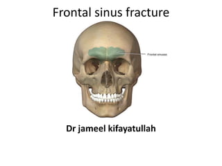
- 2. Frontal sinus anatomy. The anterior table of the frontal sinus is thick bone and provides forehead contour. The posterior table is thinner and constitutes a portion of the anterior cranial fossa. The floor or the sinus makes up a portion of the orbital roof. The frontal sinus ostia is located in the medial, posterior, and inferior portion of the sinus floor
- 3. Frontal sinus fracture patterns. (a) Normal relationship. (b) Anterior table. (c) Comminuted anterior and posterior table.
- 4. Management • Treatment Goals • Surgical access • Osseous Recovery And Access • Intraoperative Evaluation of Nasofrontal outflow • Anterior Table fracture • Posterior Table fracture • Orbital roof and supraorbital Bar reconstructon • Nasofrontal outflow tract obstruction • Sinus obliteration • Endoscopy in Management of frontal fracture
- 5. Classification of frontal sinus fracture • Anterior Table fracture • Posterior Table fracture • Combination of posterior table fracture and anterior table fracture • Combinations of fracture that compromise the NFOT Fractures of the anterior table and posterior table Fractures of anterior table and NOE Fractures of anterior table and medial superior orbital rim
- 6. Anatomic parameters that need to be assessed when developing a treatment plan for frontal sinus fractures. Yellow—anterior table; Red—posterior table; Blue— frontal recess; Green—dural integrity.
- 7. Indication of isolated Anterior Table fracture • To prevent cosmetic deformities
- 8. Indication of posterior table fracture alone or in combination with anterior table fracture • To avoid neurologic sequelae including meningitis and brain abscess
- 9. Combination of fractures that compromise the NFOT Indications • Prevention of mucoceles and pyoceles
- 10. Treatment Goals The primary goals of treating frontal sinus fractures • creation of a safe sinus so that risk of long term complications is minimized and protection of the brain
- 11. Surgical Access • Coronal Approach: provides best access to frontal bone and sinus • Gull wing or spectacle incisions: Unattractive scars • “Open sky” Approach : H-shaped scar over brows and nasion • Butterfly: combination of gull wing and open sky • Sewall : a single side medial orbital incision • Existing laceration
- 12. Surgical Access
- 13. Osseous recovery and Access • Reflection of coronal flap • Release of fragments of anterior table from perisoteum • Organising the fragments • Cleaning of bone fragments • Anterior table removed • Sinus exploration • Posterior table inspection • Sinus floor (NF outflow) evaluation • More extensive neurosurgical procedure:osseous recovery performed in concert with a craniotomy flap
- 14. Intraoperative Evaluation of NFOT • Condition of the frontal sinus floor and nasofrontal outflow-assessed by direct visualisation • Evaluaton of patency of nasofrontal duct by placing an angiocatheter and injection of methylene blue or flourescein—emergence of methylene blue observed beneath the medial turbinate or its collection in posterior pharynx
- 16. Anterior Table Fractures • Simple green stick or non-displaced anterior wall fracture: donot require operative treatment • Displaced anterior table fracture: require reduction • Rule of thumb: displacement> thickness of anterior table
- 18. Anterior Table Fractures • Closely inspect the sinus floor,the posterior wall and patency of nasofrontal duct • If posterior wall and floor free of injury-Fix the pieces of anterior wall with bone plates voids remaining in anterior wall after reconstruction closed by Titanium mesh,MMA or other bone sustitutes • Soft tissue injury repair
- 19. Posterior Table fracture • Three categories 1) Non displaced 2) Displaced 3) Displaced with gross neurologic injury
- 20. Posterior Table fracture • Joint management with neurosurgeon • Antibiotic coverage Surgeon should check for • Any displacement of the fracture • CSF leak • Entrapment of sinus membrane • Dural tears
- 21. Posterior Table fracture • Injury not substantial Nasofrontal duct patent Anterior Table replaced and fixed and soft tissue injuries repaired
- 22. Posterior Table fracture • Comminution of posterior table penetrating injury CSF leak with extensive dural damage or frontal lobe damage : Frontal sinus cranialization –removal of posterior table-dura repair with primary closure,a fascia or synthetic patch or galeal/pericranial flap • Wound closure in layers: Meticulous removal of all the mucosal elements from the walls,cul-de- sacs,and septa of the sinus and from all bone fragments • Failure to remove such elements:mucocele or pyocele
- 23. Posterior Table fracture • Mucosa then reflected down into the nasofrontal outflow • Orifice obstructed by local bone or muscle • Harvested fat placed into sinus and packed until sinus is full • Anterior table reassembled(ORIF)
- 24. Orbital Roof and Supraorbital Bar Reconstruction • Once posterior wall and sinus floor explored,inspected and evaluated for damage; any orbital roof and supraorbital bar fracture reconstructed with titanium mesh
- 25. NFOT OBSTRUCTION • One of the methods of isolating the sinus(or brain) from nasal contmination,by plugging it ith another material • NFOT obstruction necessary to seal off the frontal sinus from nasal contaminants • If NFOT is obstructed sinusitis,meningitis,osteomyelitis may develop • Consider condition of Nasofrontal outflow in fractures of NOE Complex, supraorbital rim or the sinus floor
- 26. OUTFLOW NOT PATENT • Thorough removal of every possible remnant of sinus mucosa by curettage • Any remaining remnants of nasofrontal mucosa then inverted into nose
- 27. Materials used to obstruct NF outflow • Temporal fascia • Temporalis muscle • both • Tensor fascia lata Bone graft material • Sinus septum,inner table or any elsewhere on cranium • Fibrin sealants • Autologous platelet gel • Autologous fibrin glue
- 28. Sinus Obliteration • Adds one more layer to NFOT Seal • Also eliminates the “dead space” or air within the sinus that may permit fluids to accumulate causing a seroma or hematoma
- 29. Methods of obliterating frontal sinus • Inserting no substance or object • Insertion of hydroxylapatite, glass wool, bone, cartilage, muscle, absorbable gelatin sponge , absorbable knitted fabric ,acrylic or fat • Use of pericranial flap or galeal flap for obliteration of frontal sinus
- 30. Endoscopy in Management of frontal sinus fracture • For reduction and fixation of anterior table fracture • Management of frontal sinus drainage system • Secondary management of contour defects of frontal bone
- 31. Aim in treatment of frontal sinus fracture • To create a safe sinus(ie minimize the risk of complications related to injury)
- 32. Concerns in frontal sinus fracture 1) infections particularly Meningitis 6% mortality 2) acute and chronic frontal sinusitis 3) mucocele 4) Mucopyolocele 5) Osteomyelitis
- 33. Classification • Anterior wall fracture • Posterior wall fracture • The floor of the frontal sinus • Combination of the three
- 36. Anterior wall fracture • Undisplaced- treated conservatively • Displced,depressed anterior wall fractures- reduction and fixtion plus sinus preservation
- 38. Fractures of the floor of the frontal sinus • Aim of management: reduce the possibility of development of a frontal mucocele and its sequelae • Two views 1) obliteration of sinus and sealing the frontonasal duct(renoval of sinus mucosa and filling with autogenous material) 2) reconstruction of floor of the sinus and attempting to re-establish drainage by placing a stent
- 39. Posterior wall fractures • Posterior table fracture with significant comminution ,displacement,or CSF leaks : cranialization of the frontal sinus indicated • Cranialization: persistent CSF leak with comminuted displaced posterior wall fracture
- 40. Cranialization procedural steps 1) coronal approach with frontal craniotomy 2) Preservation of anterior pericardial flap 3) Treat intracranial injury /dural repair 4) Remove all sinus mucosa 5) Obliterate frontonasal duct 6) Remove posterior wall and septum 7) Pericardial flap to floor of sinus
- 41. Alternative Strategies • Re-establish fronto-nasal duct patency if the posterior wall is intact(Gerbino et al) • Dural repair,posterior wall reconstruction,sinus obliteration with lyocartilage(Sialer et al)
- 42. Summary • Undisplaced fractures should be managed conservatively • Uncomplicated anterior wall fractures should be managed with reduction and fixation plus sinus preservation • Frontonasal duct involvement should be managed with sinus obliteration • Comminuted ,displaced anterior and posterior wall fractures with CSF leak and frontonasal duct involvement best managed with frontal sinus cranialization
- 43. Summary • Displaced ,comminuted fractures of the posterior wall of the frontal sinus are associated with a high incidence of dural tears • The sinus is cranialised by removing the posterior wall and sinus mucosa,sealing the frontonasal ducts, and using a vascularised galeal frontalis or pericranial flap to cover the sinus floor • Separating the anterior cranial fossa from nasal cavity
- 44. The posterior table bone is removed with a rongeur; cottonoids protect the frontal lobes.
- 45. Mucosa is removed from the anterior table using a diamond burr drill under loupe magnification.
- 46. A sagittal view of layered closure employed in cranialization.
- 47. Anterior table frontal sinus fracture • Comminuted segments reduced and fixated
- 48. Isolated posterior table frontal sinus fracture • Posterior table # with no brain injury or dural tear :conservative management including non treatment is recommended
- 49. Posterior table fracture with concomitant anterior table # • Treatment objectives: determined by displacement and potential for cosmetic deformity
- 50. Posterior table fracture • Significant posterior table fracture leading to brain injury or patients with suspected or established NF duct injury-additional surgical procedures required
- 51. Anterior Table and nasofrontal duct injury • Debridement of sinus membrane and obliteration of sinus and ducts • Reconstruction of anterior table
- 52. Material used for frontal sinus obliteration • Pericranial flap • Temporalis muscle • Abdominal fat
- 53. Materials used for obliteration of NF ducts • Abdominal fat • Temporalis muscle • Bone fragments
- 54. Reconstruction of Anterior Table Options Original fragments pieced together for reconstruction Severe comminution/avulsion:autogenous bone/mesh
- 55. Cranialization • Posterior and anterior tables of the frontal sinus are removed • Removal allows edematous brain to expand within volume of frontal sinus thus decreasing intracranial pressure and further brain injury • Severely fractured posterior table: cranialisation
- 56. OBJECTIVES OF TREATMENT OF FRONTAL SINUS FRACTURE a) Elimination of any factors predisposing to infection b) Preservation of normal sinus infection or,if this is not possible,obliteration of sinus cavity c) The repair of any cosmetic defect
- 57. Classification of frontal sinus fracture A) Anterior wall fracture i) frontonasal drainage intact ii) frontonasal drainage compromised B) Combined anterior and posterior wall fracture C) Posterior Wall fracture B and C almost certainly involve NF duct with the need for active management
- 58. Frontal sinus fracture • Isolated posterior wall # is rare • Occur in association with Naso- ethmoidal,orbital roof or other anterior cranial base fracture.
- 59. Anterior wall fracture • Undisplaced fracture: do not need treatment • Simple depressed fracture: managed by elevation of fragments and fixation with microplates or low profile plates • Significant gaps in frontal contour following bone loss in compound injuries: reconstructed with outer table calvarial grafts
- 60. Anterior Wall fracture • Inspect the sinus by removing a loose fragment • Irrigate it and excise damaged or contaminated mucosa • Inspect NF duct : if intact left alone • If NF duct not patent :treat it (patency of NF duct checked by instilling methylene blue or flourescein into it checking the dye by swabbing the nasal cavity)
- 61. Fractures with Frontonasal duct involvement • Duct injury should be suspected when there are associated NOE or orbital roof fracture • Combined fracture of anterior and posterior wall almost always extend into the sinus floor to involve the duct • Treatment must either i) Re-establish the drainage or ii) eliminate the sinus as a functional unit
- 62. Nasofrontal duct drainage • How to re-establish the drainage: Placement of a silicon drainage tube following the removal of obviously damaged mucosa • Tube placed for several weeks • Problem with tube: scarring and stenosis common
- 63. Obliteration of sinus More reliable procedure a) Complete stripping of sinus mucosa to prevent mucocele formation b) Removal of the surface of cortical bone with a suitable bur to eliminate any remnants of mucosa c) Occlusion of nasofrontal duct with muscle,pericranium or bone chips to prevent ingrowth of mucosa from the nose d) Obliteration of sinus cavity with fat, muscle,bone chips,lyophilised cartilage or alloplastic material
- 64. Posterior wall fracture • May involve the underlying dura leading to a CSF leak • Particularly likely in comminuted or displaced fractures • Undisplaced or minimally displaced fracture with no evidence of CSF leak
- 65. Displaced and comminuted posterior wall fracture • Cranialization of sinus • a) coronal flap raised with a separate pericranial flap • B) Frontal craniotomy performed to expose the surface of the frontal lobes • C) If the anterior wall is fractured loose fragments of bone are carefully removed • D) Any intracranial injury is treated and dural tear repaired with a pericranial graft • E) the posterior wall of sinus removed as well as the intersinus septum and other septa
- 66. Displaced and comminuted posterior wall fracture • F) The sinus mucosa is stripped from the anterior wall fragments and any intact anterior wall and the inner cortex debrided as before • G) The Nasofrontal duct is occluded with calvarial bone dust or chips • H) the pericranial flap is turned back to line the sinus floor and floor of anterior cranial fossa.This gives additional protection to the exposed dura and separation from nasal cavity
- 67. Displaced and comminuted posterior wall fracture • Fractures of the anterior wall are repaired with bone plates.The frontal craniotomy is replaced and plated in position.
- 68. Displaced and comminuted posterior wall fracture • i) Fractures of the anterior wall are repaired with bone plates.The frontal craniotomy is replaced and plated in position.
- 69. Frontal sinus fracture • Prophylactic antibiotics indicated aqueous penicilliin G+ antistaphylococcal penicillin • Nondisplaced anterior wall # : Requires no TX • Displaced anterior wall # :reduction and fixation
- 70. Management of nasofrontal duct • Controversial • Two options • 1) Lynch frontoethmoidectomy procedure and attempts at splinting the duct with a drainage tube –accompanied by significant complications • 2) complete removal of mucous membrane followed by obliteration with a fat graft should be considered
- 71. • Nondisplaced posterior wall fracture:require observation for signs of CSF fluid leak • Displaced and/or comminuted fracture:should be managed with the neurosurgical service • Coronal approach • Management of posterior wall fracture controversial : varies from repair of duramater and reduction of wall fractures to complete cranialization and obliteration of ducts
- 72. Classification of frontal sinus fracture • Anterior Table fracture • Posterior Table fracture • Status of Nasofrontal ducts
- 73. Isolated anterior wall fracture • Displacement greater than width of anterior table:depressed segment carefully reduced and fixation with titanium miniplates or resorbable plates • Nondisplaced or minimally displaced isolated anterior table fracture(less than the width of anterior table) :don’t require surgical repair
- 74. Anterior wall and NFD injury • Decision made based on the health of NFDs • Fractured anterior wall segments must be carefully removed and saved . • Status of NFDS is uncertain:confirm patency by injecting a dye directly into ducts at the base of the frontal sinus • If dye visualised in middle meatus or in nasopharynx-NFDS intact:donot require obliteration
- 75. Anterior wall fracture and NFD injury • NFD grossly involved in fracture or results of dye injection equivocal:NFDs obliteration • Procedure: remove the sinus lining from the opening of NFDS • Plugging the ducts with autogenous material such as fat,pericranium,temporalis muscle,fascia or bone • Sinus obliteration done at the same time
- 76. Sinus obliteration • Remove all remnants of respiratory epithelium from the injured sinus • Remove all sinus epithelium from fractured anterior wall as well. • Purpose of sinus obliteration • i) to close the dead space created by occluding the NFDS • ii) Offers one more layer of protection against retrograde nasal contaminants
- 77. Common materials used for sinus obliteration • Abdominal fat • Bone • Pedicled pericranial flap • Fibrin sealants
- 78. Management of posterior wall fractures • Poses the greatest risk of complications because of potential for brain injury • If posterior wall fracture present first ascertain dural tears and CSF leaks • Most non-displaced posterior wall fracture will not cause injury to dura • Grossly comminuted and displaced fragments of posterior wall : cause dural tears in frontal lobe and initiate a CSF leak
- 79. Posterior wall fracture • Intraoperative evaluation is necessary to ensure absence of CSF leakage • CSF can be seen as a clear fluid leaving through the fractures segments of posterior wall • If there is no CSF leakage ,one can proceed as stated previously with any NFD injuries or anterior wall injury • Fibrin sealants :excellent option to seal any small fractures in the posterior wall
- 80. Posterior wall fracture • Posterior wall displacement or gross contamination significant dural tears can occur • Frontal sinus fractures with displaced or grossly comminuted posterior wall fracture: cranialization procedure required • Entire damaged posterior wall removed,all dural tears are repaired and the frontal lobe is allowed to expand into sinus
- 81. Craniotomy • Required depending on extent of dural injury • The anterior cranial fossa essentially increased • If Cranialisation is undertaken,The NFDS and sinus are obliterated • If gross comminution of the anterior wall:large titanium mesh or split calvarial bone to reconstruct the anterior wall
- 82. Two separate frontal sinuses • Instances in which patient has two separate frontal sinuses separated by a thin bony septum • If only one side injured,and there is no radiographic or clinical evidence of involvement of other sinus,surgically repair only the injured sinus
- 84. • The appropriate treatment strategy for the management of frontal sinus fractures can be made by assessing four anatomic parameters . These parameters include the presence of: (a) an anterior table fracture, (b) a posterior table fracture, (c) a nasofrontal recess fracture, (d) a dural tear (cerebrospinal fluid leak). These findings can be applied to the algorithm presented to determine appropriate treatment . The treatment options include: observation, endoscopic repair, open reduction and internal fixation, sinus obliteration, sinus cranialization, and rarely sinus ablation (Reidel procedure). The indications and techniques for each of these procedures are discussed below.