
Recommended
Recommended
More Related Content
What's hot
What's hot (20)
Similar to 2011. hyperbilirubinemia in the newborn. pediatrics in review
Similar to 2011. hyperbilirubinemia in the newborn. pediatrics in review (20)
Recently uploaded
Recently uploaded (20)
2011. hyperbilirubinemia in the newborn. pediatrics in review
- 1. Hyperbilirubinemia in the Newborn Bryon J. Lauer and Nancy D. Spector Pediatrics in Review 2011;32;341 DOI: 10.1542/pir.32-8-341 The online version of this article, along with updated information and services, is located on the World Wide Web at: http://pedsinreview.aappublications.org/content/32/8/341 Pediatrics in Review is the official journal of the American Academy of Pediatrics. A monthly publication, it has been published continuously since 1979. Pediatrics in Review is owned, published, and trademarked by the American Academy of Pediatrics, 141 Northwest Point Boulevard, Elk Grove Village, Illinois, 60007. Copyright © 2011 by the American Academy of Pediatrics. All rights reserved. Print ISSN: 0191-9601. Downloaded from http://pedsinreview.aappublications.org/ at Health Internetwork on July 3, 2014
- 2. Article gastroenterology Hyperbilirubinemia in the Newborn Bryon J. Lauer, MD,* Objectives After completing this article, readersshould be able to: Nancy D. Spector, MD† 1. List the risk factors for severe hyperbilirubinemia. 2. Distinguish between physiologic jaundice and pathologic jaundice of the newborn. Author Disclosure 3. Recognize the clinical manifestations of acute bilirubin encephalopathy and the Drs Lauer and Spector permanent clinical sequelae of kernicterus. have disclosed no 4. Describe the evaluation of hyperbilirubinemia from birth through 3 months of age. financial relationships 5. Manage neonatal hyperbilirubinemia, including referral to the neonatal intensive care relevant to this unit for exchange transfusion. article. This commentary does not Introductioncontain a discussion For centuries,neonataljaundice (icterus neonatorum)has beenobservedin newborns. As of an unapproved/ early as 1724, Juncker,in the Conspectus Medicinae Theoreticopraticae, begandistinguish- investigative use of a ing between “true jaundice” and“the icteric tinge which may be observedin infants, commercial immediately afterbirth.” In 1875, Orth noticed duringautopsies thepresence ofbilirubin product/device. in the basalganglia ofinfants who hadsevere jaundice,which was labeled kernicterus by Schmorl in 1903. (1) In 1958, however,a nurse in the nurseryofthe GeneralHospitalin Rothford,Essex,Great Britain, reported “an apparent fadingaway ofthe yellowpigmen- tation in the skin of the jaundiced babies when they hadbeen a short time in sunlight.” (2) Icterus neonatorumoccurs in approximately two thirds ofall newborns in the first postnatalweek.Jaundice results frombilirubin depositionin the skin and mucous membranes.For most newborns,suchdepositionis oflittle consequence,butthe potential remains for kernicterus fromhigh bilirubin concentrations orlowerbilirubin concentra- tions in preterminfants.(3) Althoughrare,kernicterus is a preventable cause ofcerebral palsy. Hyperbilirubinemia was treated aggressively in the 1950s to 1970s because ofa high rate of Rh hemolytic diseaseand kernicterus.However,data fromthe 1980s and 1990s showed that pediatricians may have beentooaggressive in theirapproach,almost making kernicterus a disease ofthe past.Pediatricians subsequently became less aggressive, dischargingnewborns earlierfromnurseries before bilirubin concentrations peaked. These factors helped lead to an increase in kernicterus in the 1990s. (4) Because ofthese events, an American Academy ofPediatrics (AAP)Subcommittee on Hyperbilirubinemia estab- lished guidelines forthe approach to neonataljaundice. (5) Bilirubin Metabolism When red blood cells undergo hemolysis,hemoglobin is released. Within the reticuloen- dothelialsystem,heme oxygenasedegrades heme into biliverdin and carbon monoxide. Biliverdin reductase reduces biliverdin to unconjugated(indirect)bilirubin. Unconjugated bilirubin binds to albumin and is transportedto the liver. Unconjugated bilirubin can become unboundifalbumin is saturatedorif bilirubin is displaced fromalbumin by medications (eg,sulfisoxazole,streptomycin,chloramphenicol,ceftriaxone,ibuprofen). The unboundunconjugated bilirubin can cross the blood-brain barrierand is toxic to the centralnervoussystem.(5)(6) Once unconjugatedbilirubin reaches the liver,it is conjugatedby uridine diphosphate glucuronosyltransferase(UGT1A1). Hepatic UGT1A1 increases dramatically in the first few weeks afterbirth. At 30 to 40 weeks’gestation,UGT1A1values are approximately 1% *Assistant Professor of Pediatrics, Drexel University College of Medicine, St. Christopher’ s Hospital for Children, Philadelphia, PA. † Professor of Pediatri cs, Drexel Universi ty College of Medici ne, St. Christopher’ s Hospi tal for Children, Philadelphi a, PA. Pediatrics in Review Vol.32 No.8 August 2011 341 Downloaded from http://pedsinreview.aappublications.org/ at Health Internetwork on July 3, 2014
- 3. gastroenterology hyperbilirubinemia of adult values,rising to adult concentrations by 14 weeks of age. (7) Conjugated (direct) bilirubin is excreted into the intestine via the gallbladder and bile duct. Bacteria in the intestine can deconjugate bilirubin, allowing it to be reabsorbed into the blood. The rest of the bilirubin is excreted with the stool. (5)(6) Causes of Neonatal Hyperbilirubinemia Nonpathologic PHYSIOLOGIC JAUNDICE Physiologic jaundice is an unconjugated hyperbilirubinemia that occurs after the first postnatalday and can last up to 1 week. Total serum bilirubin (TSB) concentrations peak in the first 3 to 5 postnatal days and decline to adult values over the next several weeks. The TSB concentrations vary greatly in infants, depending on race, type of feeding, and genetic factors. (8) Initially, the cord TSB concentration in term newborns is approximately 1.5 mg/dL (25.7 mmol/L). The TSB concentration peaks at approximately 5.5 mg/dL (94.1 mmol/L) by the third postnatal day in white and African American infants. The mean TSB concentration peaks are higher in Asian infants at approx-imately 10 mg/dL (171.0 mmol/L). (9) By 96 hours of age, 95% of infants have TSB concentrations of less than 17 mg/dL (290.8 mmol/L). Therefore,bilirubinemia above this value is no longer considered physiologic jaundice. Physiologic jaundice occurs in infants for a number of reasons.They have a high rate ofbilirubin production and an impaired ability to extract bilirubin from the body. Bilirubin production also is increased as a result of elevated hematocrit and red blood cell volume per body weight and a shorterlife span of the red blood cells (70 to 90 days). (10) Finally, infants have immature hepatic glucuronosyl transferase, a key enzyme involved in the conjugation of bilirubin that facilitates excretion fromthe body. (5)(10) BREASTFEEDING/HUMAN MILK JAUNDICE. Early-onset breastfeeding jaundice is the most common cause of unconjugated hyperbilirubinemia. (6)(8) Breastfeeding exaggerates physiologic jaundice in the first postnatal week because of caloric deprivation, leading to an in-crease in enterohepatic circulation.Mild dehydration and delayed passage of meconium also play roles. Suc- cessful breastfeeding decreases the risk of hyperbiliru- binemia. Infants need to be fed at least 8 to 12 times in the first few days afterbirth to help improve the mother’s milk supply.The best way to judge successfulbreastfeed-ing is to monitor infant urine output, stool output, and weight. Newborns should have four to sixwet diapers 342 Pediatrics in Review Vol.32 No.8 August 2011 and three to fouryellow,seedy stools perday by the fourth day afterbirth.Breastfed infants should lose no more than 10% of their body weight by the third or fourth postnatal day. Formula supplementation may be necessary if the infant has significantweight loss,poor urine output, poor caloric intake, or delayed stooling. (4)(7) Water and dextrose solutions should not be used to supplement breastfeeding because they do not prevent hyperbilirubinemia and may lead to hyponatremia. Late-onset human milk jaundice usually occurs from the sixth through the fourteenth day after birth and may persist for 1 to 3 months. A few theories hypothesize the cause ofhuman milk jaundice,but the exact mechanismis not entirely clear. It is believed that human milk contains beta-glucuronidases and nonesterified fatty ac-ids that inhibit enzymes that conjugate bilirubin in the liver. Human milk jaundice is the most likely cause of unconjugated hyperbilirubinemia in this age group, but rarely, conjugation defects can occur.Ifthe diagnosis is in question,breastfeeding canbe discontinued for48hours to observe whethera decrease in TSB concen-tration occurs. During this time, the mother should continue to express milk to maintain her supply and supplement the infant with formula. TSB concentrations usually peak between 12 and 20 mg/dL (205.2 and 342.1 mmol/L) and should decrease 3 mg/dL (51.3 mmol/L) per day. If this decrease occurs, breast-feeding should be restarted. (6) PREMATURITY. Although preterm infants develop hy-perbilirubinemia by the same mechanisms as termin- fants, it is more common and more severe in preterm infants and lasts longer. This outcome is related to the relative immaturity of the red blood cells, hepatic cells, and gastrointestinaltract.Sick pretermnewborns are more likely to have a delay in initiating enteral nutrition, resulting in an increase in enterohepatic circulation. De- spite the prevalence of hyperbilirubinemia in preterm newborns,kernicterus is extremely uncommon.How-ever, kernicterus does occurat lowerTSB concentra-tions,even without acute neurologic signs.(11) It is unclear,however, at what value of bilirubin central ner-vous systeminjury occurs. TSB values as low as 10 to 14 mg/dL (171.0 to 239.5 mmol/L) have resulted in milder forms of bilirubin- induced neurologic dysfunc-tion (BIND) in preterm infants. (11)(12) Pathologic UNCONJUGATED HYPERBILIRUBINEMIA. Pathologic hyperbilirubinemia in a newborn can be separated into four categories: increased bilirubin production, defi- Downloaded from http://pedsinreview.aappublications.org/ at Health Internetwork on July 3, 2014
- 4. gastroenterology hyperbilirubinemia Table. Risk Factors for Hyperbilirubinemia IncreasedBilirubin Production ● Hemolytic disease –Isoantibodies ABO Rh Minor antibodies –Enzyme defects Glucose-6-phosphate deficiency Pyruvate kinase deficiency –Structural defects Spherocytosis Elliptocytosis ● Birth trauma – Cephalohematoma – Excessive bruising ● Polycythemia Impaired Bilirubin Conjugation ● Gilbert syndrome ● Crigler-Najjar syndrome I and II ● Human milk jaundice DecreasedBilirubin Excretion ● Biliaryobstruction –Biliary atresia –Choledochal cyst –Dubin-Johnson syndrome –Rotor syndrome Other/Combination ● Asian ethnicity ● Prematurity ● Metabolic disorder –Hypothyroidism – Galactosemia ● Maternal diabetes mellitus ● Infection –Urinary tract infection –Sepsis ● Breastfeeding ● Drugs – Sulfisoxazole – Streptomycin– Benzyl alcohol – Chloramphenicol ciency of hepatic uptake, impaired conjugation of biliru- bin, and increased enterohepatic circulation (Table 1). (5) Increased production occurs in infants who have erythrocyte-enzyme deficiencies, blood group incompat- ibility, or structural defects in erythrocytes. ABO incom- patibility may cause anemia in the first-born child, but Rh incompatibility rarely does. Pediatricians also should consider glucose-6-phosphate dehydrogenase (G6PD) deficiency, especially in African American infants. G6PD deficiency is a sex-linked disorder occurring in 11% to 13% of African American newborns in the United States and is a significant risk factor for kernicterus. (8) Multiple conditions can cause hyperbilirubinemia through impaired bilirubin conjugation.Gilbert syn-drome is an autosomal recessive condition in which UGT1A1 activity decreases mildly in hepatocytes, typi-cally resulting in a benign unconjugated hyperbiliru-binemia. The likelihood of severe hyperbilirubinemia is increased if the infant also has G6PD deficiency. In Crigler-Najjar syndrome type I, severe deficiency of UGT1A1 results in bilirubin encephalopathy in the first few days or month after birth. In Crigler-Najjar syn-drome type II, the incidence of bilirubin encephalopathy is low. (5) CONJUGATED HYPERBILIRUBINEMIA. Conjugated hy-perbilirubinemia is defined by a conjugated bilirubin concentration greater than 1 mg/dL (17.1 mmol/L) when the TSB concentration is 5 mg/dL (85.6 mmol/L) orless.If the TSB concentrationis greaterthan 5 mg/dL (85.6 mmol/L), conjugated hyperbilirubinemia is de-fined when the value is 20% or greater of the TSB concentration. Elevated conjugated hyperbilirubinemia may be related to a urinary tract infection or sepsis. In an infant older than 3 weeks of age, total and conjugated bilirubin should be measured to rule out cholestasis and biliary atresia, which are associated with elevated conju- gated bilirubin concentrations. The newborn screen also should be reviewed because thyroid abnormalities and galactosemia are additional causes of conjugated hyper- bilirubinemia. Kernicterus The term kernicterus was used originally for staining of the brainstem nuclei and cerebellum. Acute bilirubin encephalopathy describes the neurologic changes that occur in the first postnatal weeks frombilirubin toxicity. Kernicterus is the chronic or permanent neurologic se- quela of bilirubin toxicity. (13) The level at which biliru- bin toxicity occurs is not completely known, and multiple factors influence whether bilirubin toxicity does occur. Bilirubin can cross the blood-brain barrier and enter the brain tissue ifit is unconjugated and unbound to albumin or if there is damage to the blood-brain barrier. Asphyxia, acidosis, hypoxia, hypoperfusion, hyperosmolarity, and sepsis can damage the blood-brain barrier, allowing bili- rubin bound to albumin to enter the brain tissue. Pedi- Pediatrics in Review Vol.32 No.8 August 2011 343 Downloaded from http://pedsinreview.aappublications.org/ at Health Internetwork on July 3, 2014
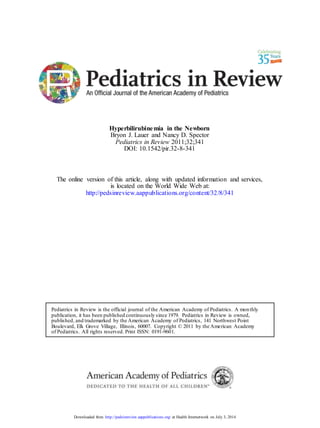
- 5. gastroenterology hyperbilirubinemia atricians should consider acute bilirubin toxicity in a term infant if there are no signs of hemolysis and the TSB concentration is greater than 25 mg/dL (427.6 mmol/ L). If the TSB concentration is above 20 mg/dL (342.1 mmol/L) in a terminfant who has hemolysis,the physi-cian should be concerned. (6) Acute bilirubin toxicity occurs in three phases during the first few weeks after birth. Phase 1 occurs during the first 1 to 2 days andresultsin poorsuck,high-pitched cry, stupor,hypotonia,and seizures.Phase2occurs during the middle of the first postnatalweekand resultsin hypertonia of extensormuscles,opisthotonus, retro-collis, and fever. Phase 3occurs afterthe first postnatal week and presents with hypertonia. If bilirubin concen-trations are not reduced, long-term morbidity can result in BIND. Neuronal injury occurs primarily in the basal ganglia and brainstem nuclei, but the hippocampus and cerebellum also may be affected. (12) BIND or ker-nicterus occurs in two phases. The first phase is seen during the first postnatal year and is characterized by hypotonia, active deep-tendon reflexes, obligatory tonic neck reflexes, and delayed motorskills.The secondphase,which occurs after the first postnatalyear,results in choreoathetotic cerebral palsy, ballismus, tremor, up-ward gaze, dental dysplasia, sensorineural hearing loss, and cognitive impairment. (6) Evaluation The following recommendations are basedon informa-tion from the AAP Subcommittee on Hyperbiliru-binemia. Evaluation for hyperbilirubinemia should occur before birth and extend through the first few postnatal weeks. Hemolytic anemia caused by isoantibodies in the infant is a major risk factor for severe hyperbilirubinemia and bilirubin neurotoxicity. (13) ABO incompatibility may occur if the mother’s blood type is O and the infant’s blood type is A or B. (13) Mother-infant ABO incom- patibility occurs in approximately 15% of all pregnancies, but symptomatic hemolytic disease occurs in only 5% of these infants. Hyperbilirubinemia in infants who have symptomatic ABO hemolytic disease usually is detected within the first 12 to 24 hours after birth. (14) Hence, ABO and Rh (D) blood types and a screen for unusual isoimmune antibodies should be evaluated for all preg- nant women. If such testing is not performed or if the motheris Rh-negative, the infant’s cord blood should be evaluated fora direct antibody (Coombs)test, blood type, and Rh determination. If the newborn is assessed adequately and the mother’s bloodtypeis not O and is Rh positive, cord blood does not need to be tested. (13) Afterbirth,the infant should be assessed forjaundice 344 Pediatrics in Review Vol.32 No.8 August 2011 at a minimum of every 8 to 12 hours. Jaundice can be detectedon a physicalexamination,but darker skin makes for a harder assessment. Jaundice has a cephalo-caudal progression, but visual assessment has been shown to predict the TSB concentration unreliably. Jaundice in an infant is best assessedby a windowin daylight; otherwise, a well-lit room is adequate. The sclera and mucous membranes are assessed for icterus, and the color of the skin and subcutaneous tissues can be revealed by blanching the skin with digital pressure. For any infants who develop jaundice in the first 24 hours after birth, the clinician should assess whether it seems excessive forgestational age. If there is any doubt in the visualevaluation,transcutaneous bilirubin (TcB) or TSB should be assessed. Newer devices used to detect TcB have been shown to correlate well with TSB. (15) Once a TcB orTSB has been measured, the result should be interpreted based on the nomogram in Figure 1. Reassessment should be based on the zone in which the bilirubin falls on the nomogram. It is important to realize that the nomogramis based on infants of greater than 35 weeks’ gestation who had no evidence of hemolytic disease. Preterm infants or infants who have risk factors for bilirubin toxicity are at higher risk of bilirubin toxicity at lower TSB concentrations. Therefore, the nomogram may not accurately predict the infant’s riskbased solely on the degree ofhyperbilirubinemia in these high-riskinfants. (13) Sometimes furtherlaboratory evaluation is required to determine the cause of hyperbilirubinemia. If the cause is not evident aftera thorough historyassessing current risk factors or significant hyperbilirubinemia oc-curred in siblings, evaluation is appropriate for any infant who is receiving phototherapy or when the TSB crosses percentiles on the nomogram. A complete blood count with smear and direct bilirubin concentration should be checked in these instances. A reticulocyte count, G6PD measurement, and end-tidal carbon monoxide (ETCO) determination (if available)can be considered. (12) ETCO is a good indicator of ongoing bilirubin produc-tion. As noted previously,biliverdin and carbon monox-ide are the byproducts ofbilirubin breakdown.Measur-ing the ETCO allows identification of infants experiencing increased bilirubin production and possibly infants who have hemolytic disease. (5) The TSB con-centration should be rechecked in 4 to 24 hours,depend-ing onthe infant’sage, TSB value, and risk factors. If the TSB is increasing despite phototherapy or if the infant is being considered for exchange transfusion, a reticulocyte count, bilirubin/albumin ratio, G6PD concentration, and ETCO should be checked. Urinalysis and urine Downloaded from http://pedsinreview.aappublications.org/ at Health Internetwork on July 3, 2014
- 6. gastroenterology hyperbilirubinemia born screen is obtained.Otherau- thors argue that data are insufficient to justify screeningallinfants at dis- charge.(16) Exclusive breastfeeding,photo- therapy in a sibling,gestationalage less than 37weeks,jaundice in the first 24 hours,hemolytic disease, East Asian race,cephalohematoma or significant bruising,and a TSB or TcB in the high riskzone before discharge are the most common clinically relevant riskfactors forse- vere hyperbilirubinemia.Each risk factorindividually has little predic- tive value,but the greaterthe num- berof risk factors,the greaterthe Figure 1. Nomogram for designation of risk for hyperbilirubinemia in 2,840 well newborns at likelihood ofthe baby developing severe hyperbilirubinemia.In gen-36 or more weeks’ gestational age whose birthweights were 2,000 g or more or 35 or more eral, a term infant who is fed pre-weeks’ gestational age whose birthweights were 2,500 g or more, based on the hour-specific serum bilirubin values. The serum bilirubin was measured before discharge, and the zone in dominately formula has a very low which the value fell predicted the likelihood of a subsequent bilirubin value exceeding the likelihood ofdeveloping severe hy- 95th percentile (high-risk zone). Reproduced with permission from Bhutani VK, Johnson L, perbilirubinemia. (13) Sivieri EM. Predictive ability ofa predischarge hour-specific serum bilirubin for subsequent The timeframe for following up significant hyperbilirubinemia in healthy term and near-term newborns. Pediatrics. 1999;103: with a pediatrician once infantsare 6 –14. © 1999 by the American Academy of Pediatrics. dischargedfromthe hospitalde- pends on the baby’s age at the time culture are appropriate ifthe infant has an elevated direct of discharge.A newborndischarged at 48to 72 hours of bilirubin value.If indicated by the history andphysical age should be evaluated forjaundice,weight gain orloss, examination, a sepsisevaluationshould be completed. stoolpatterns,voidingpatterns,and adequacyoforal (13) intake by 120 hours ofage.The child should be evalu- Althoughhuman milk jaundice is a common cause of ated at 96 hours ofage if dischargedbetween 24to prolonged jaundice in breastfedinfants,more concern- 48 hours and at 72hours ifdischargedbefore 24hours of ing conditionsshould be ruled out first. Totaland direct age.Infants dischargedbefore 48hours ofage may need bilirubin should be measured forthe infant who develops a secondvisit to ensure evaluationduring the time when jaundice orwhen jaundice persistsafter3weeks ofage. In the TSB peaks.Infants who have more riskfactors may addition,the newborn screen should be reviewedspecif- need more frequent follow-up evaluations. Also, if ically to rule out galactosemia and congenitalhypothy- follow-up cannot be ensured,delaying discharge is ap- roidism. An elevated direct bilirubin value should propriate untilfollow-up is determined oruntilthe infant prompt an evaluation forcholestasis. (13) is olderthan 72 to 96 hours ofage. One ofthe most Because TSBconcentrationspeakat 3 to 5 days of important measures is educating allparents on the risks age,aftermany infants haveleft the nursery,it is impor- and assessment ofhyperbilirubinemia as well as necessary tant to performa risk assessment onallinfants before follow-up evaluations.(13) they leave the hospital,and appropriatefollow-up evalu- Treatmentations should be stressed.Althoughsome controversy surroundsscreening andriskassessment,basedon insuf- Helping mothers breastfeedappropriately can decrease ficient evidence,the AAPSubcommittee has recom- the likelihood of severe hyperbilirubinemia. Mothers mended assessing TSBorTcB on all newborns before should breastfeedat least 8to 12 times in the first few discharge.(16)The value should be plottedon the days afterbirth to aid in bringing in the milk supply. nomogramto assessthe risklevel. (13) Some authors Mothershould be asked about anydifficulties and lacta- suggest checking a TSBon all newbornswhen the new- tion consultants involved whenneeded. The stoolpat- Pediatrics in Review Vol.32 No.8 August 2011 345 Downloaded from http://pedsinreview.aappublications.org/ at Health Internetwork on July 3, 2014
- 7. therapy. (17) Although sunlight has been shown to decrease biliru-bin concentrations, it is not recom-mended because it is difficult to determine a timeframe that is safe to expose a naked infant to sunlight without getting sunburned. (13) The totaldose delivered, or spectral irradiance, is affected sig-nificantly by the distance the infant is fromthe light and the surface area to which he or she is exposed.Therefore,infants should be placed as close as possible to the light. Placing an infant in a bassinet rather than an incubatorallows the light to be closer to the infant. When using fluorescenttubes,it is possi-ble to bring the light source to within 10 cmof the infant without overheating himor her. Halogen lights can burn the infant, so the manufacturer’s instructions should be followed to determine the cor- rect distance between the light source and the baby. (13)(17) Exposing the infant as much as possible while covering his or her eyes results in a faster decline in bilirubin concentrations. It is probably unnecessary to remove the diaper unless the bilirubin concentrations are approaching the level requiring an exchange transfusion. The bassinet should be lined with aluminum foil or white cloth when nearing the point of an exchange transfusion.In most instances,it is acceptable to interrupt photother-apy to feed the infant or for brief parentalvisits.Contin-uous phototherapy should be used if exchange transfu-sion is likely. (13) While an infant is receiving phototherapy, his or her temperature and hydration status should be monitored. Because bilirubin is excreted in the urine and the stool,it is important to assure good urine output. If the infant is dehydrated, intravenous fluids should be started, Oral nutrition is sufficient for the infant who is not dehy-drated. Supplementing breastfeeding with formula is an option to reduce enterohepatic circulation and decrease the TSB faster. (13) Initiation ofphototherapyshould be based on the TSB concentration, age in hours, and risk factors, as recommended in guidelines from the AAP (Fig. 2). The TSB value should be used, and the direct bilirubin value should notbe subtracted fromthe total when determin-ing when to initiate therapy. There are no guidelines Downloadedfrom http://pedsinreview.aappublications.org/ at HealthInternetwork onJuly 3, 2014 346 Pediatrics in Rev iew Vol.32 No.8 August 2011 terns, voiding patterns, and weight of newborns are good indicators of whether the baby is receiving adequate milk. (6)(8) Phototherapy Since the discovery of the effects of sunlight on lowering bilirubin concentrations in 1958 (2), the need for ex- change transfusions because of severe hyperbilirubinemia has decreased significantly. (17) Phototherapy works by converting bilirubin into a water-soluble compound called lumirubin, which is excreted in the urine or bile without requiring conjugation in the liver. The two biggest factors in the conversion of bilirubin to lumiru-bin are the spectrum of light and the total dose of light delivered. Bilirubin is a yellow pigment, so it most strongly absorbs blue light in the 460-nm wavelength. (5) Also, a phototherapeutic effect is seen only when the wavelength can penetrate tissue and absorb bilirubin. Lamps with output in the 460- to 490-nm range are the most effective in treating hyperbilirubinemia. Multiple types of phototherapy units are used today that contain daylight, cool white, blue, or “special blue” fluorescent tubes or tungsten-halogen lamps. Fiberoptic blankets are also available that provide light in the blue-green region. The special blue fluorescent lights are the most effective and should be used when intensive phototherapy is re-quired. (5)(17) Ultraviolet light is not usedfor photo- Figure 2. Guidelines for phototherapy in hospitalized infants of 35 or more weeks’ gestation. Reproduced with permission from Subcommittee on Hyperbilirubinemia. Pediatrics. 2004;114:297–316. © 2004 by the American Academy of Pediatrics. gastroenterology hyperbilirubinemia
- 8. gastroenterology hyperbilirubinemia infant syndrome. Affected infants develop a dark,grayish-brown color of the skin, serum, and urine. Generally, the syndrome is of little clinical significance. The only true contraindication to phototherapy is congenital porphyria or a family history of porphyria. Phototherapy in these patients could result in se- vere blistering and photosensitivity. (17) Exchange Transfusion Exchange transfusion was the first successful treatment for severe hy- perbilirubinemia. These procedures should be performed only in a neo- natalintensive care unit by a trained physician.An exchange transfusion for an infant is a medical emer- gency, and the patient should be admitted directly to the neonatal Figure 3. Guidelines for exchange transfusion in infants 35 or more weeks’gestation. intensive care unit, bypassing the Reproduced with permission from Subcommittee on Hyperbilirubinemia. Pediatrics. emergency department.(9)Basi- 2004;114:297–316. © 2004 by the American Academy of Pediatrics. cally, the physicianrapidly removes from the circulation bilirubin and publishedforinfants bornearlierthan 35 weeks’gesta- any antibodies that may be contributingto ongoing tion.When usingintensive phototherapy,a decrease of hemolysis.The procedure involves takingsmallaliquots 0.5 mg/dL (8.6 mmol/L) perhourcan be expected in of the infant’s bloodand replacingthemwith the same the first 4 to 8 hours.Whenthe TSBdoes not decline or quantity ofdonorred cells via one to two centralcathe- rises during phototherapy,ongoing hemolysisis likely. ters untilthe infant’s bloodvolume has been replaced Discontinuationofphototherapy is not standardized. twice. (5) An infusion ofalbumin 1 to 4 hours before the Therefore,clinical judgment is recommended. (5)Some procedure can increase the amount ofbilirubin that is authorssuggest stoppingonce the bilirubin decreases removed.Intravenous gamma globulin is recommended 4 to 5 mg/dL (68.4 to 85.5 mmol/L). (5) Others state for infants who have isoimmune hemolytic disease ifthe that the value should decrease to 13to 14 mg/dL TSB is rising despite phototherapyorthe TSBis within (222.4 to 239.5 mmol/L) if the child is readmitted for 2 to 3 mg/dL (34.2 to 51.3 mmol/L) of the level for an hyperbilirubinemia.A common misconceptionis that exchange transfusionin hopes ofavoidingan exchange discontinuationofphototherapyresultsin a rebound transfusion. Another dose can be administered in hyperbilirubinemia.Reboundis a rare event in an infant 12 hours,ifneeded.(13) who weighs more than 1,800 g and has no evidence of Figure 3 shows guidelines forinitiating an exchange hemolysis.(5) Whetherthis observationholdstrue for transfusion.Exchange transfusionshould be startedim- smaller infants orthose who have evidence ofhemolysis is mediately in a jaundiced infant demonstrating signsof uncertain.A reboundbilirubin determination is not rec- acute bilirubin encephalopathy,even ifthe TSB value is ommended,but if an infant is readmitted,a repeat TSB falling. Risk factors forsevere hyperbilirubinemia and the measurement orclinical follow-up in 24 hours is op- albumin/bilirubin ratio should be taken into account tional.(13) when considering when to start an exchange transfusion. Phototherapy is performed safely formillions of in- (13) fants,but rare adverse effectsdo occur. The infant who Althoughexchange transfusions are successfulin in- has cholestatic jaundice with elevated conjugatedhyper- fants who have severe hyperbilirubinemia,there are many bilirubinemia has the potentialfordevelopingbronze complications,includinginfection,portalvenous throm- Pediatrics in Review Vol.32 No.8 August 2011 347 Downloaded from http://pedsinreview.aappublications.org/ at Health Internetwork on July 3, 2014
- 9. gastroenterology hyperbilirubinemia Summary • Based on strong research evidence, breastfeeding, prematurity, significant jaundice in a previous sibling, and jaundice noted before discharge from the nurseryare the most common risk factors associated with severe hyperbilirubinemia. (13) • Based on research evaluating benefit versusharm, jaundice in the first 24 hours after birth is not physiologic jaundice and needs further evaluation. • All newborns should undergo a risk assessment for hyperbilirubinemia before discharge from the newbornnursery and have appropriate follow-up evaluation after discharge. • Visual assessment of jaundice does not assess the TSB reliably; clinicians should check either a TSB or TcB when in doubt. • The infant’s age in hours is used when evaluating and managing bilirubin concentrations. bosis, thrombocytopenia, necrotizing enterocolitis, elec- trolyte imbalances, graft versus host disease, and even death. The complication rate is reported to be approxi- mately 12%. (5) Because of these risk factors, photother- apy should be maximized to reduce the need for an exchange transfusion. (13) Conclusion Kernicterus,although a rare event, is a preventable cause of cerebralpalsy.Nowthat infantsare being discharged at earlier ages, it is important to consider screening with a TcB or TSB before dischargebecause visualassessment is not always reliable. It is equally important to arrange for follow-up evaluation after discharge, ideally within 48 hours, for additional screening. Mothers should be educated about feeding to ensure that the infants are receiving adequate caloric intake and monitoringstooland urine output.Weightcan be checked at the follow-up visit. When evaluating bilirubin concentra-tions, nomograms can be used to guide initiation of phototherapy and exchange transfusions. Guidelines and published nomograms can support clinical judgment and individualize the approach to the infant who has hyper- bilirubinemia. References 1. Gartner, LM. Historical Review and Recent Advances in Neona- tal and Perinatal Medicine. Evansville, IN: Mead Johnson Nutri- tional Division; 1980 2. Cremer RJ, Perryman PW, Richards DH. Influenceof light on the hyperbilirubinemia of infants. Lancet. 1958;1:1094 – 1097 3. Maisels MJ, Bhutani VK, Bogen D, Newman TB, Stark AR, Watchko JF. Hyperbilirubinemia in the newborn infant 35 weeks gestation:an updatewith clarifications. Pediatrics. 2009;124: 1193– 1198 4. Johnson L, Bhutani VK, Karp K, Sivieri EM, Shapiro SM. Clinical report from the pilot USA kernicterus registry (1992 to 2004). J Perinatol. 2009;29:S25–S45 5. Dennery PA, Seidman DS, Stevenson DK. Neonatal hyperbili- rubinemia. N Engl J Med. 2001;344:581–590 6. Watchko JF, Maisels MJ. Jaundice in low birthweight infants: pathobiology and outcome. Arch Dis Child Fetal Neonatal Ed. 2003;88:F455–F458 7. Watchko JF, Lin Z. Exploringthegenetic architecture ofneo-natal hyperbilirubinemia. Semin Fetal Neonatal Med. 2010;15: 169 –175 8. Maisels MJ. Jaundice in a newborn: answers to questions about a common clinical problem. First of two parts. Contemp Pediatr. 2005;22(5) 9. PorterML,Dennis BL. Hyperbilirubinemia in the termnew-born. Am Fam Physician. 2002;65:599 – 606 10. Gartner LM, Herschel M. Jaundice and breastfeeding. Pediatr Clin North Am. 2001;48:389 –399 11. Bhutani VK, JohnsonLH, KerenR. Diagnosis andmanage-ment of hyperbilirubinemia in the term neonate: for a safer first week. Pediatr Clin North Am. 2004;51:843– 861 12. Smitherman H, Stark AR, Bhutani VK. Early recognition of neonatal hyperbilirubinemia and its emergent management. Semin Fetal Neonatal Med. 2006;11:214 –224 13. American Academy ofPediatrics Subcommittee on Hyperbili- rubinemia. Management of hyperbilirubinemiain the newborninfant 35 or more weeks of gestation. Pediatrics. 2004;114: 297–316 14. Watchko JF. Identification of neonates at risk for hazardous hyperbilirubinemia: emerging clinical insights. Pediatr Clin North Am. 2009;56:671– 687 15. Fouzas S, MantagouL, Skylogianni E,Mantagos S, Varvarigou A. Transcutaneous bilirubin levels forthe first 120postnatal hours in healthy neonates. Pediatrics. 2010;125:e52– e57 16. Newman TB. Universal bilirubin screening, guidelines, and evidence. Pediatrics. 2009;124:1199 –1202 17. Maisels MJ, McDonagh AF. Phototherapy for neonatal jaun- dice. N Engl J Med. 2008;358:920 –928 HealthyChildren.org Parent Resources From the AAP The reader is likely to find material to share with parents that is relevant to this article by visiting this link: http://www.healthychildren.org/English/ages-stages/baby/Pages/ Jaundice.aspx. 348 Pediatrics in Review Vol.32 No.8 August 2011 Downloaded from http://pedsinreview.aappublications.org/ at Health Internetwork on July 3, 2014
- 10. PIR Quiz Quiz also available online at: http://pedsinreview.aappublications.org. 15. You are evaluating a 3-day-old term infant who has jaundice. His neonatal course was unremarkable and he is breastfed exclusively. He has approximately seven wet diapers and three stools per day. Findings on his physical examination are normal except for jaundice. His bilirubin measures 12 mg/dL (205.2 mol/L) and is all unconjugated. Of the following, the most appropriate management is to A. Admit the baby for phototherapy. B. Continue breastfeeding. C. Discontinue breastfeeding for 3 days, then resume. D. Supplement the human milk with water. E. Supplement the human milk with cow milk-based formula. 16. A 4-day-old girl is brought to your office for evaluation of jaundice. She was born at 39 weeks’ gestation and had no complications. Her and her mother’s blood types both are A . She is breastfeeding well and has normal stools and urine output. She has significant jaundice on examination but is vigorous and well hydrated. At what total serum bilirubin value should phototherapy be initiated for this infant? A. 10 mg/dL (171.0 mol/L). B. 12 mg/dL (205.2 mol/L). C. 15 mg/dL (256.5 mol/L). D. 17 mg/dL (290.8 mol/L). E. 20 mg/dL (342.1 mol/L). 17. Which of the following is most likely to be present during the initial phase of acute bilirubin toxicity? A. Cerebral palsy. B. Chorea. C. Opisthotonus. D. Retrocollis. E. Seizures. 18. Which of the following statements regarding the optimal use of phototherapy is true? A. Infants receiving phototherapy should be placed in an incubator. B. Infants should wear full clothing during phototherapy to prevent burns. C. Intravenous fluids are required for all infants receiving phototherapy. D. The light source should be 30 cm from the infant’s skin. E. Stopping phototherapy to allow for breastfeeding is acceptable in most cases.