SlideShare une entreprise Scribd logo
1  sur  9
Télécharger pour lire hors ligne
PETSAVERS PAPER
Diagnostic accuracy of positive
contrast computed tomography
arthrography for the detection of
injuries to the medial meniscus in
dogs with naturally occurring cranial
cruciate ligament insufficiency
OBJECTIVE: To assess the usefulness of computed tomography
arthrography of the stifle in diagnosing meniscal tears in dogs with
cranial cruciate ligament insufficiency.
METHODS: A prospective clinical study was performed. Dogs were
included if they had evidence of cranial cruciate ligament
insufficiency or persistent or recurrent lameness following surgery
for cranial cruciate ligament insufficiency. Dogs were sedated for
a computed tomography scan of the affected stifle, orientated in the
dorsal plane. A survey computed tomography scan was followed by
a computed tomography arthrogram. A stifle arthrotomy was
performed, and the surgical findings were recorded. The computed
tomography scans were reviewed by three blinded reviewers, and the
results were compared to the surgical findings.
RESULTS: Twenty-one computed tomography arthrograms from 20
dogs were included. At surgery, damage to the medial meniscus was
identified in 14 stifles. Initial interpretation of computed tomography
arthrography images was 57 to 64 per cent sensitive and 71 to 100
per cent specific for diagnosing medial meniscal injuries.
Interpretation of the images on retrospective analysis was 71 per
cent sensitive and 100 per cent specific, with an accuracy of 0857.
CLINICAL SIGNIFICANCE: The accuracy of stifle computed tomography
arthrography for the diagnosis of tears to the medial meniscus was
found to be good. It is a minimally invasive and repeatable technique,
which does not require general anaesthesia or specialist training to
obtain the images. The ability to reliably diagnose meniscal injury
without the need for surgery may be advantageous, particularly in
dogs which had previously had surgery for cranial cruciate ligament
insufficiency.
M. S. TIVERS, P. N. MAHONEY, E. A.
BAINES AND S. A. CORR
Journal of Small Animal Practice (2009)
50, 324–332
DOI: 10.1111/j.1748-5827.2009.00780.x
Accepted: 12 May 2009
Department of Veterinary Clinical Sciences, Royal
Veterinary College, Hawkshead Lane, North
Mymms, Hatfield, Hertfordshire AL9 7TA
324 Journal of Small Animal Practice Á Vol 50 ÁJuly 2009 ÁÓ 2009 British Small Animal Veterinary Association
INTRODUCTION
Rupture of the cranial cruciate ligament
(CCL) is one of the most common causes
of canine pelvic limb lameness (Johnson
and others 1994, Ness and others 1996).
Stifle instability secondary to cranial cruci-
ate ligament insufficiency (CCLI) leaves
the menisci vulnerable to damage. Menis-
cal injuries are seen in 49 to 70 per cent of
dogs with concurrent CCLI (Flo and
DeYoung 1978, Bennett and May 1991,
Elkins and others 1991, Flo 1993, Metel-
man and others 1995). A variety of surgical
treatments are advocated for the manage-
ment of CCLI. Stifle arthrotomy or
arthroscopy is recommended to allow
inspection of the menisci as a neglected
meniscal injury will result in a poor out-
come (Bennett and May 1991, Flo 1993,
Ralphs and Whitney 2002).
Following CCL surgery, dogs can suffer
prolonged or incomplete recoveries for
a variety of reasons including rupture of
the contralateral CCL, joint sepsis or
implant failure (Metelman and others
1995, Moore and Read 1995, Hill and
others 1999, Innes and others 2000,
Pacchiana and others 2003). So-called
‘‘latemeniscal’’injury,whereanapparently
normalmeniscusatthetimeofsurgerysub-
sequently becomes damaged, is reported in
6Á3 to 21Á7 per cent of dogs, regardless of
the surgery performed (Metelman and
others 1995, Thieman and others 2006,
Lafaver and others 2007, Case and others
2008, Stein and Schmoekel 2008). Diag-
nosis of a late meniscal injury can be chal-
lenging and traditionally relies upon
arthrotomy or arthroscopy. The ability to
diagnose a meniscal injury without surgery
would have significant advantages, guiding
decision-making and potentially avoiding
an unnecessary procedure. Ultrasonogra-
phy and magnetic resonance imaging
(MRI) have been suggested as useful in
the diagnosis of meniscal injury in the
dog (Banfield and Morrison 2000, Mahn
and others 2005, Martig and others
2006, Blond and others 2008). However,
both techniques have limitations with
ultrasound being highly operator depen-
dent (Mahn and others 2005) and MRI
being expensive and requiring a general
anaesthetic.
MRI is the modality of choice for imag-
ingthemenisciinhumanpatientswithvery
high sensitivity and specificity (Crues and
others1987,Minkandothers1988).Com-
puted tomography arthrography (CTA) is
also used for the detection of meniscal inju-
ries in human beings and has been reported
to have similar sensitivity and specificity to
those of MRI (Ghelman 1985, Vande Berg
and others 2000, 2002).
There are several published studies
describing the use of stifle CTA in canine
cadavers. These have described the normal
intra-articular anatomy as seen on CTA
(Samii and Dyce 2004, Soler and others
2007) and also demonstrated that CTA
can be used to diagnose CCL rupture
(Han and others 2008) and medial menis-
cal injury (Tivers and others 2008) in
cadavers. The cadaver study by Tivers
and others (2008) showed that dorsal
CTA images had a sensitivity of 90 per cent
and a specificity of 100 per cent for the
diagnosis of simulated injuries of the cau-
dal horn of the medial meniscus. A more
recent study investigated the use of CTA
using transverse images for the detection
of tears to the cruciate ligaments and
menisci (Samii and others 2009). This
study reported a low accuracy of interpre-
tation of CTA images for the diagnosis of
meniscal injuries with sensitivities of 13Á3
to 73Á3 per cent and specificities of 57Á1 to
100 per cent.
The aim of this current study was to
assess dorsal plane computed tomography
(CT) arthrograms for the diagnosis of inju-
ries to the menisci in dogs suffering from
CCLI. The hypothesis was that the inter-
pretation of dorsal plane CT arthrograms
would have high sensitivity and specificity
for the diagnosis of meniscal injuries in
dogs. We also hypothesised that an abnor-
mal outline to the meniscus asseenon a CT
arthrogram would be consistent with
a meniscal injury.
MATERIALS AND METHODS
Dogs were prospectively recruited between
August 2006 and March 2008 with full
informed owner consent. Dogs were eligi-
ble for inclusion if they had pelvic limb
lameness localised to the stifle joint with
a provisional diagnosis of CCLI or sus-
pected late meniscal injury and were des-
tined to have an exploratory arthrotomy
as part of their investigation or treatment.
Recruitment of dogs was dependent on
owner and clinician variables, and there-
fore, not all eligible dogs were recruited
during the study period. Two populations
of dogs were recruited: those which had
undergone previous surgery for CCLI
and those which had not had previous sur-
gery but had a preoperative diagnosis of
CCLI.
All dogs were examined by a board-
certified orthopaedic surgeon or by a sur-
gery resident under direct supervision. A
diagnosis of CCLI was made on the basis
of physical examination findings, assess-
ment of craniocaudal stifle stability (cranial
draw and/or tibial thrust), stifle radiogra-
phy and arthrocentesis for cytology and
culture (as necessary). Following the diag-
nostic workup, each dog had a CT scan
made of the affected stifle before surgery.
Dogs were sedated with a dose of 0Á01
mg/kg intravenous medetomidine (Domi-
tor; Pfizer)and 0Á1 to 0Á2 mg/kg butorpha-
nol (Torbugesic; Fort Dodge). They were
positioned in the CT scanner in lateral
recumbency with the affected leg depen-
dent (Fig 1). A fourth-generation single-
slice spiral CT scanner (Picker PQ 5000
CT Scanner; Universal Systems) was used
for all scans. A survey CT scan was made of
the affected limb, as previously described
(Tivers and others 2008). The limb was
positioned so that the tibial plateau was
in line with the long axis of the CT couch.
Contiguous, dorsal 1Á5-mm sections of the
stifle were made starting cranial to the pa-
tella and finishing just caudal to the fabel-
lae. All scans were made using a sharp
algorithm, 100Á0 mA and 120Á0 kV; the
scan time was 90 seconds. Following the
survey scan, routine arthrocentesis was per-
formed on the stifle joint. Samples of joint
fluid were submitted for diagnostic testing
as appropriate. Sodium and meglumine
amidotrizoate, 370 mg I/ml, (Urografin
370; Schering Health Care Limited) was
diluted in a 1:1 ratio with sterile saline
(0Á9 per cent NaCl), creating a concentra-
tion of 185 mg iodine/ml as previously
reported (Tivers and others 2008). A vol-
ume of 0Á2 ml/kg of this diluted contrast
agent was injected into the stifle, and
the needle was removed. This volume of
Journal of Small Animal Practice Á Vol 50 ÁJuly 2009 ÁÓ 2009 British Small Animal Veterinary Association 325
Diagnostic accuracy of stifle computed tomography arthrography
contrast was extrapolated from a previous
study and some unpublished data (Tivers
and others 2008). The stifle was repeatedly
flexed and extended to ensure good contrast
distribution throughout the joint, and the
dogwasrepositionedasbefore.TheCTscan
was repeated resulting in a positive contrast
CT arthrogram. If the dog was not having
surgery immediately, the contrast was
removed from the joint througharthrocent-
esis to prevent any possible discomfort asso-
ciated with distention of the synovium. The
medetomidine was antagonised with atipa-
mazole (Antisedan; Pfizer), given intramus-
cularly at the same volume.
Dogs were subsequently anaesthetised
for surgery within 24 hours of the CT scan.
All surgeries involved a standard lateral or
medial parapatellar arthrotomy (Piermat-
tei and Johnson 2004) with inspection of
the cruciate ligaments and both menisci.
The surgeons were blinded to the CT
results and inspected the medial and lateral
menisci both visually and using a probe.
They were provided with a printed form
and asked to record whether the CCL
was intact, partiallyruptured or completely
ruptured. They also recorded whether
there was damage to either of the menisci
and the precise nature of the injury, includ-
ing the area damaged and whether it was
displaced.
Following completion of the study, the
CT arthrograms were reviewed, indepen-
dently, by two board-certified radiologists
andaboard-certifiedsurgeon.Thereviewers
had access to the CTA images of beagle
cadaver stifles with simulated meniscal
injuries from a previous study, but they
had no experience of the interpretation
of stifle CT arthrograms from dogs with
clinical disease. The scans were analysed
as Digital Imaging and Communications
in Medicine (DICOM) images using
Image Viewer (Visbion Ltd). They were
viewedinarandomorder,andthereviewers
were blinded to the dogs’ history, physical
examination findings and surgical findings.
The reviewers recorded whether the CT
arthrogram had sufficient contrast to be
of diagnostic quality and whether the pres-
ence of metal implants affected the inter-
pretation. They then recorded whether
the meniscus was intact or abnormal. A
meniscus was considered abnormal if there
was disruption of the normal outline.
Where the meniscus was abnormal, the
reviewers recorded how the image of the
meniscus differed from the normal appear-
ance. The reviewers also recorded whether
the CT arthrogram had sufficient contrast
to be of diagnostic quality and whether the
presence of metal implants affected the
interpretation.
The results were subsequently com-
pared with the surgical findings to deter-
mine the sensitivity, specificity and
positive and negative predictive values of
interpretation of CTA for the detection
of meniscal pathology. The measure of
agreement between the three reviewers
was assessed using a multi-rater variation
of Brennan and Prediger’s (1981) free-
marginal kappa statistic as described by
Randolph (2005) using an online calcula-
tor (Randolph 2008). In addition, the
accuracy of the test for each reviewer was
evaluated using receiver operating charac-
teristic (ROC) curve analysis using an
online calculator (Eng 2009).
Approximately five months following
the blinded analysis, a retrospective inter-
pretation was performed by one of the
authors (P. N. M). The images were re-
viewed, and the findings were recorded
as described above. Sensitivity, specificity,
positive and negative predictive values and
ROC curveanalysiswere then recalculated.
RESULTS
Twenty dogs were recruited. Twenty-one
stifle CT arthrograms and arthrotomies
were performed. One dog (dog 12) was
originally presented for investigation and
treatment of suspected CCLI. A CT
arthrogram was performed, and no menis-
cal injury was detected at arthrotomy. The
stifle was stabilised with a fabellotibial
suture. The dog was re-presented for per-
sistent lameness following the surgery,
and repeat CTA and arthrotomy were
performed.
Breeds comprised four Labradors, four
boxers, two golden retrievers, one rottwei-
ler, one bullmastiff, one German shepherd
dog, one Jack Russell terrier and six cross-
breeds. There were five entire male dogs,
five neutered male dogs, four entire female
dogs and six neutered female dogs. Age
ranged from one year to nine years with
a median of five years. The weight of the
dogs ranged from 8Á5 to 63 kg with
a median of 35Á1 kg.
Seven of the CT arthrograms were ofsti-
fles which had not had surgery, and the
remaining 14 scans were of joints which
had previously undergone surgery. For
the dogs which had not had previous sur-
gery, the duration of lameness ranged from
0Á5 to 12 months with a median of 1Á5
months. Of the stifles which had had pre-
vious surgery, 10 had been stabilised with
a fabellotibial suture,three witha tibial pla-
teau levelling osteotomy (TPLO) and one
with an over-the-top (OTT) fascia lata
graft. The time interval between the origi-
nal surgery and the subsequent CT arthro-
gram and arthrotomy ranged from 0Á75 to
23 months with a median of 4Á25 months.
These results are summarised in Table 1.
FIG 1. Dog positioned for computed tomography (CT) scan of the left stifle. The dog is positioned in
lateral recumbency with the affected leg underneath. The leg is positioned approximately
perpendicular to the long axis of the CT couch with the tibial plateau in line with the couch
326 Journal of Small Animal Practice Á Vol 50 ÁJuly 2009 ÁÓ 2009 British Small Animal Veterinary Association
M. S. Tivers and others
The arthrotomy findings were recorded
for all the stifles (Table 2). There were 15
full CCL ruptures and six partial ruptures
(dog 14 had an intra-articular graft in the
location of the CCL). There were 14 inju-
ries to the medial meniscus. The remaining
seven medial menisci were considered nor-
mal. There were no injuries to the lateral
meniscus. Meniscal injuries varied in type
and severity.
Twenty-one CT arthrograms were
available for interpretation. The diagnostic
quality of the images was considered good
in all but one CTA (dog 15). The lateral
and medial menisci were visible as
wedge-shaped structures, highlighted by
the contrast (Fig 2). Meniscal injuries were
identified by an abnormal outline to the
meniscus. Typically, this was a bulge or
contour at the axial part of the meniscal
outline (Fig 3).
In the dog treated with an OTT, the
graft could be seen, mimicking the
CCL. The TPLO plates and screws and
the crimps used to secure fabellotibial
sutures were also clearly visible (Fig 4).
Both types of metal implants caused streak
artefacts, although that caused by the
crimps did not obscure the view of the
intra-articular structures. The artefact
caused by the TPLO implants did inter-
fere with the quality of the images; how-
ever, it was possible to see the intra-
articular structures (particularly the
menisci) on sufficient slices that the stud-
ies were considered diagnostic. It was still
possible to successfully identify meniscal
injuries in dogs which had TPLO im-
plants, despite the considerable artefact
created by the implants.
Theresultsoftheblindedreviewarepre-
sented in Table 2. The sensitivity of inter-
pretation of the images for meniscal injury
was57percentforreviewers1and3and64
per cent for reviewer 2, and the specificity
was 100 per cent for reviewers 1 and 3 and
71per cent for reviewer 2.Thepositive pre-
dictivevaluewas82to100percent,andthe
negative predictive value was 50 to 54 per
cent. Agreement between the reviewers was
0Á37 (free-marginal kappa). The ROC
curve analysis resulted in an estimated area
under the curve of 0Á786 for reviewers
1 and 3 and 0Á679 for reviewer 2.
The results of the retrospective interpre-
tation are also included in Table 2. The
reviewer correctly identified 10 of 14
meniscalinjuriesandsevenofsevennormal
medial menisci. This gave a sensitivity of
71 per cent, a specificity of 100 per cent,
a positive predictive value of 100 per cent
and a negative predictive value of 64 per
cent. ROC curve analysis resulted in an
estimated area under the curve of 0Á857.
DISCUSSION
This study has shown that stifle dorsal
plane CTA has potential for the diagnosis
of injuries to the medial meniscus in dogs.
Before this study, there was little informa-
tion on how a damaged meniscus would
appear on a CT arthrogram. The normal
menisci are wedge shaped, and the axial
part of the medial meniscus, in particular,
has a very long and thin profile in cross-
section on CTA images (Tivers and others
2008). We therefore hypothesised that
a change in the normal outline of the
meniscus would be consistent with an
injury such as a displaced tear or fold.
The images from stifles with meniscal inju-
ries support this hypothesis with an abnor-
mal outline,in particular at theaxial border
of the meniscus being consistent with an
injury. This was most commonly seen at
the mid part of the meniscus and in the
axial part of the joint. The more obvious
lesions resulted in an abnormal outline
of contrast at the axial border of the menis-
cus described as an ‘‘axial bulge’’ (Figs 3, 5
and 6). Comparison with the surgical find-
ings confirmed that these areas corre-
sponded with displacement of a torn part
of the meniscus or folding of the caudal
horn. There was considerable variation
Table 1. Signalment and significant historical information
Dog Age
(years)
Sex Breed Weight
(kg)
Affected
leg
Previous
cruciate
surgery
Duration of clinical signs
or time interval since
original surgery (months)
1 5 FE Labrador 27Á8 Right None 3
2 9 ME Crossbreed 63 Left TPLO 2Á5
3 6 MN Crossbreed 35Á2 Left FT Suture 5
4 9 FN Labrador 36Á8 Left None 1Á5
5 3 ME Jack Russell terrier 8Á5 Left None 12
6 3 MN Bullmastiff 57Á5 Left FT suture 23
7 2 FN Boxer 25 Right FT suture 3
8 2 FN Boxer 33Á4 Left FT suture 3
9 3 MN Boxer 35Á2 Right FT suture 3Á5
10 2 MN Crossbreed 39Á2 Left FT suture 8
11 7 ME Labrador 36Á5 Left None 1
12 3 ME Labrador 35 Right None 2
12* 3 ME Labrador 35 Right FT suture 1Á5
13 7 FN Crossbreed 43Á3 Right FT suture 7
14 1 FE Rottweiler 34Á5 Right OTT 10
15 7 FN Crossbreed 25 Right None 0Á5
16 5 FE Golden retriever 32Á2 Right TPLO 0Á75
17 5 FE Golden retriever 36Á5 Left TPLO 5Á5
18 5 FN German shepherd dog 37 Right FT suture 2Á5
19 2 ME Boxer 29Á5 Left FT suture 7
20 7 MN Crossbreed 32 Left None 0Á5
FE Female entire, ME Male entire, MN Male neutered, FN Female neutered, TPLO tibial plateau levelling osteotomy, FT Fabellotibial, OTT Over the top
*Dog 12 had two computed tomography scans and subsequent surgeries at separate time points
Journal of Small Animal Practice Á Vol 50 ÁJuly 2009 ÁÓ 2009 British Small Animal Veterinary Association 327
Diagnostic accuracy of stifle computed tomography arthrography
betweenthesizeandtheshapeoftheabnor-
mal contrast outline, and this resulted in
some lesions being missed on initial inter-
pretation. At retrospective analysis, 10 of
14 meniscal injuries were correctly diag-
nosed (Table 2). It was not possible to
see any evidence of injury in the four dam-
aged menisci, which appeared normal on
the CT arthrogram. One of these scans
(dog 15) had poor contrast filling, which
resulted in poor diagnostic quality which
was considered to have hindered interpre-
tation. The three other dogs (dogs 5, 9 and
10) had scans of diagnostic quality, but no
meniscal injury was evident at retrospective
review. Dogs 5 and 9 had tears which were
non-displaced in association with damage
to the meniscus which had not resulted in
a tear (crushing or thickening). Although
the crushing or thickening was seen at sur-
gery, we would suggest that these sorts of
injury do not affect the outline of the
meniscus and would therefore be very dif-
ficultorimpossibletodiagnoseusingCTA.
The tears present in these two dogs were
most likely not seen as they were not dis-
placed. Dog 10 was unusual, in that a small
bucket handle tear, along the axial margin
ofthemeniscus,had beenremovedatapre-
vious surgery. Therefore, the outline of the
meniscus was intact, and no lesion was seen
on the CT arthrogram.
These results suggest that meniscal
lesions which are large or displaced are
readily seen on CT arthrograms, whereas
injuries not resulting in a tear or those
tears which are not displaced are not
apparent.
Atinitialinterpretation,CTAwasfound
to have a sensitivity of 57 to 64 per cent and
a specificity of 71 to 100 per cent with fair
agreement between the reviewers. The
accuracy as determined by ROC curve
analysis was fair to good. A previous study
Table 2. Arthrotomy findings and the results of the blinded and retrospective reviews
Dog Degree of
cruciate
rupture
Meniscal
injury?
Surgical findings including
injury to medial meniscus
Reviewer identification
of meniscal injury
Identification of
meniscal injury on
retrospective review?
1 2 3
1 Partial No No No No No
2 Full No Tibial plateau levelling osteotomy implants removed No No No No
3 Full Yes Longitudinal tear to caudal pole. Cranial pole
thickened and abnormal
Yes No No Yes
4 Full Yes Longitudinal tear to caudal pole Yes No Yes Yes
5 Full Yes Bucket handle tear and crushing of caudal pole No Yes No No
6 Full No Synovial inflammation, marked periarticular fibrosis No No No No
7 Partial No Inflammation and fibrosis around nylon suture No No No No
8 Full Yes Multiple tears in caudal pole. Caudal pole folded forward Yes Yes Yes Yes
9 Partial Yes Caudal pole torn but non-displaced. Cranial pole thickened Yes No No No
10 Full Yes Previous bucket handle tear removed No Yes No No
11 Partial Yes Small transverse tear No Yes Yes Yes
12 Full No No No No No
12 Full Yes Caudal pole torn and folded cranially Yes Yes Yes Yes
13 Partial No Mineralised fragment in meniscus. Patellar luxation No Yes No No
14 Full No Marked periarticular fibrous reaction and fibrous reaction
associated with graft
No Yes No No
15 Full Yes Very small longitudinal tear No Yes No No
16 Full Yes Caudal pole torn and folded cranially Yes Yes Yes Yes
17 Full Yes Caudal pole torn and folded cranially Yes Yes Yes Yes
18 Full Yes Whole medial meniscus crushed. Caudal pole folded cranially Yes Yes Yes Yes
19 Partial Yes Longitudinal tear to caudal pole. Caudal pole folded cranially No No Yes Yes
20 Full Yes Bucket handle tear to mid meniscus. Caudal pole detached
but non-displaced
No No No Yes
Bold denotes correct interpretation of computed tomographic arthrogram by reviewer
FIG 2. Dorsal plane computed tomography arthrograms of the stifle from dog 1 (a) and dog 13 (b)
showing normal medial and lateral menisci. The lateral meniscus is highlighted with a white arrow,
and the medial meniscus is highlighted with a black arrow in the first image. The menisci are wedge
shaped and surrounded by white contrast
328 Journal of Small Animal Practice Á Vol 50 ÁJuly 2009 ÁÓ 2009 British Small Animal Veterinary Association
M. S. Tivers and others
onsimulatedmeniscalinjuryhadsuggested
that the accuracy of CTA would be greater
(Tivers and others 2008). There was also
some variation between the reviewers’
results. None of the reviewers had previous
experience in the interpretation of CT
arthrograms in dogs with CCLI or late
meniscal injury. It is likely that this inexpe-
rience contributed to variation in interpre-
tation between reviewers. The effectiveness
of interpretation of CT arthrograms im-
proved at retrospective review with a sensi-
tivity of 71 per cent, a specificity of 100 per
cent and good accuracy as determined by
ROC curve analysis. This suggests that
the interpretation of CT arthrograms has
a steep learning curve but that with some
experience, it is an accurate technique for
the diagnosis of medial meniscal injury
in the dog.
CTA of the human knee is reported to
have a sensitivity of 92 to 100 per cent
anda specificityof78to94percent(Vande
Bergandothers2000,2002,Mutschlerand
others 2003), varying with the type of
meniscal injury and whether previous sur-
gery has been performed. These studies
used a dual detector helical CT unit, which
allowed reformatting of images with min-
imal loss of quality. Similarly to our study,
‘‘a meniscal abnormality was defined bythe
presence of contour irregularity, peripheral
separation, or tear of a meniscus’’ (Vande
Berg and others 2000). Although the accu-
racy reported in our study compares well
with that in the human literature, it is clear
that CTA is able to detect much more sub-
tle lesions in people. This discrepancy
could be related to the quality of the scans
produced by the CT machine used; 1Á5-
mm thick slices were obtained which
resultedinasignificantlossofimagequality
whenreformattingfromthedorsalplane.It
is likely that the use of a 16-slice multi-
detectorCTscannerwouldproduceimages
of greater quality and diagnostic potential.
The image quality could also be improved
byusingsmallerslicethicknessandoverlap-
ping slices, allowing reconstruction of sag-
ittal or transverse images of much higher
quality. This would facilitate the detection
of smaller and more subtle lesions.
A previous study investigated the use of
transverse CTA scans for the diagnosis of
injuries to the cruciate ligaments and
menisci (Samii and others 2009). They
found a sensitivity of 13Á3 to 73Á3 per cent
and a specificity of 57Á1 to 100 per cent for
the diagnosis of meniscal tears. Sensitivity
was 64Á3 per cent and specificity was 73Á3
per cent on consensus evaluation. The
results in our current study compare
favourably with these findings and would
support the use of dorsal plane scans over
FIG 3. Dorsal plane computed tomography arthrograms of the stifle from dog 12 before surgery (a)
and six weeks after surgery (b) for rupture of the cranial cruciate ligament. In image (a), the medial
meniscus has a normal outline. In image (b), the medial meniscus has an axial bulge and has lost its
normal wedge-shaped appearance (red ring). At surgery, the caudal pole of the meniscus was torn
and folded cranially
FIG 4. Dorsal plane computed tomography arthrograms of the stifle from dog 7 (a) showing the
fabellotibial suture crimps and dog 2 (b) showing the tibial plateau levelling osteotomy (TPLO) plate
and screws. The crimps produce a minimal amount of streak artefact, which does not obscure the
intra-articular structures. The TPLO implants produce a much greater amount of artefact, which
interferes with the view of the joint. However, the artefact is inconsistent on the different slices, and
it is possible to still achieve a diagnostic scan of the joint. In this example, the medial meniscus is
clearly seen, despite the artefact (red ring)
Journal of Small Animal Practice Á Vol 50 ÁJuly 2009 ÁÓ 2009 British Small Animal Veterinary Association 329
Diagnostic accuracy of stifle computed tomography arthrography
transverse scans for the investigation of
meniscal injuries. It is likely that a combi-
nation of both scan orientations would
produce even more accurate results,
although this is unproven.
One of the potential limitations of CTA
for the diagnosis of late meniscal injuries is
image distortion as a result of streak arte-
facts from metal implants. Metal crimps
were present in 10 dogs but did not inter-
fere with the diagnostic quality of the
images produced. Three dogs had TPLO
implants in situ, and although these pro-
duced much greater distortion which inter-
fered with the view of the intra-articular
structures on some scan slices, images of
diagnostic quality could still be obtained.
If artefact were a significant problem in
stifles with TPLO implants, then a trans-
verse scan could be performed and the
images reformatted into the dorsal plane.
Visual inspection of the menisci
through arthroscopy or arthrotomy is rec-
ommended in all dogs with CCLI to assess
for meniscal injury (Bennett and May
1991, Flo 1993, Ralphs and Whitney
2002). It is also useful to probe the menisci
todetectincompleteorsubtlelesions.With
arthrotomy or arthroscopy, it can be diffi-
cult to inspect the most caudal aspect of the
joint, and hence, caudal lesions can be
missed. Arthrotomy is associated with con-
siderable morbidity, and while arthroscopy
is associated with much less morbidity, it
does require specialist equipment and
training (Hoelzler and others 2004). A
method of diagnosing meniscal injury
without the need for surgery would there-
fore be advantageous. CTA is minimally
invasive and requires no specialist training
to obtain the images, which are produced
using a standard protocol and are easily
reproducible. In many dogs with stifle dis-
ease, arthrocentesis is routinely performed
as part of their investigation, and this could
be combined with the arthrogram.
Although contrast is injected into the joint,
no complications related to this were seen,
and the authors are unaware of any studies
reporting complications associated with
this procedure.
Studies have shown that MRI and MRI
arthrography can be used to diagnose cru-
ciate ligament rupture and meniscal injury
in experimental and naturally occurring
CCLI in dogs (Banfield and Morrison
2000, Martig and others 2006, Blond
and others 2008). In a study of 11 dogs,
which had not had previous surgery,
MRI was reported to have a sensitivity of
100 per cent and a specificity of 94 per cent
(Blond and others 2008). However, it is
suggested that interpretation of images
from previously operated stifles may be
more difficult because of increased signal
of healing or healed tissues, which could
be mistaken for meniscal damage (Blond
and others 2008). In addition, two studies
reported image distortion as a result of
either metallic microfragments from
instruments used during surgery (Banfield
and Morrison 2000) or the ferromagnetic
properties of coloured polydioxanone
suture (Martig and others 2006). This
FIG 5. Dorsal plane computed tomography arthrogram (CTA) of the stifle and an intra-operative
image from dog 8. The CTA shows that the medial meniscus has an abnormal outline with an axial
bulge (red ring). The photograph shows a medial arthrotomy with the medial femoral condyle
highlighted with an asterix. The caudal pole of the medial meniscus (arrow) is torn and displaced
cranially, consistent with the CTA findings
FIG 6. Dorsal plane computed tomography arthrogram (CTA) of the stifle and an intra-operative
image from dog 18. The CTA shows a marked axial bulge of the medial meniscus (red ring). The
photograph shows a medial arthrotomy with the medial femoral condyle highlighted with an asterix.
The caudal pole of the medial meniscus is folded forward, consistent with the CTA findings
330 Journal of Small Animal Practice Á Vol 50 ÁJuly 2009 ÁÓ 2009 British Small Animal Veterinary Association
M. S. Tivers and others
would not be a concern with CTA. CTA
has several potential advantages over
MRI, in that it is considerably cheaper,
the scan times are much faster and the
dog need only be sedated rather than
anaesthetised.
Ultrasonography has been shown to
have great potential for investigating
meniscal lesions in dogs, with a sensitivity
of 90 per cent and a specificity of 92Á9
percent (Mahn and others 2005). Ultra-
sonography is a completely non-invasive
technique and has the advantages that
it is widely available and it can be per-
formed in a conscious animal. However,
musculoskeletal ultrasonography is highly
specialised, and the results are largely
dependent on the skill and experience of
the operator (Mahn and others 2005).
Whereas Mahn and others (2005) reported
excellent results, they included only cases
with significant meniscal pathology and
commented that more subtle pathologies
may be more difficult to detect. Equally,
these authors stated that it may not be pos-
sible to obtain diagnostic images when
there is marked periarticular fibrosis or os-
teophytosis,potentiallylimitingtheuseful-
ness in dogs with chronic disease or
following surgery. Unlike ultrasonogra-
phy, stifle CTA requires no specialist train-
ing to produce diagnostic images, although
image interpretation is associated with
a steep learning curve as discussed above.
This study has shown that stifle CTA
canbeusedtosuccessfullydiagnoseinjuries
to the medial meniscus in association with
CCLI and that it has good accuracy. Image
interpretation is potentially challenging,
particularly for those inexperienced with
CTA. Hopefully, the information in this
paper will aid clinicians who use this tech-
nique in the future. We would anticipate
that with advances in equipment and expe-
riencewithimageinterpretation,thiscould
become an extremely reliable method of
diagnosing meniscal injuries. It could then
be used as an alternative to arthrotomy or
arthroscopy and hence eliminate the mor-
bidity for those animals undergoing either
as a purely diagnostic procedure.
Acknowledgements
This study was funded by a BSAVA Petsa-
vers grant, and the authors are very grateful
for this support. We would also like to
acknowledge themembers oftheorthopae-
dic and radiology services at our institution
for their help with this study.
References
BANFIELD, C. M.  MORRISON, W. B. (2000) Magnetic
resonance arthrography of the canine stifle joint:
technique and applications in eleven military
dogs. Veterinary Radiology  Ultrasound 41,
200-213
BENNETT, D.  MAY, C. (1991) Meniscal damage asso-
ciated with cruciate disease in the dog. Journal of
Small Animal Practice 32, 111-117
BLOND, L., THRALL, D. E., ROE, S. C., CHAILLEUX, N. 
ROBERTSON, I. D. (2008) Diagnostic accuracy of
magnetic resonance imaging for meniscal tears
in dogs affected with naturally occurring cranial
cruciate ligament rupture. Veterinary Radiology
 Ultrasound 49, 425-431
BRENNAN, R. L.  PREDIGER, D. J. (1981) Coefficient
kappa: some uses, misuses, and alternatives.
Educational and Psycholoical Measurement 41,
687-699
CASE, J. B., HULSE, D., KERWIN, S. C.  PEYCKE, L. E.
(2008) Meniscal injury following initial cranial cru-
ciate ligament stabilization surgery in 26 dogs (29
stifles). Veterinary and Comparative Orthopae-
dics and Traumatology 21, 365-367
CRUES, J. V., III, MINK, J., LEVY, T. L., LOTYSCH, M. 
STOLLER, D. W. (1987) Meniscal tears of the knee:
accuracy of MR imaging. Radiology 164, 445-
448
ELKINS, A. D., PECHMAN, R., KEARNEY, M. T.  HERRON, M.
(1991) A retrospective study evaluating the
degree of degenerative joint disease in the stifle
joint of dogs following surgical repair of anterior
cruciate ligament rupture. Journal of the Ameri-
can Animal Hospital Association 27, 533-540
ENG, J. (2009) ROC analysis: web-based calculator for
ROC curves. http://www.jrocfit.org. Accessed
April 7, 2009
FLO, G. L. (1993) Meniscal injuries. Veterinary Clin-
ics of North America: Small Animal Practice 23,
831-843
FLO, G. L.  DEYOUNG, D. (1978) Meniscal injuries and
medial meniscectomy in the canine stifle. Journal
of the American Animal Hospital Association 14,
683-689
GHELMAN, B. (1985) Meniscal tears of the knee:
evaluation by high-resolution CT combined with
arthrography. Radiology 157, 23-27
HAN, S., CBEON, H., CHO, H., KIM, J., KANG, J. H., YANG,
M. P., LEE, Y., LEE, H.  CHANG, D. (2008) Evalua-
tion of partial cranial cruciate ligament rupture
with positive contrast computed tomographic
arthrography in dogs. Journal of Veterinary Sci-
ence 9, 395-400
HILL, C. R., CONZEMIUS, M. G., SMITH, G. K., MCMANUS,
P. M.  MALONEY, D. (1999) Bacterial culture of the
canine stifle joint following surgical repair of rup-
tured cranial cruciate ligament. Veterinary and
Comparative Orthopaedics and Traumatology
12, 1-5
HOELZLER, M. G., MILLIS, D. L., FRANCIS, D. A.  WEIGEL,
J. P. (2004) Results of arthroscopic versus open
arthrotomy for surgical management of cranial
cruciate ligament deficiency in dogs. Veterinary
Surgery 33, 146-153
INNES, J. F., BACON, D., LYNCH, C.  POLLARD, A. (2000)
Long-term outcome of surgery for dogs with cra-
nial cruciate ligament deficiency. Veterinary
Record 147, 325-328
JOHNSON, J. A., AUSTIN, C.  BREUR, G. J. (1994) Inci-
dence of canine appendicular musculoskeletal
disorders in 16 veterinary teaching hospitals from
1980 through 1989. Veterinary and Comparative
Orthopaedics and Traumatology 7, 56-69
LAFAVER, S., MILLER, N. A., STUBBS, W. P., TAYLOR, R. A. 
BOUDRIEAU, R. J. (2007) Tibial tuberosity advance-
ment for stabilization of the canine cranial cruciate
ligament-deficient stifle joint: surgical technique,
early results, and complications in 101 dogs. Vet-
erinary Surgery 36, 573-586
MAHN, M. M., COOK, J. L., COOK, C. R.  BALKE, M. T.
(2005) Arthroscopic verification of ultrasono-
graphic diagnosis of meniscal pathology in dogs.
Veterinary Surgery 34, 318-323
MARTIG, S., KONAR, M., SCHMOKEL, H. G., RYTZ, U., SPRENG,
D., SCHEIDEGGER, J., HOHL, B., KIRCHER, P. R., BOISCLAIR,
J.  LANG, J. (2006) Low-field MRI and arthroscopy
of meniscal lesions in ten dogs with experimentally
induced cranial cruciate ligament insufficiency.
Veterinary Radiology  Ultrasound 47, 515-522
METELMAN, L. A., SCHWARZ, P. D., SALMAN, M.  ALVIS,
M. R. (1995) An evaluation of 3 different cranial
cruciate ligament surgical stabilization procedures
as they relate to postoperative meniscal injuries –
a retrospective study of 665 stifles. Veterinary and
Comparative Orthopaedics and Traumatology 8,
118-123
MINK, J. H., LEVY, T.  CRUES, J. V., III (1988) Tears of
the anterior cruciate ligament and menisci of the
knee: MR imaging evaluation. Radiology 167,
769-774
MOORE, K. W.  READ, R. A. (1995) Cranial cruciate
ligament rupture in the dog – a retrospective study
comparing surgical techniques. Australian Veter-
inary Journal 72, 281-285
MUTSCHLER, C., VANDE BERG, B. C., LECOUVET, F. E.,
POILVACHE, P., DUBUC, J. E., MALDAGUE, B.  MALGHEM,
J. (2003) Postoperative meniscus: assessment at
dual-detector row spiral CT arthrography of the
knee. Radiology 228, 635-641
NESS, M. G., ABERCROMBY, R. H., MAY, C., TURNER, B. M.
 CARMICHAEL, S. (1996) A survey of orthopaedic
conditions in small animal veterinary practice in
Britain. Veterinary and Comparative Orthopaedics
and Traumatology 9, 43-52
PACCHIANA, P. D., MORRIS, E., GILLINGS, S. L., JESSEN,
C. R.  LIPOWITZ, A. J. (2003) Surgical and postop-
erative complications associated with tibial pla-
teau leveling osteotomy in dogs with cranial
cruciate ligament rupture: 397 cases (1998-
2001). Journal of the American Veterinary Medi-
cal Association 222, 184-193
PIERMATTEI, D. L.  JOHNSON, K. A. (2004) Approach to
the stifle joint. In: An Atlas of Surgical
Approaches to the Bones and Joints of the Dog
and Cat. 4th edn. Saunders, Philadelphia, PA,
USA. pp 342-349
RALPHS, S. C.  WHITNEY, W. O. (2002) Arthroscopic
evaluation of menisci in dogs with cranial cruciate
ligament injuries: 100 cases (1999-2000). Jour-
nal of the American Veterinary Medical Associa-
tion 221, 1601-1604
RANDOLPH, J. J. (2005) Free-marginal multirater kappa:
an alternative to Fleiss’ fixed-marginal multirater
kappa. Proceedings of the Joensuu Learning and
Instruction Symposium. Joensuu, Finland, October
14 to 15, 2005. pp 1-20
RANDOLPH, J. J. (2008) Online kappa calculator. http://
justus.randolph.name/kappa. Accessed October
18, 2008
SAMII, V. F.  DYCE, J. (2004) Computed tomographic
arthrography of the normal canine stifle. Veteri-
nary Radiology  Ultrasound 45, 402-406
SAMII, V. F., DYCE, J., POZZI, A., DROST, W. T.,
MATTOON, J. S., GREEN, E. M., KOWALESKI, M. P. 
LEHMAN, A. M. (2009) Computed tomographic
arthrography of the stifle for detection of cranial
and caudal cruciate ligament and meniscal tears
in dogs. Veterinary Radiology  Ultrasound 50,
144-150
SOLER, M., MURCIANO, J., LATORRE, R., BELDA, E., RODRIGUEZ,
M. J.  AGUT, A. (2007) Ultrasonographic, com-
puted tomographic and magnetic resonance imag-
ing anatomy of the normal canine stifle joint.
Veterinary Journal 174, 351-361
STEIN, S.  SCHMOEKEL, H. (2008) Short-term and eight
to 12 months results of a tibial tuberosity
advancement as treatment of canine cranial cru-
ciate ligament damage. Journal of Small Animal
Practice 49, 398-404
Journal of Small Animal Practice Á Vol 50 ÁJuly 2009 ÁÓ 2009 British Small Animal Veterinary Association 331
Diagnostic accuracy of stifle computed tomography arthrography
THIEMAN, K. M., TOMLINSON, J. L., FOX, D. B., COOK, C. 
COOK, J. L. (2006) Effect of meniscal release on
rate of subsequent meniscal tears and owner-
assessed outcome in dogs with cruciate disease
treated with tibial plateau levelling osteotomy.
Veterinary Surgery 35, 705-710
TIVERS, M. S., MAHONEY, P.  CORR, S. A. (2008) Canine
stifle positive contrast computed tomography
arthrography for assessment of caudal horn
meniscal injury: a cadaver study. Veterinary Sur-
gery 37, 269-277
VANDE BERG, B. C., LECOUVET, F. E., POILVACHE, P., DUBUC, J.
E., BEDAT, B., MALDAGUE, B., ROMBOUTS, J. J.  MALGHEM,
J. (2000) Dual-detector spiral CT arthrography of
the knee: accuracy for detection of meniscal abnor-
malities and unstable meniscal tears. Radiology
216, 851-857
VANDE BERG, B. C., LECOUVET, F. E., POILVACHE, P., DUBUC,
J. E., MALDAGUE, B.  MALGHEM, J. (2002) Anterior
cruciate ligament tears and associated meniscal
lesions: assessment at dual-detector spiral CT
arthrography. Radiology 223, 403-409
332 Journal of Small Animal Practice Á Vol 50 ÁJuly 2009 ÁÓ 2009 British Small Animal Veterinary Association
M. S. Tivers and others

Contenu connexe

Tendances

Femoroplasty for Hip Fractures
Femoroplasty for Hip FracturesFemoroplasty for Hip Fractures
Femoroplasty for Hip FracturesArun Shanbhag
 
Hammer Toe Correction Comparative Study
Hammer Toe Correction Comparative StudyHammer Toe Correction Comparative Study
Hammer Toe Correction Comparative StudyWenjay Sung
 
Piis0968016001001363
Piis0968016001001363Piis0968016001001363
Piis0968016001001363Bruno Franco
 
A Geometrical Heuristic Image Processing Approach For The Automatic Detection...
A Geometrical Heuristic Image Processing Approach For The Automatic Detection...A Geometrical Heuristic Image Processing Approach For The Automatic Detection...
A Geometrical Heuristic Image Processing Approach For The Automatic Detection...CSCJournals
 
CT-Guided Percutaneous Radiofrequency Thermal Ablation of Osteoid Osteoma-Cri...
CT-Guided Percutaneous Radiofrequency Thermal Ablation of Osteoid Osteoma-Cri...CT-Guided Percutaneous Radiofrequency Thermal Ablation of Osteoid Osteoma-Cri...
CT-Guided Percutaneous Radiofrequency Thermal Ablation of Osteoid Osteoma-Cri...CrimsonPublishersOPROJ
 
Pelvic fractures in racehorses
Pelvic fractures in racehorsesPelvic fractures in racehorses
Pelvic fractures in racehorsesSimon Hennessy
 
Rotator cuff-repair
Rotator cuff-repairRotator cuff-repair
Rotator cuff-repairSoulderPain
 
Guidelines for DVT Prophylaxis
Guidelines for DVT ProphylaxisGuidelines for DVT Prophylaxis
Guidelines for DVT ProphylaxisArun Shanbhag
 
Revision Total Ankle Arthroplasty to Tibial Stemmed Implant: A Case Report
Revision Total Ankle Arthroplasty to Tibial Stemmed Implant: A Case ReportRevision Total Ankle Arthroplasty to Tibial Stemmed Implant: A Case Report
Revision Total Ankle Arthroplasty to Tibial Stemmed Implant: A Case Reportskisnfeet
 
Comparison Results between Patients with Developmental Hip Dysplasia Treated ...
Comparison Results between Patients with Developmental Hip Dysplasia Treated ...Comparison Results between Patients with Developmental Hip Dysplasia Treated ...
Comparison Results between Patients with Developmental Hip Dysplasia Treated ...CrimsonPublishersOPROJ
 
31 title pagewithauthordetails-724-1-10-20210129
31 title pagewithauthordetails-724-1-10-2021012931 title pagewithauthordetails-724-1-10-20210129
31 title pagewithauthordetails-724-1-10-20210129buatdownload6
 
12. Meniscal flounce
12. Meniscal flounce12. Meniscal flounce
12. Meniscal flouncedrajun
 
THA for Femoral Neck Fractures
THA for Femoral Neck FracturesTHA for Femoral Neck Fractures
THA for Femoral Neck FracturesArun Shanbhag
 
Treating Injuries from the War Zone
Treating Injuries from the War ZoneTreating Injuries from the War Zone
Treating Injuries from the War ZoneArun Shanbhag
 
Management of the Patella on Total Knee Arthroplasty - ISAKOS NEWSLETTER
Management of the Patella on Total Knee Arthroplasty - ISAKOS NEWSLETTERManagement of the Patella on Total Knee Arthroplasty - ISAKOS NEWSLETTER
Management of the Patella on Total Knee Arthroplasty - ISAKOS NEWSLETTERDavid Sadigursky
 

Tendances (20)

Limb salvage surgery
Limb salvage surgery Limb salvage surgery
Limb salvage surgery
 
Femoroplasty for Hip Fractures
Femoroplasty for Hip FracturesFemoroplasty for Hip Fractures
Femoroplasty for Hip Fractures
 
Hammer Toe Correction Comparative Study
Hammer Toe Correction Comparative StudyHammer Toe Correction Comparative Study
Hammer Toe Correction Comparative Study
 
Piis0968016001001363
Piis0968016001001363Piis0968016001001363
Piis0968016001001363
 
A Geometrical Heuristic Image Processing Approach For The Automatic Detection...
A Geometrical Heuristic Image Processing Approach For The Automatic Detection...A Geometrical Heuristic Image Processing Approach For The Automatic Detection...
A Geometrical Heuristic Image Processing Approach For The Automatic Detection...
 
CT-Guided Percutaneous Radiofrequency Thermal Ablation of Osteoid Osteoma-Cri...
CT-Guided Percutaneous Radiofrequency Thermal Ablation of Osteoid Osteoma-Cri...CT-Guided Percutaneous Radiofrequency Thermal Ablation of Osteoid Osteoma-Cri...
CT-Guided Percutaneous Radiofrequency Thermal Ablation of Osteoid Osteoma-Cri...
 
Pelvic fractures in racehorses
Pelvic fractures in racehorsesPelvic fractures in racehorses
Pelvic fractures in racehorses
 
Rotator cuff-repair
Rotator cuff-repairRotator cuff-repair
Rotator cuff-repair
 
Surgery in myeloma
Surgery in myelomaSurgery in myeloma
Surgery in myeloma
 
Guidelines for DVT Prophylaxis
Guidelines for DVT ProphylaxisGuidelines for DVT Prophylaxis
Guidelines for DVT Prophylaxis
 
Revision Total Ankle Arthroplasty to Tibial Stemmed Implant: A Case Report
Revision Total Ankle Arthroplasty to Tibial Stemmed Implant: A Case ReportRevision Total Ankle Arthroplasty to Tibial Stemmed Implant: A Case Report
Revision Total Ankle Arthroplasty to Tibial Stemmed Implant: A Case Report
 
Management of displaced_patella_fracture
Management of displaced_patella_fractureManagement of displaced_patella_fracture
Management of displaced_patella_fracture
 
Comparison Results between Patients with Developmental Hip Dysplasia Treated ...
Comparison Results between Patients with Developmental Hip Dysplasia Treated ...Comparison Results between Patients with Developmental Hip Dysplasia Treated ...
Comparison Results between Patients with Developmental Hip Dysplasia Treated ...
 
Bone metastasis
Bone metastasisBone metastasis
Bone metastasis
 
31 title pagewithauthordetails-724-1-10-20210129
31 title pagewithauthordetails-724-1-10-2021012931 title pagewithauthordetails-724-1-10-20210129
31 title pagewithauthordetails-724-1-10-20210129
 
12. Meniscal flounce
12. Meniscal flounce12. Meniscal flounce
12. Meniscal flounce
 
Palmar dislocation of the proximal interphalangeal joint of the little fing...
  Palmar dislocation of the proximal interphalangeal joint of the little fing...  Palmar dislocation of the proximal interphalangeal joint of the little fing...
Palmar dislocation of the proximal interphalangeal joint of the little fing...
 
THA for Femoral Neck Fractures
THA for Femoral Neck FracturesTHA for Femoral Neck Fractures
THA for Femoral Neck Fractures
 
Treating Injuries from the War Zone
Treating Injuries from the War ZoneTreating Injuries from the War Zone
Treating Injuries from the War Zone
 
Management of the Patella on Total Knee Arthroplasty - ISAKOS NEWSLETTER
Management of the Patella on Total Knee Arthroplasty - ISAKOS NEWSLETTERManagement of the Patella on Total Knee Arthroplasty - ISAKOS NEWSLETTER
Management of the Patella on Total Knee Arthroplasty - ISAKOS NEWSLETTER
 

Similaire à CT Arthrography Accuracy for Detecting Meniscal Tears

Maximum intensity projection (mip) (2)
Maximum intensity projection (mip) (2)Maximum intensity projection (mip) (2)
Maximum intensity projection (mip) (2)hazem youssef
 
[5]Isotope_Scan_Surgical_Diseases
[5]Isotope_Scan_Surgical_Diseases[5]Isotope_Scan_Surgical_Diseases
[5]Isotope_Scan_Surgical_DiseasesSanjoy Sanyal
 
Dental 3D Cone Beam CT Imaging: Part IV Anterior Extension of IAN from Menta...
Dental 3D Cone Beam CT Imaging:  Part IV Anterior Extension of IAN from Menta...Dental 3D Cone Beam CT Imaging:  Part IV Anterior Extension of IAN from Menta...
Dental 3D Cone Beam CT Imaging: Part IV Anterior Extension of IAN from Menta...Alan Winter
 
A Prospective Comparative Study Correlating Arthroscopic Findings And Magneti...
A Prospective Comparative Study Correlating Arthroscopic Findings And Magneti...A Prospective Comparative Study Correlating Arthroscopic Findings And Magneti...
A Prospective Comparative Study Correlating Arthroscopic Findings And Magneti...Dr.Avinash Rao Gundavarapu
 
Use of coronal reconstructions of multi slice ct in ed pacs to rule out arti...
Use of coronal reconstructions of multi slice ct  in ed pacs to rule out arti...Use of coronal reconstructions of multi slice ct  in ed pacs to rule out arti...
Use of coronal reconstructions of multi slice ct in ed pacs to rule out arti...All India Institute of Medical Sciences
 
Swine Spine Presentation Patricia 10_28_16
Swine Spine Presentation Patricia 10_28_16Swine Spine Presentation Patricia 10_28_16
Swine Spine Presentation Patricia 10_28_16Patricia Stan
 
A comparative study on the clinical and functional outcome of limb salvage su...
A comparative study on the clinical and functional outcome of limb salvage su...A comparative study on the clinical and functional outcome of limb salvage su...
A comparative study on the clinical and functional outcome of limb salvage su...NAAR Journal
 
Prediction of Plantar Plate Injury using MRI
Prediction of Plantar Plate Injury using MRIPrediction of Plantar Plate Injury using MRI
Prediction of Plantar Plate Injury using MRIWenjay Sung
 
Radiographic aids in dx of periodontol ds
Radiographic aids in dx of periodontol dsRadiographic aids in dx of periodontol ds
Radiographic aids in dx of periodontol dsDrIbrahim Shaikh
 
Poster - STIR vs FAT SAT_1009_2015 (1) (1)
Poster - STIR vs  FAT SAT_1009_2015 (1) (1)Poster - STIR vs  FAT SAT_1009_2015 (1) (1)
Poster - STIR vs FAT SAT_1009_2015 (1) (1)Louise Meincke
 
Van schie2010
Van schie2010Van schie2010
Van schie2010physiovj
 

Similaire à CT Arthrography Accuracy for Detecting Meniscal Tears (20)

sj%2Fdmfr%2F4600626
sj%2Fdmfr%2F4600626sj%2Fdmfr%2F4600626
sj%2Fdmfr%2F4600626
 
Maximum intensity projection (mip) (2)
Maximum intensity projection (mip) (2)Maximum intensity projection (mip) (2)
Maximum intensity projection (mip) (2)
 
[5]Isotope_Scan_Surgical_Diseases
[5]Isotope_Scan_Surgical_Diseases[5]Isotope_Scan_Surgical_Diseases
[5]Isotope_Scan_Surgical_Diseases
 
Df w recon
Df w reconDf w recon
Df w recon
 
Iej12076
Iej12076Iej12076
Iej12076
 
Dental 3D Cone Beam CT Imaging: Part IV Anterior Extension of IAN from Menta...
Dental 3D Cone Beam CT Imaging:  Part IV Anterior Extension of IAN from Menta...Dental 3D Cone Beam CT Imaging:  Part IV Anterior Extension of IAN from Menta...
Dental 3D Cone Beam CT Imaging: Part IV Anterior Extension of IAN from Menta...
 
Osteoid Osteomas
Osteoid OsteomasOsteoid Osteomas
Osteoid Osteomas
 
A Prospective Comparative Study Correlating Arthroscopic Findings And Magneti...
A Prospective Comparative Study Correlating Arthroscopic Findings And Magneti...A Prospective Comparative Study Correlating Arthroscopic Findings And Magneti...
A Prospective Comparative Study Correlating Arthroscopic Findings And Magneti...
 
Use of coronal reconstructions of multi slice ct in ed pacs to rule out arti...
Use of coronal reconstructions of multi slice ct  in ed pacs to rule out arti...Use of coronal reconstructions of multi slice ct  in ed pacs to rule out arti...
Use of coronal reconstructions of multi slice ct in ed pacs to rule out arti...
 
Evidence based medicine dr. saumya
Evidence based medicine dr. saumyaEvidence based medicine dr. saumya
Evidence based medicine dr. saumya
 
Alberto Bazzocchi , today multiparametric imaging of bone and soft tissue tum...
Alberto Bazzocchi , today multiparametric imaging of bone and soft tissue tum...Alberto Bazzocchi , today multiparametric imaging of bone and soft tissue tum...
Alberto Bazzocchi , today multiparametric imaging of bone and soft tissue tum...
 
Swine Spine Presentation Patricia 10_28_16
Swine Spine Presentation Patricia 10_28_16Swine Spine Presentation Patricia 10_28_16
Swine Spine Presentation Patricia 10_28_16
 
Diagnosis of Anterolateral Knee Injury
Diagnosis of Anterolateral Knee InjuryDiagnosis of Anterolateral Knee Injury
Diagnosis of Anterolateral Knee Injury
 
A comparative study on the clinical and functional outcome of limb salvage su...
A comparative study on the clinical and functional outcome of limb salvage su...A comparative study on the clinical and functional outcome of limb salvage su...
A comparative study on the clinical and functional outcome of limb salvage su...
 
Prediction of Plantar Plate Injury using MRI
Prediction of Plantar Plate Injury using MRIPrediction of Plantar Plate Injury using MRI
Prediction of Plantar Plate Injury using MRI
 
Radiographic aids in dx of periodontol ds
Radiographic aids in dx of periodontol dsRadiographic aids in dx of periodontol ds
Radiographic aids in dx of periodontol ds
 
1 sk jain
1 sk jain1 sk jain
1 sk jain
 
Imaging in sports injury
Imaging in sports injuryImaging in sports injury
Imaging in sports injury
 
Poster - STIR vs FAT SAT_1009_2015 (1) (1)
Poster - STIR vs  FAT SAT_1009_2015 (1) (1)Poster - STIR vs  FAT SAT_1009_2015 (1) (1)
Poster - STIR vs FAT SAT_1009_2015 (1) (1)
 
Van schie2010
Van schie2010Van schie2010
Van schie2010
 

Dernier

Vision and reflection on Mining Software Repositories research in 2024
Vision and reflection on Mining Software Repositories research in 2024Vision and reflection on Mining Software Repositories research in 2024
Vision and reflection on Mining Software Repositories research in 2024AyushiRastogi48
 
ECG Graph Monitoring with AD8232 ECG Sensor & Arduino.pptx
ECG Graph Monitoring with AD8232 ECG Sensor & Arduino.pptxECG Graph Monitoring with AD8232 ECG Sensor & Arduino.pptx
ECG Graph Monitoring with AD8232 ECG Sensor & Arduino.pptxmaryFF1
 
User Guide: Orion™ Weather Station (Columbia Weather Systems)
User Guide: Orion™ Weather Station (Columbia Weather Systems)User Guide: Orion™ Weather Station (Columbia Weather Systems)
User Guide: Orion™ Weather Station (Columbia Weather Systems)Columbia Weather Systems
 
Citronella presentation SlideShare mani upadhyay
Citronella presentation SlideShare mani upadhyayCitronella presentation SlideShare mani upadhyay
Citronella presentation SlideShare mani upadhyayupadhyaymani499
 
Pests of Blackgram, greengram, cowpea_Dr.UPR.pdf
Pests of Blackgram, greengram, cowpea_Dr.UPR.pdfPests of Blackgram, greengram, cowpea_Dr.UPR.pdf
Pests of Blackgram, greengram, cowpea_Dr.UPR.pdfPirithiRaju
 
Dubai Calls Girl Lisa O525547819 Lexi Call Girls In Dubai
Dubai Calls Girl Lisa O525547819 Lexi Call Girls In DubaiDubai Calls Girl Lisa O525547819 Lexi Call Girls In Dubai
Dubai Calls Girl Lisa O525547819 Lexi Call Girls In Dubaikojalkojal131
 
Servosystem Theory / Cybernetic Theory by Petrovic
Servosystem Theory / Cybernetic Theory by PetrovicServosystem Theory / Cybernetic Theory by Petrovic
Servosystem Theory / Cybernetic Theory by PetrovicAditi Jain
 
FREE NURSING BUNDLE FOR NURSES.PDF by na
FREE NURSING BUNDLE FOR NURSES.PDF by naFREE NURSING BUNDLE FOR NURSES.PDF by na
FREE NURSING BUNDLE FOR NURSES.PDF by naJASISJULIANOELYNV
 
REVISTA DE BIOLOGIA E CIÊNCIAS DA TERRA ISSN 1519-5228 - Artigo_Bioterra_V24_...
REVISTA DE BIOLOGIA E CIÊNCIAS DA TERRA ISSN 1519-5228 - Artigo_Bioterra_V24_...REVISTA DE BIOLOGIA E CIÊNCIAS DA TERRA ISSN 1519-5228 - Artigo_Bioterra_V24_...
REVISTA DE BIOLOGIA E CIÊNCIAS DA TERRA ISSN 1519-5228 - Artigo_Bioterra_V24_...Universidade Federal de Sergipe - UFS
 
Observational constraints on mergers creating magnetism in massive stars
Observational constraints on mergers creating magnetism in massive starsObservational constraints on mergers creating magnetism in massive stars
Observational constraints on mergers creating magnetism in massive starsSérgio Sacani
 
ALL ABOUT MIXTURES IN GRADE 7 CLASS PPTX
ALL ABOUT MIXTURES IN GRADE 7 CLASS PPTXALL ABOUT MIXTURES IN GRADE 7 CLASS PPTX
ALL ABOUT MIXTURES IN GRADE 7 CLASS PPTXDole Philippines School
 
trihybrid cross , test cross chi squares
trihybrid cross , test cross chi squarestrihybrid cross , test cross chi squares
trihybrid cross , test cross chi squaresusmanzain586
 
Pests of jatropha_Bionomics_identification_Dr.UPR.pdf
Pests of jatropha_Bionomics_identification_Dr.UPR.pdfPests of jatropha_Bionomics_identification_Dr.UPR.pdf
Pests of jatropha_Bionomics_identification_Dr.UPR.pdfPirithiRaju
 
Pests of soyabean_Binomics_IdentificationDr.UPR.pdf
Pests of soyabean_Binomics_IdentificationDr.UPR.pdfPests of soyabean_Binomics_IdentificationDr.UPR.pdf
Pests of soyabean_Binomics_IdentificationDr.UPR.pdfPirithiRaju
 
Pests of Bengal gram_Identification_Dr.UPR.pdf
Pests of Bengal gram_Identification_Dr.UPR.pdfPests of Bengal gram_Identification_Dr.UPR.pdf
Pests of Bengal gram_Identification_Dr.UPR.pdfPirithiRaju
 
CHROMATOGRAPHY PALLAVI RAWAT.pptx
CHROMATOGRAPHY  PALLAVI RAWAT.pptxCHROMATOGRAPHY  PALLAVI RAWAT.pptx
CHROMATOGRAPHY PALLAVI RAWAT.pptxpallavirawat456
 
Radiation physics in Dental Radiology...
Radiation physics in Dental Radiology...Radiation physics in Dental Radiology...
Radiation physics in Dental Radiology...navyadasi1992
 
Pests of castor_Binomics_Identification_Dr.UPR.pdf
Pests of castor_Binomics_Identification_Dr.UPR.pdfPests of castor_Binomics_Identification_Dr.UPR.pdf
Pests of castor_Binomics_Identification_Dr.UPR.pdfPirithiRaju
 
Base editing, prime editing, Cas13 & RNA editing and organelle base editing
Base editing, prime editing, Cas13 & RNA editing and organelle base editingBase editing, prime editing, Cas13 & RNA editing and organelle base editing
Base editing, prime editing, Cas13 & RNA editing and organelle base editingNetHelix
 
Microphone- characteristics,carbon microphone, dynamic microphone.pptx
Microphone- characteristics,carbon microphone, dynamic microphone.pptxMicrophone- characteristics,carbon microphone, dynamic microphone.pptx
Microphone- characteristics,carbon microphone, dynamic microphone.pptxpriyankatabhane
 

Dernier (20)

Vision and reflection on Mining Software Repositories research in 2024
Vision and reflection on Mining Software Repositories research in 2024Vision and reflection on Mining Software Repositories research in 2024
Vision and reflection on Mining Software Repositories research in 2024
 
ECG Graph Monitoring with AD8232 ECG Sensor & Arduino.pptx
ECG Graph Monitoring with AD8232 ECG Sensor & Arduino.pptxECG Graph Monitoring with AD8232 ECG Sensor & Arduino.pptx
ECG Graph Monitoring with AD8232 ECG Sensor & Arduino.pptx
 
User Guide: Orion™ Weather Station (Columbia Weather Systems)
User Guide: Orion™ Weather Station (Columbia Weather Systems)User Guide: Orion™ Weather Station (Columbia Weather Systems)
User Guide: Orion™ Weather Station (Columbia Weather Systems)
 
Citronella presentation SlideShare mani upadhyay
Citronella presentation SlideShare mani upadhyayCitronella presentation SlideShare mani upadhyay
Citronella presentation SlideShare mani upadhyay
 
Pests of Blackgram, greengram, cowpea_Dr.UPR.pdf
Pests of Blackgram, greengram, cowpea_Dr.UPR.pdfPests of Blackgram, greengram, cowpea_Dr.UPR.pdf
Pests of Blackgram, greengram, cowpea_Dr.UPR.pdf
 
Dubai Calls Girl Lisa O525547819 Lexi Call Girls In Dubai
Dubai Calls Girl Lisa O525547819 Lexi Call Girls In DubaiDubai Calls Girl Lisa O525547819 Lexi Call Girls In Dubai
Dubai Calls Girl Lisa O525547819 Lexi Call Girls In Dubai
 
Servosystem Theory / Cybernetic Theory by Petrovic
Servosystem Theory / Cybernetic Theory by PetrovicServosystem Theory / Cybernetic Theory by Petrovic
Servosystem Theory / Cybernetic Theory by Petrovic
 
FREE NURSING BUNDLE FOR NURSES.PDF by na
FREE NURSING BUNDLE FOR NURSES.PDF by naFREE NURSING BUNDLE FOR NURSES.PDF by na
FREE NURSING BUNDLE FOR NURSES.PDF by na
 
REVISTA DE BIOLOGIA E CIÊNCIAS DA TERRA ISSN 1519-5228 - Artigo_Bioterra_V24_...
REVISTA DE BIOLOGIA E CIÊNCIAS DA TERRA ISSN 1519-5228 - Artigo_Bioterra_V24_...REVISTA DE BIOLOGIA E CIÊNCIAS DA TERRA ISSN 1519-5228 - Artigo_Bioterra_V24_...
REVISTA DE BIOLOGIA E CIÊNCIAS DA TERRA ISSN 1519-5228 - Artigo_Bioterra_V24_...
 
Observational constraints on mergers creating magnetism in massive stars
Observational constraints on mergers creating magnetism in massive starsObservational constraints on mergers creating magnetism in massive stars
Observational constraints on mergers creating magnetism in massive stars
 
ALL ABOUT MIXTURES IN GRADE 7 CLASS PPTX
ALL ABOUT MIXTURES IN GRADE 7 CLASS PPTXALL ABOUT MIXTURES IN GRADE 7 CLASS PPTX
ALL ABOUT MIXTURES IN GRADE 7 CLASS PPTX
 
trihybrid cross , test cross chi squares
trihybrid cross , test cross chi squarestrihybrid cross , test cross chi squares
trihybrid cross , test cross chi squares
 
Pests of jatropha_Bionomics_identification_Dr.UPR.pdf
Pests of jatropha_Bionomics_identification_Dr.UPR.pdfPests of jatropha_Bionomics_identification_Dr.UPR.pdf
Pests of jatropha_Bionomics_identification_Dr.UPR.pdf
 
Pests of soyabean_Binomics_IdentificationDr.UPR.pdf
Pests of soyabean_Binomics_IdentificationDr.UPR.pdfPests of soyabean_Binomics_IdentificationDr.UPR.pdf
Pests of soyabean_Binomics_IdentificationDr.UPR.pdf
 
Pests of Bengal gram_Identification_Dr.UPR.pdf
Pests of Bengal gram_Identification_Dr.UPR.pdfPests of Bengal gram_Identification_Dr.UPR.pdf
Pests of Bengal gram_Identification_Dr.UPR.pdf
 
CHROMATOGRAPHY PALLAVI RAWAT.pptx
CHROMATOGRAPHY  PALLAVI RAWAT.pptxCHROMATOGRAPHY  PALLAVI RAWAT.pptx
CHROMATOGRAPHY PALLAVI RAWAT.pptx
 
Radiation physics in Dental Radiology...
Radiation physics in Dental Radiology...Radiation physics in Dental Radiology...
Radiation physics in Dental Radiology...
 
Pests of castor_Binomics_Identification_Dr.UPR.pdf
Pests of castor_Binomics_Identification_Dr.UPR.pdfPests of castor_Binomics_Identification_Dr.UPR.pdf
Pests of castor_Binomics_Identification_Dr.UPR.pdf
 
Base editing, prime editing, Cas13 & RNA editing and organelle base editing
Base editing, prime editing, Cas13 & RNA editing and organelle base editingBase editing, prime editing, Cas13 & RNA editing and organelle base editing
Base editing, prime editing, Cas13 & RNA editing and organelle base editing
 
Microphone- characteristics,carbon microphone, dynamic microphone.pptx
Microphone- characteristics,carbon microphone, dynamic microphone.pptxMicrophone- characteristics,carbon microphone, dynamic microphone.pptx
Microphone- characteristics,carbon microphone, dynamic microphone.pptx
 

CT Arthrography Accuracy for Detecting Meniscal Tears

  • 1. PETSAVERS PAPER Diagnostic accuracy of positive contrast computed tomography arthrography for the detection of injuries to the medial meniscus in dogs with naturally occurring cranial cruciate ligament insufficiency OBJECTIVE: To assess the usefulness of computed tomography arthrography of the stifle in diagnosing meniscal tears in dogs with cranial cruciate ligament insufficiency. METHODS: A prospective clinical study was performed. Dogs were included if they had evidence of cranial cruciate ligament insufficiency or persistent or recurrent lameness following surgery for cranial cruciate ligament insufficiency. Dogs were sedated for a computed tomography scan of the affected stifle, orientated in the dorsal plane. A survey computed tomography scan was followed by a computed tomography arthrogram. A stifle arthrotomy was performed, and the surgical findings were recorded. The computed tomography scans were reviewed by three blinded reviewers, and the results were compared to the surgical findings. RESULTS: Twenty-one computed tomography arthrograms from 20 dogs were included. At surgery, damage to the medial meniscus was identified in 14 stifles. Initial interpretation of computed tomography arthrography images was 57 to 64 per cent sensitive and 71 to 100 per cent specific for diagnosing medial meniscal injuries. Interpretation of the images on retrospective analysis was 71 per cent sensitive and 100 per cent specific, with an accuracy of 0857. CLINICAL SIGNIFICANCE: The accuracy of stifle computed tomography arthrography for the diagnosis of tears to the medial meniscus was found to be good. It is a minimally invasive and repeatable technique, which does not require general anaesthesia or specialist training to obtain the images. The ability to reliably diagnose meniscal injury without the need for surgery may be advantageous, particularly in dogs which had previously had surgery for cranial cruciate ligament insufficiency. M. S. TIVERS, P. N. MAHONEY, E. A. BAINES AND S. A. CORR Journal of Small Animal Practice (2009) 50, 324–332 DOI: 10.1111/j.1748-5827.2009.00780.x Accepted: 12 May 2009 Department of Veterinary Clinical Sciences, Royal Veterinary College, Hawkshead Lane, North Mymms, Hatfield, Hertfordshire AL9 7TA 324 Journal of Small Animal Practice Á Vol 50 ÁJuly 2009 ÁÓ 2009 British Small Animal Veterinary Association
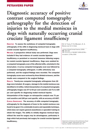
  • 2. INTRODUCTION Rupture of the cranial cruciate ligament (CCL) is one of the most common causes of canine pelvic limb lameness (Johnson and others 1994, Ness and others 1996). Stifle instability secondary to cranial cruci- ate ligament insufficiency (CCLI) leaves the menisci vulnerable to damage. Menis- cal injuries are seen in 49 to 70 per cent of dogs with concurrent CCLI (Flo and DeYoung 1978, Bennett and May 1991, Elkins and others 1991, Flo 1993, Metel- man and others 1995). A variety of surgical treatments are advocated for the manage- ment of CCLI. Stifle arthrotomy or arthroscopy is recommended to allow inspection of the menisci as a neglected meniscal injury will result in a poor out- come (Bennett and May 1991, Flo 1993, Ralphs and Whitney 2002). Following CCL surgery, dogs can suffer prolonged or incomplete recoveries for a variety of reasons including rupture of the contralateral CCL, joint sepsis or implant failure (Metelman and others 1995, Moore and Read 1995, Hill and others 1999, Innes and others 2000, Pacchiana and others 2003). So-called ‘‘latemeniscal’’injury,whereanapparently normalmeniscusatthetimeofsurgerysub- sequently becomes damaged, is reported in 6Á3 to 21Á7 per cent of dogs, regardless of the surgery performed (Metelman and others 1995, Thieman and others 2006, Lafaver and others 2007, Case and others 2008, Stein and Schmoekel 2008). Diag- nosis of a late meniscal injury can be chal- lenging and traditionally relies upon arthrotomy or arthroscopy. The ability to diagnose a meniscal injury without surgery would have significant advantages, guiding decision-making and potentially avoiding an unnecessary procedure. Ultrasonogra- phy and magnetic resonance imaging (MRI) have been suggested as useful in the diagnosis of meniscal injury in the dog (Banfield and Morrison 2000, Mahn and others 2005, Martig and others 2006, Blond and others 2008). However, both techniques have limitations with ultrasound being highly operator depen- dent (Mahn and others 2005) and MRI being expensive and requiring a general anaesthetic. MRI is the modality of choice for imag- ingthemenisciinhumanpatientswithvery high sensitivity and specificity (Crues and others1987,Minkandothers1988).Com- puted tomography arthrography (CTA) is also used for the detection of meniscal inju- ries in human beings and has been reported to have similar sensitivity and specificity to those of MRI (Ghelman 1985, Vande Berg and others 2000, 2002). There are several published studies describing the use of stifle CTA in canine cadavers. These have described the normal intra-articular anatomy as seen on CTA (Samii and Dyce 2004, Soler and others 2007) and also demonstrated that CTA can be used to diagnose CCL rupture (Han and others 2008) and medial menis- cal injury (Tivers and others 2008) in cadavers. The cadaver study by Tivers and others (2008) showed that dorsal CTA images had a sensitivity of 90 per cent and a specificity of 100 per cent for the diagnosis of simulated injuries of the cau- dal horn of the medial meniscus. A more recent study investigated the use of CTA using transverse images for the detection of tears to the cruciate ligaments and menisci (Samii and others 2009). This study reported a low accuracy of interpre- tation of CTA images for the diagnosis of meniscal injuries with sensitivities of 13Á3 to 73Á3 per cent and specificities of 57Á1 to 100 per cent. The aim of this current study was to assess dorsal plane computed tomography (CT) arthrograms for the diagnosis of inju- ries to the menisci in dogs suffering from CCLI. The hypothesis was that the inter- pretation of dorsal plane CT arthrograms would have high sensitivity and specificity for the diagnosis of meniscal injuries in dogs. We also hypothesised that an abnor- mal outline to the meniscus asseenon a CT arthrogram would be consistent with a meniscal injury. MATERIALS AND METHODS Dogs were prospectively recruited between August 2006 and March 2008 with full informed owner consent. Dogs were eligi- ble for inclusion if they had pelvic limb lameness localised to the stifle joint with a provisional diagnosis of CCLI or sus- pected late meniscal injury and were des- tined to have an exploratory arthrotomy as part of their investigation or treatment. Recruitment of dogs was dependent on owner and clinician variables, and there- fore, not all eligible dogs were recruited during the study period. Two populations of dogs were recruited: those which had undergone previous surgery for CCLI and those which had not had previous sur- gery but had a preoperative diagnosis of CCLI. All dogs were examined by a board- certified orthopaedic surgeon or by a sur- gery resident under direct supervision. A diagnosis of CCLI was made on the basis of physical examination findings, assess- ment of craniocaudal stifle stability (cranial draw and/or tibial thrust), stifle radiogra- phy and arthrocentesis for cytology and culture (as necessary). Following the diag- nostic workup, each dog had a CT scan made of the affected stifle before surgery. Dogs were sedated with a dose of 0Á01 mg/kg intravenous medetomidine (Domi- tor; Pfizer)and 0Á1 to 0Á2 mg/kg butorpha- nol (Torbugesic; Fort Dodge). They were positioned in the CT scanner in lateral recumbency with the affected leg depen- dent (Fig 1). A fourth-generation single- slice spiral CT scanner (Picker PQ 5000 CT Scanner; Universal Systems) was used for all scans. A survey CT scan was made of the affected limb, as previously described (Tivers and others 2008). The limb was positioned so that the tibial plateau was in line with the long axis of the CT couch. Contiguous, dorsal 1Á5-mm sections of the stifle were made starting cranial to the pa- tella and finishing just caudal to the fabel- lae. All scans were made using a sharp algorithm, 100Á0 mA and 120Á0 kV; the scan time was 90 seconds. Following the survey scan, routine arthrocentesis was per- formed on the stifle joint. Samples of joint fluid were submitted for diagnostic testing as appropriate. Sodium and meglumine amidotrizoate, 370 mg I/ml, (Urografin 370; Schering Health Care Limited) was diluted in a 1:1 ratio with sterile saline (0Á9 per cent NaCl), creating a concentra- tion of 185 mg iodine/ml as previously reported (Tivers and others 2008). A vol- ume of 0Á2 ml/kg of this diluted contrast agent was injected into the stifle, and the needle was removed. This volume of Journal of Small Animal Practice Á Vol 50 ÁJuly 2009 ÁÓ 2009 British Small Animal Veterinary Association 325 Diagnostic accuracy of stifle computed tomography arthrography
  • 3. contrast was extrapolated from a previous study and some unpublished data (Tivers and others 2008). The stifle was repeatedly flexed and extended to ensure good contrast distribution throughout the joint, and the dogwasrepositionedasbefore.TheCTscan was repeated resulting in a positive contrast CT arthrogram. If the dog was not having surgery immediately, the contrast was removed from the joint througharthrocent- esis to prevent any possible discomfort asso- ciated with distention of the synovium. The medetomidine was antagonised with atipa- mazole (Antisedan; Pfizer), given intramus- cularly at the same volume. Dogs were subsequently anaesthetised for surgery within 24 hours of the CT scan. All surgeries involved a standard lateral or medial parapatellar arthrotomy (Piermat- tei and Johnson 2004) with inspection of the cruciate ligaments and both menisci. The surgeons were blinded to the CT results and inspected the medial and lateral menisci both visually and using a probe. They were provided with a printed form and asked to record whether the CCL was intact, partiallyruptured or completely ruptured. They also recorded whether there was damage to either of the menisci and the precise nature of the injury, includ- ing the area damaged and whether it was displaced. Following completion of the study, the CT arthrograms were reviewed, indepen- dently, by two board-certified radiologists andaboard-certifiedsurgeon.Thereviewers had access to the CTA images of beagle cadaver stifles with simulated meniscal injuries from a previous study, but they had no experience of the interpretation of stifle CT arthrograms from dogs with clinical disease. The scans were analysed as Digital Imaging and Communications in Medicine (DICOM) images using Image Viewer (Visbion Ltd). They were viewedinarandomorder,andthereviewers were blinded to the dogs’ history, physical examination findings and surgical findings. The reviewers recorded whether the CT arthrogram had sufficient contrast to be of diagnostic quality and whether the pres- ence of metal implants affected the inter- pretation. They then recorded whether the meniscus was intact or abnormal. A meniscus was considered abnormal if there was disruption of the normal outline. Where the meniscus was abnormal, the reviewers recorded how the image of the meniscus differed from the normal appear- ance. The reviewers also recorded whether the CT arthrogram had sufficient contrast to be of diagnostic quality and whether the presence of metal implants affected the interpretation. The results were subsequently com- pared with the surgical findings to deter- mine the sensitivity, specificity and positive and negative predictive values of interpretation of CTA for the detection of meniscal pathology. The measure of agreement between the three reviewers was assessed using a multi-rater variation of Brennan and Prediger’s (1981) free- marginal kappa statistic as described by Randolph (2005) using an online calcula- tor (Randolph 2008). In addition, the accuracy of the test for each reviewer was evaluated using receiver operating charac- teristic (ROC) curve analysis using an online calculator (Eng 2009). Approximately five months following the blinded analysis, a retrospective inter- pretation was performed by one of the authors (P. N. M). The images were re- viewed, and the findings were recorded as described above. Sensitivity, specificity, positive and negative predictive values and ROC curveanalysiswere then recalculated. RESULTS Twenty dogs were recruited. Twenty-one stifle CT arthrograms and arthrotomies were performed. One dog (dog 12) was originally presented for investigation and treatment of suspected CCLI. A CT arthrogram was performed, and no menis- cal injury was detected at arthrotomy. The stifle was stabilised with a fabellotibial suture. The dog was re-presented for per- sistent lameness following the surgery, and repeat CTA and arthrotomy were performed. Breeds comprised four Labradors, four boxers, two golden retrievers, one rottwei- ler, one bullmastiff, one German shepherd dog, one Jack Russell terrier and six cross- breeds. There were five entire male dogs, five neutered male dogs, four entire female dogs and six neutered female dogs. Age ranged from one year to nine years with a median of five years. The weight of the dogs ranged from 8Á5 to 63 kg with a median of 35Á1 kg. Seven of the CT arthrograms were ofsti- fles which had not had surgery, and the remaining 14 scans were of joints which had previously undergone surgery. For the dogs which had not had previous sur- gery, the duration of lameness ranged from 0Á5 to 12 months with a median of 1Á5 months. Of the stifles which had had pre- vious surgery, 10 had been stabilised with a fabellotibial suture,three witha tibial pla- teau levelling osteotomy (TPLO) and one with an over-the-top (OTT) fascia lata graft. The time interval between the origi- nal surgery and the subsequent CT arthro- gram and arthrotomy ranged from 0Á75 to 23 months with a median of 4Á25 months. These results are summarised in Table 1. FIG 1. Dog positioned for computed tomography (CT) scan of the left stifle. The dog is positioned in lateral recumbency with the affected leg underneath. The leg is positioned approximately perpendicular to the long axis of the CT couch with the tibial plateau in line with the couch 326 Journal of Small Animal Practice Á Vol 50 ÁJuly 2009 ÁÓ 2009 British Small Animal Veterinary Association M. S. Tivers and others
  • 4. The arthrotomy findings were recorded for all the stifles (Table 2). There were 15 full CCL ruptures and six partial ruptures (dog 14 had an intra-articular graft in the location of the CCL). There were 14 inju- ries to the medial meniscus. The remaining seven medial menisci were considered nor- mal. There were no injuries to the lateral meniscus. Meniscal injuries varied in type and severity. Twenty-one CT arthrograms were available for interpretation. The diagnostic quality of the images was considered good in all but one CTA (dog 15). The lateral and medial menisci were visible as wedge-shaped structures, highlighted by the contrast (Fig 2). Meniscal injuries were identified by an abnormal outline to the meniscus. Typically, this was a bulge or contour at the axial part of the meniscal outline (Fig 3). In the dog treated with an OTT, the graft could be seen, mimicking the CCL. The TPLO plates and screws and the crimps used to secure fabellotibial sutures were also clearly visible (Fig 4). Both types of metal implants caused streak artefacts, although that caused by the crimps did not obscure the view of the intra-articular structures. The artefact caused by the TPLO implants did inter- fere with the quality of the images; how- ever, it was possible to see the intra- articular structures (particularly the menisci) on sufficient slices that the stud- ies were considered diagnostic. It was still possible to successfully identify meniscal injuries in dogs which had TPLO im- plants, despite the considerable artefact created by the implants. Theresultsoftheblindedreviewarepre- sented in Table 2. The sensitivity of inter- pretation of the images for meniscal injury was57percentforreviewers1and3and64 per cent for reviewer 2, and the specificity was 100 per cent for reviewers 1 and 3 and 71per cent for reviewer 2.Thepositive pre- dictivevaluewas82to100percent,andthe negative predictive value was 50 to 54 per cent. Agreement between the reviewers was 0Á37 (free-marginal kappa). The ROC curve analysis resulted in an estimated area under the curve of 0Á786 for reviewers 1 and 3 and 0Á679 for reviewer 2. The results of the retrospective interpre- tation are also included in Table 2. The reviewer correctly identified 10 of 14 meniscalinjuriesandsevenofsevennormal medial menisci. This gave a sensitivity of 71 per cent, a specificity of 100 per cent, a positive predictive value of 100 per cent and a negative predictive value of 64 per cent. ROC curve analysis resulted in an estimated area under the curve of 0Á857. DISCUSSION This study has shown that stifle dorsal plane CTA has potential for the diagnosis of injuries to the medial meniscus in dogs. Before this study, there was little informa- tion on how a damaged meniscus would appear on a CT arthrogram. The normal menisci are wedge shaped, and the axial part of the medial meniscus, in particular, has a very long and thin profile in cross- section on CTA images (Tivers and others 2008). We therefore hypothesised that a change in the normal outline of the meniscus would be consistent with an injury such as a displaced tear or fold. The images from stifles with meniscal inju- ries support this hypothesis with an abnor- mal outline,in particular at theaxial border of the meniscus being consistent with an injury. This was most commonly seen at the mid part of the meniscus and in the axial part of the joint. The more obvious lesions resulted in an abnormal outline of contrast at the axial border of the menis- cus described as an ‘‘axial bulge’’ (Figs 3, 5 and 6). Comparison with the surgical find- ings confirmed that these areas corre- sponded with displacement of a torn part of the meniscus or folding of the caudal horn. There was considerable variation Table 1. Signalment and significant historical information Dog Age (years) Sex Breed Weight (kg) Affected leg Previous cruciate surgery Duration of clinical signs or time interval since original surgery (months) 1 5 FE Labrador 27Á8 Right None 3 2 9 ME Crossbreed 63 Left TPLO 2Á5 3 6 MN Crossbreed 35Á2 Left FT Suture 5 4 9 FN Labrador 36Á8 Left None 1Á5 5 3 ME Jack Russell terrier 8Á5 Left None 12 6 3 MN Bullmastiff 57Á5 Left FT suture 23 7 2 FN Boxer 25 Right FT suture 3 8 2 FN Boxer 33Á4 Left FT suture 3 9 3 MN Boxer 35Á2 Right FT suture 3Á5 10 2 MN Crossbreed 39Á2 Left FT suture 8 11 7 ME Labrador 36Á5 Left None 1 12 3 ME Labrador 35 Right None 2 12* 3 ME Labrador 35 Right FT suture 1Á5 13 7 FN Crossbreed 43Á3 Right FT suture 7 14 1 FE Rottweiler 34Á5 Right OTT 10 15 7 FN Crossbreed 25 Right None 0Á5 16 5 FE Golden retriever 32Á2 Right TPLO 0Á75 17 5 FE Golden retriever 36Á5 Left TPLO 5Á5 18 5 FN German shepherd dog 37 Right FT suture 2Á5 19 2 ME Boxer 29Á5 Left FT suture 7 20 7 MN Crossbreed 32 Left None 0Á5 FE Female entire, ME Male entire, MN Male neutered, FN Female neutered, TPLO tibial plateau levelling osteotomy, FT Fabellotibial, OTT Over the top *Dog 12 had two computed tomography scans and subsequent surgeries at separate time points Journal of Small Animal Practice Á Vol 50 ÁJuly 2009 ÁÓ 2009 British Small Animal Veterinary Association 327 Diagnostic accuracy of stifle computed tomography arthrography
  • 5. betweenthesizeandtheshapeoftheabnor- mal contrast outline, and this resulted in some lesions being missed on initial inter- pretation. At retrospective analysis, 10 of 14 meniscal injuries were correctly diag- nosed (Table 2). It was not possible to see any evidence of injury in the four dam- aged menisci, which appeared normal on the CT arthrogram. One of these scans (dog 15) had poor contrast filling, which resulted in poor diagnostic quality which was considered to have hindered interpre- tation. The three other dogs (dogs 5, 9 and 10) had scans of diagnostic quality, but no meniscal injury was evident at retrospective review. Dogs 5 and 9 had tears which were non-displaced in association with damage to the meniscus which had not resulted in a tear (crushing or thickening). Although the crushing or thickening was seen at sur- gery, we would suggest that these sorts of injury do not affect the outline of the meniscus and would therefore be very dif- ficultorimpossibletodiagnoseusingCTA. The tears present in these two dogs were most likely not seen as they were not dis- placed. Dog 10 was unusual, in that a small bucket handle tear, along the axial margin ofthemeniscus,had beenremovedatapre- vious surgery. Therefore, the outline of the meniscus was intact, and no lesion was seen on the CT arthrogram. These results suggest that meniscal lesions which are large or displaced are readily seen on CT arthrograms, whereas injuries not resulting in a tear or those tears which are not displaced are not apparent. Atinitialinterpretation,CTAwasfound to have a sensitivity of 57 to 64 per cent and a specificity of 71 to 100 per cent with fair agreement between the reviewers. The accuracy as determined by ROC curve analysis was fair to good. A previous study Table 2. Arthrotomy findings and the results of the blinded and retrospective reviews Dog Degree of cruciate rupture Meniscal injury? Surgical findings including injury to medial meniscus Reviewer identification of meniscal injury Identification of meniscal injury on retrospective review? 1 2 3 1 Partial No No No No No 2 Full No Tibial plateau levelling osteotomy implants removed No No No No 3 Full Yes Longitudinal tear to caudal pole. Cranial pole thickened and abnormal Yes No No Yes 4 Full Yes Longitudinal tear to caudal pole Yes No Yes Yes 5 Full Yes Bucket handle tear and crushing of caudal pole No Yes No No 6 Full No Synovial inflammation, marked periarticular fibrosis No No No No 7 Partial No Inflammation and fibrosis around nylon suture No No No No 8 Full Yes Multiple tears in caudal pole. Caudal pole folded forward Yes Yes Yes Yes 9 Partial Yes Caudal pole torn but non-displaced. Cranial pole thickened Yes No No No 10 Full Yes Previous bucket handle tear removed No Yes No No 11 Partial Yes Small transverse tear No Yes Yes Yes 12 Full No No No No No 12 Full Yes Caudal pole torn and folded cranially Yes Yes Yes Yes 13 Partial No Mineralised fragment in meniscus. Patellar luxation No Yes No No 14 Full No Marked periarticular fibrous reaction and fibrous reaction associated with graft No Yes No No 15 Full Yes Very small longitudinal tear No Yes No No 16 Full Yes Caudal pole torn and folded cranially Yes Yes Yes Yes 17 Full Yes Caudal pole torn and folded cranially Yes Yes Yes Yes 18 Full Yes Whole medial meniscus crushed. Caudal pole folded cranially Yes Yes Yes Yes 19 Partial Yes Longitudinal tear to caudal pole. Caudal pole folded cranially No No Yes Yes 20 Full Yes Bucket handle tear to mid meniscus. Caudal pole detached but non-displaced No No No Yes Bold denotes correct interpretation of computed tomographic arthrogram by reviewer FIG 2. Dorsal plane computed tomography arthrograms of the stifle from dog 1 (a) and dog 13 (b) showing normal medial and lateral menisci. The lateral meniscus is highlighted with a white arrow, and the medial meniscus is highlighted with a black arrow in the first image. The menisci are wedge shaped and surrounded by white contrast 328 Journal of Small Animal Practice Á Vol 50 ÁJuly 2009 ÁÓ 2009 British Small Animal Veterinary Association M. S. Tivers and others
  • 6. onsimulatedmeniscalinjuryhadsuggested that the accuracy of CTA would be greater (Tivers and others 2008). There was also some variation between the reviewers’ results. None of the reviewers had previous experience in the interpretation of CT arthrograms in dogs with CCLI or late meniscal injury. It is likely that this inexpe- rience contributed to variation in interpre- tation between reviewers. The effectiveness of interpretation of CT arthrograms im- proved at retrospective review with a sensi- tivity of 71 per cent, a specificity of 100 per cent and good accuracy as determined by ROC curve analysis. This suggests that the interpretation of CT arthrograms has a steep learning curve but that with some experience, it is an accurate technique for the diagnosis of medial meniscal injury in the dog. CTA of the human knee is reported to have a sensitivity of 92 to 100 per cent anda specificityof78to94percent(Vande Bergandothers2000,2002,Mutschlerand others 2003), varying with the type of meniscal injury and whether previous sur- gery has been performed. These studies used a dual detector helical CT unit, which allowed reformatting of images with min- imal loss of quality. Similarly to our study, ‘‘a meniscal abnormality was defined bythe presence of contour irregularity, peripheral separation, or tear of a meniscus’’ (Vande Berg and others 2000). Although the accu- racy reported in our study compares well with that in the human literature, it is clear that CTA is able to detect much more sub- tle lesions in people. This discrepancy could be related to the quality of the scans produced by the CT machine used; 1Á5- mm thick slices were obtained which resultedinasignificantlossofimagequality whenreformattingfromthedorsalplane.It is likely that the use of a 16-slice multi- detectorCTscannerwouldproduceimages of greater quality and diagnostic potential. The image quality could also be improved byusingsmallerslicethicknessandoverlap- ping slices, allowing reconstruction of sag- ittal or transverse images of much higher quality. This would facilitate the detection of smaller and more subtle lesions. A previous study investigated the use of transverse CTA scans for the diagnosis of injuries to the cruciate ligaments and menisci (Samii and others 2009). They found a sensitivity of 13Á3 to 73Á3 per cent and a specificity of 57Á1 to 100 per cent for the diagnosis of meniscal tears. Sensitivity was 64Á3 per cent and specificity was 73Á3 per cent on consensus evaluation. The results in our current study compare favourably with these findings and would support the use of dorsal plane scans over FIG 3. Dorsal plane computed tomography arthrograms of the stifle from dog 12 before surgery (a) and six weeks after surgery (b) for rupture of the cranial cruciate ligament. In image (a), the medial meniscus has a normal outline. In image (b), the medial meniscus has an axial bulge and has lost its normal wedge-shaped appearance (red ring). At surgery, the caudal pole of the meniscus was torn and folded cranially FIG 4. Dorsal plane computed tomography arthrograms of the stifle from dog 7 (a) showing the fabellotibial suture crimps and dog 2 (b) showing the tibial plateau levelling osteotomy (TPLO) plate and screws. The crimps produce a minimal amount of streak artefact, which does not obscure the intra-articular structures. The TPLO implants produce a much greater amount of artefact, which interferes with the view of the joint. However, the artefact is inconsistent on the different slices, and it is possible to still achieve a diagnostic scan of the joint. In this example, the medial meniscus is clearly seen, despite the artefact (red ring) Journal of Small Animal Practice Á Vol 50 ÁJuly 2009 ÁÓ 2009 British Small Animal Veterinary Association 329 Diagnostic accuracy of stifle computed tomography arthrography
  • 7. transverse scans for the investigation of meniscal injuries. It is likely that a combi- nation of both scan orientations would produce even more accurate results, although this is unproven. One of the potential limitations of CTA for the diagnosis of late meniscal injuries is image distortion as a result of streak arte- facts from metal implants. Metal crimps were present in 10 dogs but did not inter- fere with the diagnostic quality of the images produced. Three dogs had TPLO implants in situ, and although these pro- duced much greater distortion which inter- fered with the view of the intra-articular structures on some scan slices, images of diagnostic quality could still be obtained. If artefact were a significant problem in stifles with TPLO implants, then a trans- verse scan could be performed and the images reformatted into the dorsal plane. Visual inspection of the menisci through arthroscopy or arthrotomy is rec- ommended in all dogs with CCLI to assess for meniscal injury (Bennett and May 1991, Flo 1993, Ralphs and Whitney 2002). It is also useful to probe the menisci todetectincompleteorsubtlelesions.With arthrotomy or arthroscopy, it can be diffi- cult to inspect the most caudal aspect of the joint, and hence, caudal lesions can be missed. Arthrotomy is associated with con- siderable morbidity, and while arthroscopy is associated with much less morbidity, it does require specialist equipment and training (Hoelzler and others 2004). A method of diagnosing meniscal injury without the need for surgery would there- fore be advantageous. CTA is minimally invasive and requires no specialist training to obtain the images, which are produced using a standard protocol and are easily reproducible. In many dogs with stifle dis- ease, arthrocentesis is routinely performed as part of their investigation, and this could be combined with the arthrogram. Although contrast is injected into the joint, no complications related to this were seen, and the authors are unaware of any studies reporting complications associated with this procedure. Studies have shown that MRI and MRI arthrography can be used to diagnose cru- ciate ligament rupture and meniscal injury in experimental and naturally occurring CCLI in dogs (Banfield and Morrison 2000, Martig and others 2006, Blond and others 2008). In a study of 11 dogs, which had not had previous surgery, MRI was reported to have a sensitivity of 100 per cent and a specificity of 94 per cent (Blond and others 2008). However, it is suggested that interpretation of images from previously operated stifles may be more difficult because of increased signal of healing or healed tissues, which could be mistaken for meniscal damage (Blond and others 2008). In addition, two studies reported image distortion as a result of either metallic microfragments from instruments used during surgery (Banfield and Morrison 2000) or the ferromagnetic properties of coloured polydioxanone suture (Martig and others 2006). This FIG 5. Dorsal plane computed tomography arthrogram (CTA) of the stifle and an intra-operative image from dog 8. The CTA shows that the medial meniscus has an abnormal outline with an axial bulge (red ring). The photograph shows a medial arthrotomy with the medial femoral condyle highlighted with an asterix. The caudal pole of the medial meniscus (arrow) is torn and displaced cranially, consistent with the CTA findings FIG 6. Dorsal plane computed tomography arthrogram (CTA) of the stifle and an intra-operative image from dog 18. The CTA shows a marked axial bulge of the medial meniscus (red ring). The photograph shows a medial arthrotomy with the medial femoral condyle highlighted with an asterix. The caudal pole of the medial meniscus is folded forward, consistent with the CTA findings 330 Journal of Small Animal Practice Á Vol 50 ÁJuly 2009 ÁÓ 2009 British Small Animal Veterinary Association M. S. Tivers and others
  • 8. would not be a concern with CTA. CTA has several potential advantages over MRI, in that it is considerably cheaper, the scan times are much faster and the dog need only be sedated rather than anaesthetised. Ultrasonography has been shown to have great potential for investigating meniscal lesions in dogs, with a sensitivity of 90 per cent and a specificity of 92Á9 percent (Mahn and others 2005). Ultra- sonography is a completely non-invasive technique and has the advantages that it is widely available and it can be per- formed in a conscious animal. However, musculoskeletal ultrasonography is highly specialised, and the results are largely dependent on the skill and experience of the operator (Mahn and others 2005). Whereas Mahn and others (2005) reported excellent results, they included only cases with significant meniscal pathology and commented that more subtle pathologies may be more difficult to detect. Equally, these authors stated that it may not be pos- sible to obtain diagnostic images when there is marked periarticular fibrosis or os- teophytosis,potentiallylimitingtheuseful- ness in dogs with chronic disease or following surgery. Unlike ultrasonogra- phy, stifle CTA requires no specialist train- ing to produce diagnostic images, although image interpretation is associated with a steep learning curve as discussed above. This study has shown that stifle CTA canbeusedtosuccessfullydiagnoseinjuries to the medial meniscus in association with CCLI and that it has good accuracy. Image interpretation is potentially challenging, particularly for those inexperienced with CTA. Hopefully, the information in this paper will aid clinicians who use this tech- nique in the future. We would anticipate that with advances in equipment and expe- riencewithimageinterpretation,thiscould become an extremely reliable method of diagnosing meniscal injuries. It could then be used as an alternative to arthrotomy or arthroscopy and hence eliminate the mor- bidity for those animals undergoing either as a purely diagnostic procedure. Acknowledgements This study was funded by a BSAVA Petsa- vers grant, and the authors are very grateful for this support. We would also like to acknowledge themembers oftheorthopae- dic and radiology services at our institution for their help with this study. References BANFIELD, C. M. MORRISON, W. B. (2000) Magnetic resonance arthrography of the canine stifle joint: technique and applications in eleven military dogs. Veterinary Radiology Ultrasound 41, 200-213 BENNETT, D. MAY, C. (1991) Meniscal damage asso- ciated with cruciate disease in the dog. Journal of Small Animal Practice 32, 111-117 BLOND, L., THRALL, D. E., ROE, S. C., CHAILLEUX, N. ROBERTSON, I. D. (2008) Diagnostic accuracy of magnetic resonance imaging for meniscal tears in dogs affected with naturally occurring cranial cruciate ligament rupture. Veterinary Radiology Ultrasound 49, 425-431 BRENNAN, R. L. PREDIGER, D. J. (1981) Coefficient kappa: some uses, misuses, and alternatives. Educational and Psycholoical Measurement 41, 687-699 CASE, J. B., HULSE, D., KERWIN, S. C. PEYCKE, L. E. (2008) Meniscal injury following initial cranial cru- ciate ligament stabilization surgery in 26 dogs (29 stifles). Veterinary and Comparative Orthopae- dics and Traumatology 21, 365-367 CRUES, J. V., III, MINK, J., LEVY, T. L., LOTYSCH, M. STOLLER, D. W. (1987) Meniscal tears of the knee: accuracy of MR imaging. Radiology 164, 445- 448 ELKINS, A. D., PECHMAN, R., KEARNEY, M. T. HERRON, M. (1991) A retrospective study evaluating the degree of degenerative joint disease in the stifle joint of dogs following surgical repair of anterior cruciate ligament rupture. Journal of the Ameri- can Animal Hospital Association 27, 533-540 ENG, J. (2009) ROC analysis: web-based calculator for ROC curves. http://www.jrocfit.org. Accessed April 7, 2009 FLO, G. L. (1993) Meniscal injuries. Veterinary Clin- ics of North America: Small Animal Practice 23, 831-843 FLO, G. L. DEYOUNG, D. (1978) Meniscal injuries and medial meniscectomy in the canine stifle. Journal of the American Animal Hospital Association 14, 683-689 GHELMAN, B. (1985) Meniscal tears of the knee: evaluation by high-resolution CT combined with arthrography. Radiology 157, 23-27 HAN, S., CBEON, H., CHO, H., KIM, J., KANG, J. H., YANG, M. P., LEE, Y., LEE, H. CHANG, D. (2008) Evalua- tion of partial cranial cruciate ligament rupture with positive contrast computed tomographic arthrography in dogs. Journal of Veterinary Sci- ence 9, 395-400 HILL, C. R., CONZEMIUS, M. G., SMITH, G. K., MCMANUS, P. M. MALONEY, D. (1999) Bacterial culture of the canine stifle joint following surgical repair of rup- tured cranial cruciate ligament. Veterinary and Comparative Orthopaedics and Traumatology 12, 1-5 HOELZLER, M. G., MILLIS, D. L., FRANCIS, D. A. WEIGEL, J. P. (2004) Results of arthroscopic versus open arthrotomy for surgical management of cranial cruciate ligament deficiency in dogs. Veterinary Surgery 33, 146-153 INNES, J. F., BACON, D., LYNCH, C. POLLARD, A. (2000) Long-term outcome of surgery for dogs with cra- nial cruciate ligament deficiency. Veterinary Record 147, 325-328 JOHNSON, J. A., AUSTIN, C. BREUR, G. J. (1994) Inci- dence of canine appendicular musculoskeletal disorders in 16 veterinary teaching hospitals from 1980 through 1989. Veterinary and Comparative Orthopaedics and Traumatology 7, 56-69 LAFAVER, S., MILLER, N. A., STUBBS, W. P., TAYLOR, R. A. BOUDRIEAU, R. J. (2007) Tibial tuberosity advance- ment for stabilization of the canine cranial cruciate ligament-deficient stifle joint: surgical technique, early results, and complications in 101 dogs. Vet- erinary Surgery 36, 573-586 MAHN, M. M., COOK, J. L., COOK, C. R. BALKE, M. T. (2005) Arthroscopic verification of ultrasono- graphic diagnosis of meniscal pathology in dogs. Veterinary Surgery 34, 318-323 MARTIG, S., KONAR, M., SCHMOKEL, H. G., RYTZ, U., SPRENG, D., SCHEIDEGGER, J., HOHL, B., KIRCHER, P. R., BOISCLAIR, J. LANG, J. (2006) Low-field MRI and arthroscopy of meniscal lesions in ten dogs with experimentally induced cranial cruciate ligament insufficiency. Veterinary Radiology Ultrasound 47, 515-522 METELMAN, L. A., SCHWARZ, P. D., SALMAN, M. ALVIS, M. R. (1995) An evaluation of 3 different cranial cruciate ligament surgical stabilization procedures as they relate to postoperative meniscal injuries – a retrospective study of 665 stifles. Veterinary and Comparative Orthopaedics and Traumatology 8, 118-123 MINK, J. H., LEVY, T. CRUES, J. V., III (1988) Tears of the anterior cruciate ligament and menisci of the knee: MR imaging evaluation. Radiology 167, 769-774 MOORE, K. W. READ, R. A. (1995) Cranial cruciate ligament rupture in the dog – a retrospective study comparing surgical techniques. Australian Veter- inary Journal 72, 281-285 MUTSCHLER, C., VANDE BERG, B. C., LECOUVET, F. E., POILVACHE, P., DUBUC, J. E., MALDAGUE, B. MALGHEM, J. (2003) Postoperative meniscus: assessment at dual-detector row spiral CT arthrography of the knee. Radiology 228, 635-641 NESS, M. G., ABERCROMBY, R. H., MAY, C., TURNER, B. M. CARMICHAEL, S. (1996) A survey of orthopaedic conditions in small animal veterinary practice in Britain. Veterinary and Comparative Orthopaedics and Traumatology 9, 43-52 PACCHIANA, P. D., MORRIS, E., GILLINGS, S. L., JESSEN, C. R. LIPOWITZ, A. J. (2003) Surgical and postop- erative complications associated with tibial pla- teau leveling osteotomy in dogs with cranial cruciate ligament rupture: 397 cases (1998- 2001). Journal of the American Veterinary Medi- cal Association 222, 184-193 PIERMATTEI, D. L. JOHNSON, K. A. (2004) Approach to the stifle joint. In: An Atlas of Surgical Approaches to the Bones and Joints of the Dog and Cat. 4th edn. Saunders, Philadelphia, PA, USA. pp 342-349 RALPHS, S. C. WHITNEY, W. O. (2002) Arthroscopic evaluation of menisci in dogs with cranial cruciate ligament injuries: 100 cases (1999-2000). Jour- nal of the American Veterinary Medical Associa- tion 221, 1601-1604 RANDOLPH, J. J. (2005) Free-marginal multirater kappa: an alternative to Fleiss’ fixed-marginal multirater kappa. Proceedings of the Joensuu Learning and Instruction Symposium. Joensuu, Finland, October 14 to 15, 2005. pp 1-20 RANDOLPH, J. J. (2008) Online kappa calculator. http:// justus.randolph.name/kappa. Accessed October 18, 2008 SAMII, V. F. DYCE, J. (2004) Computed tomographic arthrography of the normal canine stifle. Veteri- nary Radiology Ultrasound 45, 402-406 SAMII, V. F., DYCE, J., POZZI, A., DROST, W. T., MATTOON, J. S., GREEN, E. M., KOWALESKI, M. P. LEHMAN, A. M. (2009) Computed tomographic arthrography of the stifle for detection of cranial and caudal cruciate ligament and meniscal tears in dogs. Veterinary Radiology Ultrasound 50, 144-150 SOLER, M., MURCIANO, J., LATORRE, R., BELDA, E., RODRIGUEZ, M. J. AGUT, A. (2007) Ultrasonographic, com- puted tomographic and magnetic resonance imag- ing anatomy of the normal canine stifle joint. Veterinary Journal 174, 351-361 STEIN, S. SCHMOEKEL, H. (2008) Short-term and eight to 12 months results of a tibial tuberosity advancement as treatment of canine cranial cru- ciate ligament damage. Journal of Small Animal Practice 49, 398-404 Journal of Small Animal Practice Á Vol 50 ÁJuly 2009 ÁÓ 2009 British Small Animal Veterinary Association 331 Diagnostic accuracy of stifle computed tomography arthrography
  • 9. THIEMAN, K. M., TOMLINSON, J. L., FOX, D. B., COOK, C. COOK, J. L. (2006) Effect of meniscal release on rate of subsequent meniscal tears and owner- assessed outcome in dogs with cruciate disease treated with tibial plateau levelling osteotomy. Veterinary Surgery 35, 705-710 TIVERS, M. S., MAHONEY, P. CORR, S. A. (2008) Canine stifle positive contrast computed tomography arthrography for assessment of caudal horn meniscal injury: a cadaver study. Veterinary Sur- gery 37, 269-277 VANDE BERG, B. C., LECOUVET, F. E., POILVACHE, P., DUBUC, J. E., BEDAT, B., MALDAGUE, B., ROMBOUTS, J. J. MALGHEM, J. (2000) Dual-detector spiral CT arthrography of the knee: accuracy for detection of meniscal abnor- malities and unstable meniscal tears. Radiology 216, 851-857 VANDE BERG, B. C., LECOUVET, F. E., POILVACHE, P., DUBUC, J. E., MALDAGUE, B. MALGHEM, J. (2002) Anterior cruciate ligament tears and associated meniscal lesions: assessment at dual-detector spiral CT arthrography. Radiology 223, 403-409 332 Journal of Small Animal Practice Á Vol 50 ÁJuly 2009 ÁÓ 2009 British Small Animal Veterinary Association M. S. Tivers and others