1. NEURO CUTANEOUS MARKERS by Dr. Kurian Joseph
1. Café- au- lait spots
- Dark brown hypermelanotic macules with smooth or irregular borders, commonly seen in back, buttocks and trunk.
- Seen in neurofibromatosis. Also seen in Albright's synd, Fanconi anemia, Bloom syndrome.
- Axillary Freckling - similar small round spots in axilla and inguinal areas are pathognomonic in NF.
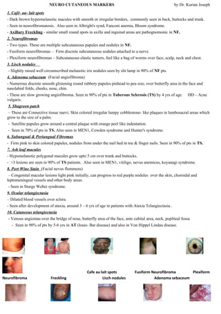
2. Neurofibromas
- Two types. These are multiple subcutaneous papules and nodules in NF.
- Fusiform neurofibromas – Firm discrete subcutaneous nodules attached to a nerve.
- Plexiform neurofibromas – Subcutaneous elastic tumors, feel like a bag of worms over face, scalp, neck and chest.
3. Lisch nodules
- Slightly raised well circumscribed melanotic iris nodules seen by slit lamp in 90% of NF pts.
4. Adenoma sebaceum (Facial angiofibroma)
- Numerous discrete smooth glistening round rubbery papules pinhead to pea size, over butterfly area in the face and
nasolabial folds, cheeks, nose, chin.
- These are slow growing angiofibroma. Seen in 90% of pts in Tuberous Sclerosis (TS) by 4 yrs of age. DD – Acne
vulgaris.
5. Shagreen patch
– These are Connective tissue naevi. Skin colored irregular lumpy cobblestone- like plaques in lumbosacral areas which
grow to the size of a palm.
- Satellite papules grow around a central plaque with orange peel like indentation.
- Seen in 70% of pts in TS. Also seen in MEN1, Cowden syndrome and Hunter's syndrome.
6. Subungual & Periungual Fibromas
- Firm pink to skin colored papules, nodules from under the nail bed in toe & finger nails. Seen in 90% of pts in TS.
7. Ash leaf macules
- Hypomelanotic polygonal macules grow upto 5 cm over trunk and buttocks.
- >3 lesions are seen in 90% of TS patients. Also seen in MEN1, vitiligo, nevus anemicus, koyanagi syndrome.
8. Port Wine Stain (Facial nevus flemmens)
– Congenital macular lesions light pink initially, can progress to red purple nodules over the skin, choroidal and
leptomeningeal vessels and other body areas.
- Seen in Sturge Weber syndrome.
9. Ocular telangiectasia
– Dilated blood vessels over sclera.
- Seen after development of ataxia, around 3 – 6 yrs of age in patients with Ataxia Telangiectasia..
10. Cutaneous telangiectasia
- Venous angiomas over the bridge of nose, butterfly area of the face, ante cubital area, neck, popliteal fossa
- Seen in 90% of pts by 5-6 yrs in AT (louis- Bar disease) and also in Von Hippel Lindau disease.
Cafe au lait spots Fusiform Neurofibroma Plexiform
Neurofibroma Freckling Lisch nodules Adenoma sebaceum
2. . Shagreen patch Subungual fibroma Ash leaf macules
Port wine stain Ocular telangiectasia Cutaneous telangiectasia
Neuro cutaneous syndromes:
1. Neurofibromatosis: Two clinical types. Chromosome 17 defect
NF1 (von Recklinghausen’s diseas e/ Peripheral NF ) - Diagnostic criteria are presence of ≥2 of the foll. ① ≥6 cafe au
lait spots each >15mm dia.m in adults. ② ≥2 Neurofibroma or one Plexiform NF. ③
Axillary / Inguinal Freckles. ④ ≥2 Lisch nodules. ⑤ Bone lesions e.g. Sphenoidal Dysplasia, Tibial pseudoarthrosis. ⑥ 1st
degree rel. with NF1.
NF 2 – (central NF) - Few or no skin lesions, B/L Acoustic Neuroma, Cerebral & optic nerve gliomas, Meningiomas,
Spinal neurofibromas.
2. Tuberous Sclerosis: (Bournville’s disease) Classic Triad of Mental Retardation + Epilepsy + Skin lesions (Ash leaf
macules, Adenoma sebaceum, Sub & Periungual fibromas, Shagreen patches). Other features – Hyperplastic gums, retinal
phakomas, renal lung & heart tumors, cerebral gliomas, calcification of basal ganglia.
3. Von Hippel–Lindau Disease: Characterized by the combination of retinal, cerebellar & spinal hemangioblastomas. Chr 3
defect
4. Sturge Weber Syndrome: Characterized by facial nevus flemmens (Port wine stain-usually along distribution of
ophthalmic br. of 5th
nerve), contra lateral focal seizures, calcification of cortical & sub cortical structures, glaucoma on the
same side of skin lesions.
5. Ataxia Telangiectasia: Progressive cerebellar ataxia, oculo-cutaneous telangiectasia, pulmonary & sinus infections,
immunodeficiency, choreo-athetosis, lymphoreticular malignancy.
All the Neuro cutaneous syndromes are inherited except SWS. And all these are inherited as Autosomal Dominant except
Ataxia Telangiectasia which is autosomal recessive.