1. 2/20/2023 1
GENERAL ANATOMY
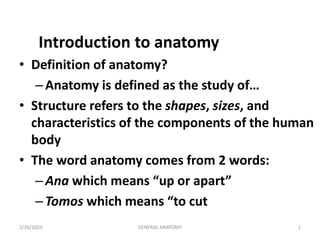
Introduction to anatomy
• Definition of anatomy?
–Anatomy is defined as the study of…
• Structure refers to the shapes, sizes, and
characteristics of the components of the human
body
• The word anatomy comes from 2 words:
–Ana which means “up or apart”
–Tomos which means “to cut
2. 2/20/2023 2 GENERAL ANATOMY
SUB DIVISION OF ANATOMY
• General anatomy – gross & microscopic structures as
well as the composition of the body, its tissues & fluids
• Special anatomy –certain definite organs or groups of
organs involved in the performance of special function
• Gross ( macroscopic) anatomy –is study of human body
or its parts without help of a microscope
• Microscopic anatomy – the study of cells, tissues &
organs of body by help of using the microscope
3. 2/20/2023 3 GENERAL ANATOMY
• Regional or Topographic anatomy- is the anatomy of
certain related parts or divisions of the body
• Systemic anatomy - the anatomy of the different systems
of the body
• Descriptive anatomy - deals with the description of the
physical structure of man
• Comparative anatomy – is the comparative study of
structures with regard to homologous organs or parts
• Applied anatomy – the practical application of
anatomical knowledge to the diagnosis & treatment of
disease.
4. 2/20/2023 4 GENERAL ANATOMY
• Ultrastructural anatomy – the ultramicroscopic study of
structures too small to be seen with light microscope
• Developmental anatomy – anatomy of the structural
changes of an individual from fertilization to adulthood. It
includes embryology, foetology & postnatal development
• Pathological anatomy – pertains to gross & microscopic
study of organs & tissues removed for biopsy or
postmortem examination & also the interrelationships of
results of such a study. Briefly , it is the study of structural
changes caused by diseases
5. 2/20/2023 5 GENERAL ANATOMY
• Radiological anatomy – the study of the body by the help
of radiographs
• Surgical anatomy – applied anatomy in reference to
surgical diagnosis & treatment
• Physiological (functional) anatomy –studied in relation to
function.
6. 2/20/2023 6 GENERAL ANATOMY
Body plane
1. Sagittalplane
• is directed from the front to the back & divides the body into right
& left parts
2. Frontal(Coronal) plane
is directed from side to side being perpendicular to the median plane
& parallel to the fore head
3. Horizontal ( Transverse)plane)
– any plane at right angle to the vertical plane Dividing the body into an
upper & lower Segment .
•
8. • Anatomy has an international vocabulary that is the foundation of
medical terminology
describe the relationship of parts of the body in the anatomical
position and compare the position of two structures relative to
each other
• Superior (cranial)
• Anterior (ventral)
• Medial
• Proximal
• Superficial
• Dorsum
• Sole /palm
• Combined terms eg:Inferomedial
Anatomicomedical Terminology
9. 2/20/2023 9 GENERAL ANATOMY
Anatomicomedical Terminology
TERMS OF POSITION & RELATION
o describe the relationship of parts of the body in the anatomical
position and compare the position of two structures relative to each
other
o Most of these terms appear in pairs of antonyms:
– Superior – above
– Inferior - below
– Cranial (rostral , cephalic ) - nearer to the head
– Caudal – away from the head
– Anterior - in front
– Posterior – behind
10. 2/20/2023 10 GENERAL ANATOMY
• Ventral – in the direction of the abdomen
• Dorsal – in the direction of the back
• Medial – nearer to the midline
• Lateral – to the side
• Median – at the median plane
• Proximal – upper
• Distal – lower
• Palmar (volar), Plantar – on the side of the palm of the hand &
sole of the foot respectively.
• Superficial - nearer to the body surface
• Deep – nearer to the center of the body
11. 2/20/2023 11 GENERAL ANATOMY
1. Cranial toward the head
2. Caudal - toward the feet
3. Medial - toward the middle
4. Lateral - toward/from the side
5. Proximal - toward the attachment of
a limb
6. Distal - toward the finger/toes
7. Superior - above
8. Inferior - below
9. Anterior - toward/from the front
10. Posterior - toward/from the back
11. Peripheral - toward the surface
12. Palmer - toward/on the palm of the
hand
13. Plantar - toward/on the sole of the
foot
12. 2/20/2023 12 GENERAL ANATOMY
DESCRIPTION OF BODY MOV’TS
• The description of body movements are based on
the following:
–Along Longitudinal ( vertical) axis
• Rotation – movement of a part of the body along
its long axis.
• Medial rotation – the movement that results in
the anterior surface of the part facing medially.
• Lateral rotation – the movement that results in
the anterior surface of the part facing laterally.
14. 2/20/2023 14 GENERAL ANATOMY
Pronation of the forearm
- Medial rotation of the forearm in such a
manner that the palm of the hand faces
posteriorly.
Supination of the forearm
- Lateral rotation of the forearm from the
pronated position. So that the hand comes to
face anteriorly.
16. 2/20/2023 16 GENERAL ANATOMY
– Along transverse plane
Flexion – flexion of the elbow joint
approximates the anterior surface of the
forearm to the arm.
Extension – means straightening the joint &
usually takes place in a posterior direction.
21. 2/20/2023 21 GENERAL ANATOMY
– Along sagittal plane
• Abduction – movement away from the
midline of the body in the coronal plane.
• Adduction – movement toward the body in
the coronal plane.
• Lateral flexion – movement of the trunk in
the coronal plane.
• Circumduction – combined movement of
flexion, extension, abduction, and
adduction.
31. • The human body can be studied according to its body
cavities and their internal organs
• A cavity is a hollow space that is surrounded by bone
– The bone supports and protects the organs and structures
within the cavity
• The cranial cavity lies within and is protected by the
skull
– The cranial cavity contains the brain, the cranial nerves, and
other structures
Body Cavities Approach
32. • The spinal cavity is a continuation of the cranial cavity as
it travels down the midline of the back
• The spinal cavity lies within and is protected by the
bones (vertebrae) of the spinal column
• The spinal cavity contains the spinal cord, the spinal
nerves, and spinal fluid
cont’d
33. • The thoracic cavity lies within the chest and is protected
by the breastbone (sternum) anteriorly, the bony ribs
laterally, and the spinal column posteriorly
• The inferior border of the thoracic cavity is the large,
muscular diaphragm that functions during respiration
• The thoracic cavity contains both lungs (pleural cavity)
and the heart (pericardial cavity)
– A smaller, central area, known as the mediastinum, contains
the trachea, esophagus, heart, and other structures
cont’d
34. • The Abdominal Cavity lies within the abdomen and is
protected by the bones of the spinal column posteriorly
– The abdominal cavity contains anterior abdominal muscles
which provide support
• The Pelvic Cavity is a continuation of the abdominal
cavity inferiorly and lies within and is protected by the
pelvis
cont’d
35. • The 4 quadrants include:
– Right upper quadrant (RUQ)
– Left upper quadrant (LUQ)
– Left lower quadrant (LLQ)
– Right lower quadrant (RLQ)
Quadrants, Regions, and Areas Approach
36. • The 9 regions include:
– Right hypochondriac region
– Epigastric region
– Left hypochondriac region
– Right lumbar region
– Umbilical region
– Left lumbar region
– Right inguinal or iliac region
– Hypogastric region
– Left inguinal region
cont’d
37. Body parts Regions
The body can generally be described to have areas of:
1.Axial body part: - It is the part of the body near the
axis of the body.
• This includes head, neck, thorax (chest),
abdomen,and pelvis.
2.Appendicular body part: - It is the part of the body
out of the axis line.
This includes the upper and lower extremities.
38.
39. The skeletal system
Introduction
• Components of the skeletal system
• The skeleton of the body is composed of bones and
cartilages
Bone, a living tissue, is a highly specialized, hard
form of connective tissue that makes up most of
the skeleton
40. • has two main parts (GROUPS)
AXIAL , AND
APPENDICULAR
• The axial skeleton consists of the bones of the head
(cranium), neck (cervical vertebrae), and trunk (ribs,
sternum, vertebrae, and sacrum).
• The appendicular skeleton consists of the bones of the
limbs, including those forming the pectoral (shoulder)
and pelvic girdles
41. • Cartilage is a resilient, semirigid, avascular form of
connective tissue that forms parts of the skeleton where
more flexibility is necessary
– Cartilage is avascular and is, therefore, nourished by
diffusion
– The proportion of bone and cartilage in the skeleton
changes as the body grows (The bones of a newborn
infant are soft and flexible because they are mostly
composed of cartilage)
articular cartilage –capps the articulating surfaces of
bones participating in a synovial joint
43. • periosteum – The fibrous connective tissue covering
that surrounds bone
• perichondrium - that surrounds cartilage elements,
excluding articular cartilage
• Functions of periosteum & perichondrium
1. nourishment ( contains vessels)
2.assist in fracture healing (capable of
laying down more cartilage or bone )
3. provide an interface for attachment of
tendons and ligaments
4. protect the bone ( grows in thickness)
44. • Long Bones
– are tubular structures
– Has a shaft and two ends
– Named according to shape and not size
• Femur, Humerus, Phalanges
• Short Bones
– Shaped like cubes
• Carpals and Tarsals
– Sesamoid bone is a special type of short bone that
forms in a tendon where there is physical stress e.g.
the patella (knee) also palms and soles.
The function of sesamoid bones is to protect tendon from excessive wear
and tear.
Classification of Bones
45. • Flat Bones
– Thin, broad and a bit curved
– Serve for protection and provide an extensive surface for
muscle attachment
• Scapula, rib, sternum, skull bone
• Irregular Bones
– Complex shape that do not fit any of the categories above
• Vertebrae and hip bones and calcaneus
47. • Support
– Structural support for the whole body
• Protection
– Vital organs like the brain and spinal cord
• Movement
– Levers for muscle to provide movement
• Mineral storage
– Calcium and phosphates, lipids in the bone marrow
• Blood cell production
– Hematopoiesis
Functions of Bone
48. • 1. Gross Anatomy ( surface features)
– Bone markings are projections, depressions, and
openings found on the surface of bones that function
as sites of muscle, ligament, and tendon attachment,
as joint surfaces, and as openings for the passage of
blood vessels and nerves.
• 2. Bone Textures: (classification on texture)
– Compact bone: All bone has a dense outer layer that
appears smooth and solid.
– Spongy bone: Internal to compact bone , which
consists of honeycomb, needle-like, or flat pieces,
called trabeculae.
Bone Structure
49. Spongy Bone vs Compact Bone
• Spongy Bone
Internal layer bone
The spaces between the
trabeculae are filled
with bone marrow
• Compact bone
External layer of bone
Provides protection and
resists the stresses
produce by weight and
movement.
50.
51. • Long bones have a tubular bone shaft:-
• diaphysis, consisting of a bone collar surrounding a hollow
medullary cavity, which is filled with yellow bone marrow
in adults.
• Epiphyses are at the ends of the bone, and consist of
internal spongy bone covered by an outer layer of
compact.
• Epiphysial plates :cartilaginous intervene between the
diaphysis and the epiphyses
Structure of a typical long bone
52. • The epiphyseal line is located between the epiphyses
and diaphysis, and is a remnant of the epiphyseal
plate.
• Endosteum : is a connective tissue membrane that
lines the internal surface of the bone.
• The medullary cavity contains triglyceride storing
yellow bone marrow in adult.
53. • Short, flat, and irregular bones consist of thin plates of
periosteum-covered compact bone on the outside, and
endosteum-covered spongy bone inside, which houses
bone marrow between the trabeculae.
Structure of Short, Flat, and
Irregular Bones
54. • Hematopoietic tissue of bones, red bone marrow, is
located within the trabecular cavities of the spongy
bone in flat bones, and in the epiphyses of long bones.
• Red bone marrow is found in all flat bones, epiphyses,
and medullary cavities of infants, but in adults,
distribution is restricted to flat bones and the proximal
epiphyses of the humerus and femur
Location of hematopoietic tissue in bones
55. • A reduction in the quantity of bone, or atrophy of
skeletal tissue.
• The bones become brittle, lose their elasticity, and
fracture easily
• Is due to :as people age, both the organic and the
inorganic components of bone decrease
Osteoporosis
56. DIVISIONS OF THE SKELETAL SYSTEM
• The adult human skeleton consists of 206 named bones, most of
which are paired, with one member of each pair on the right and left
sides of the body.
• The skeletons of infants and children have more than 206 bones
because some of their bones fuse later in life.
– Examples are the hip bones and some bones of the vertebral
column (backbone).
• Bones of the adult skeleton are grouped into two principal divisions:
– the axial skeleton 80 bones in number
– the appendicular skeleton (appendic- to hang onto) 126 bones
57. Cont…
• The axial skeleton consists of the bones that lie around the
longitudinal axis of the human body, an imaginary vertical
line that runs through the body’s center of gravity from the
head to the space between the feet:
– skull bones, auditory ossicles (ear bones), hyoid bone ,
ribs, sternum (breastbone), and bones of the vertebral
column.
– The appendicular skeleton consists of the bones of the
upper and lower limbs (extremities), plus the bones
forming the girdles that connect the limbs to the axial
skeleton.
• Functionally, the auditory ossicles in the middle ear, which
vibrate in response to sound waves that strike the eardrum,
are not part of either the axial or appendicular skeleton, but
they are grouped with the axial skeleton for convenience
58. Bones of the axial skeleton
The skull
The skull (cranium), with its 22 bones, rests on the
superior end the vertebral column (backbone).
The bones of the skull are grouped into two
categories: cranial bones and facial bones.
The cranial bones (crani- brain case) form the cranial
cavity, which encloses and protects the brain.
The eight cranial bones are the frontal bone, two
parietal bones, two temporal bones, the occipital
bone, the sphenoid bone, and the ethmoid bone.
Fourteen facial bones form the face: two nasal bones,
two maxillae (or maxillas), two zygomatic bones, the
mandible, two lacrimal bones, two palatine bones,
two inferior nasal conchae, and the vomer.
59. skull
59
Major Bones of the
Skull:
•Frontal Bone
•Parietal bone
•Occipital bone
•Temporal bone
•Mastoid process
•Sphenoid bone
•Ethmoid bone (not
seen in this view)
60. Frontal Bone
• The frontal bone forms the forehead (the
anterior part of the cranium), the roofs of the
orbits (eye sockets), and most of the anterior
part of the cranial floor.
• Soon after birth, the left and right sides of the
frontal bone are united by the metopic suture,
which usually disappears between the ages of
six and eight
61. Parietal Bones
– The two parietal bones (pa-RI¯ -e-tal; pariet- wall)
form the greater portion of the sides and roof of the
cranial cavity
– The internal surfaces of the parietal bones contain
many protrusions and depressions that accommodate
the blood vessels supplying the dura mater, the
superficial connective tissue covering of the brain.
62. Temporal Bones
The paired temporal bones (tempor- temple) form the
inferior lateral aspects of the cranium and part of the
cranial floor.
the temporal squama ( scale), the thin, flat part of the
temporal bone that forms the anterior and superior part of
the temple.
Projecting from the inferior portion of the temporal squama
is the zygomatic process, which articulates (forms a joint)
with the temporal process of the zygomatic (cheek) bone.
63. Together, the zygomatic process of the temporal
bone and the temporal process of the zygomatic
bone form the zygomatic arch.
A socket called the mandibular fossa is located
on the inferior posterior surface of the zygomatic
process of each temporal bone. Anterior to the
mandibular fossa is a rounded elevation, the
articular tubercle.
The mandibular fossa and articular tubercle
articulate with the mandible (lower jawbone) to
form the temporomandibular joint (TMJ).
64. The mastoid portion (mastoid breast-shaped); of the
temporal bone is located posterior and inferior to the
external auditory meatus (meatus passageway), or ear
canal, which directs sound waves into the ear. In an
adult, this portion of the bone contains several mastoid
air cells. These tiny air filled compartments are
separated from the brain by thin bony partitions.
In cases of mastoiditis (inflammation of the mastoid
air cells caused, for example, by a middle-ear
infection), the infection may spread to the brain.
The mastoid process is a rounded projection of the
mastoid portion of the temporal bone posterior and
inferior to the external auditory meatus.
It is the point of attachment for several neck muscles.
The internal auditory meatus is the opening through
which the facial (VII) nerve and vestibulocochlear
(VIII) nerve pass
65. Temporal bone cont…
The styloid process (styl- stake or pole) projects inferiorly
from the inferior surface of the temporal bone and serves as a
point of attachment for muscles and ligaments of the tongue
and neck.
Between the styloid process and the mastoid process is the
stylomastoid foramen, through which the facial (VII) nerve
and stylomastoid artery pass.
At the floor of the cranial cavity is the petrous portion (
petrous rock) of the temporal bone.
This triangular part, located at the base of the skull between
the sphenoid and occipital bones, houses the internal ear and
the middle ear, structures involved in hearing and equilibrium
(balance). It also contains the carotid foramen, through which
the carotid artery passes.
Posterior to the carotid foramen and anterior to the occipital
bone is the jugular foramen, a passageway for the jugular
vein.
66. Occipital Bone
The occipital bone (ok-SIP-i-tal; occipit- back of head)
forms the posterior part and most of the base of the cranium.
The foramen magnum ( large hole) is in the inferior part of
the bone.
The medulla oblongata (inferior part of the brain) connects
with the spinal cord within this foramen, and the vertebral
and spinal arteries also pass through it.
The occipital condyles, oval processes with convex surfaces
on either side of the foramen magnum, articulate with
depressions on the first cervical vertebra (atlas) to form the
atlanto-occipital joint, which allows you to nod your head
“yes.
67. Superior to each occipital condyle on the inferior
surface of the skull is the hypoglossal canal (hypo-
under; -glossal tongue).
The external occipital protuberance is the most
prominent midline projection on the posterior
surface of the bone just above the foramen
magnum.
A large fibrous, elastic ligament, the ligamentum
nuchae (nucha- nape of neck), extends from the
external occipital protuberance to the seventh
cervical vertebra to help support the head.
Extending laterally from the protuberance are two
curved ridges, the superior nuchal lines, and below
these are two inferior nuchal lines, which are
areas of muscle attachment
68. Sphenoid Bone
– The sphenoid bone (SFE¯ -noyd wedge-shaped) lies at
the middle part of the base of the skull
– This bone is the keystone of the cranial floor because it
articulates with all the other cranial bones, holding them
together.
Articulations the sphenoid:
– anteriorly with the frontal bone,
– later- ally with the temporal bones
– posteriorly with the occipital bone
The sphenoid lies posterior and slightly superior to
the nasal cavity and forms part of the floor, side
walls, and rear wall of the orbit
69. The shape of the sphenoid resembles a butterfly with
outstretched wings.
The body of the sphenoid is the hollowed cube like medial
portion between the ethmoid and occipital bones.
The space inside the body is the sphenoidal sinus, which
drains into the nasal cavity.
The sella turcica (SEL-a TUR-si-ka; sella saddle; turcica
Turkish) is a bony saddle-shaped structure on the superior
surface of the body of the sphenoid.
The anterior part of the sella turcica, which forms the horn of
the saddle, is a ridge called the tuberculum sellae.
The seat of the saddle is a depression, the hypophyseal fossa
(hı¯-po¯-FIZ-e¯ -al), which contains the pituitary gland.
The posterior part of the sella turcica, which forms the back
of the saddle, is another ridge called the dorsum sellae.
70. • The greater wings of the sphenoid project laterally from
the body and form the anterolateral floor of the cranium.
• The greater wings also form part of the lateral wall of the
skull just anterior to the temporal bone and can be viewed
externally.
• The lesser wings, which are smaller, form a ridge of bone
anterior and superior to the greater wings. They form part
of the floor of the cranium and the posterior part of the
orbit of the eye.
• Between the body and lesser wing just anterior to the sella
turcica is the optic foramen or canal (optic eye), through
which the optic (II) nerve and ophthalmic artery pass into
the orbit.
• Lateral to the body between the greater and lesser wings
is a triangular slit called the superior orbital fissure
71. Blood vessels and cranial nerves pass through this
fissure.
the pterygoid processes (TER-i-goyd winglike)
project inferiorly from the points where the body
and greater wings of the sphenoid bone unite;
they form the lateral posterior region of the nasal
cavity
Some of the muscles that move the mandible
attach to the pterygoid processes.
At the base of the lateral pterygoid process in the
greater wing is the foramen ovale ( oval hole
72. The foramen lacerum ( lacerated), covered in part by a
layer of fibrocartilage in living subjects, is bounded
anteriorly by the sphenoid bone and medially by the
sphenoid and occipital
• bones. It transmits a branch of the ascending pharyngeal
• artery. Another foramen associated with the sphenoid bone
is
• the foramen rotundum ( round hole) located at the junction
• of the anterior and medial parts of the sphenoid bone. The
• maxillary branch of the trigeminal (V) nerve passes through
• the foramen rotundum.
73. Ethmoid Bone
The ethmoid bone (ETH-moyd like a sieve) is sponge like
in appearance and is located on the midline in the anterior
part of the cranial floor medial to the orbits.
It is anterior to the sphenoid and posterior to the nasal bones.
The ethmoid bone forms
1. part of the anterior portion of the cranial floor;
2. the medial wall of the orbits;
3. the superior portion of the nasal septum, a partition
that divides the nasal cavity into right and left sides;
4. most of the superior sidewalls of the nasal cavity.
The ethmoid bone is a major superior supporting structure
of the nasal cavity.
74. The cribriform plate (cribri- sieve) of the ethmoid
bone lies in the anterior floor of the cranium and
forms the roof of the nasal cavity.
The cribriform plate contains the olfactory foramina
(olfact- to smell) through which the olfactory nerves
pass.
Projecting superiorly from the cribriform plate is a
triangular process called the crista galli (crista crest;
galli cock),
crista galli serves as a point of attachment for the
membranes that separate the two sides of the brain.
Projecting inferiorly from the cribriform plate is the
perpendicular plate, which forms the superior
portion of the nasal septum.
75. The lateral masses of the ethmoid bone compose most of
the wall between the nasal cavity and the orbits.
They contain 3 to 18 air spaces called ethmoidal cells.
The ethmoidal cells together form the ethmoidal sinuses.
The lateral masses contain two thin, scroll-shaped
projections lateral to the nasal septum. These are called
the superior nasal concha (KONG-ka shell) or turbinate
and the middle nasal concha (turbinate). The plural form is
conchae (KONG-ke¯ ).
A third pair of conchae, the inferior nasal conchae, are
separate bones (discussed shortly).
The conchae greatly increase the vascular and mucous
membrane surface area in the nasal cavity, which warms
and moistens (humidifies) inhaled air before it passes into
the lungs.
76. The conchae also cause inhaled air to swirl, and
the result is that many inhaled particles become
trapped in the mucus that lines the nasal cavity.
This action of the conchae helps cleanse inhaled
air before it passes into the rest of the
respiratory passageways.
The superior nasal conchae are near the
olfactory foramina of the cribriform plate where
the sensory receptors for olfaction (smell)
terminate in the mucous membrane of the
superior nasal conchae. Thus, they increase the
surface area for the sense of smell.
77. Facial Bones
The shape of the face changes dramatically during the first two
years after birth.
The brain and cranial bones expand, the first set of teeth form and
erupt (emerge), and the paranasal sinuses increase in size.
Growth of the face ceases at about 16 years of age.
The 14 facial bones include two nasal bones, two maxillae (or
maxillas), two zygomatic bones, the mandible, two lacrimal bones,
two palatine bones, two inferior nasal conchae, and the vomer.
Nasal Bones
The paired nasal bones meet at the midline and form the bridge of
the nose.
The rest of the supporting tissue of the nose consists of cartilage.
78. Maxillae
The paired maxillae (mak-SIL-e¯ jawbones; singular is
maxilla) unite to form the upper jawbone.
They articulate with every bone of the face except the
mandible (lower jawbone).
The maxillae form
– part of the floors of the orbits,
– part of the lateral walls and floor of the nasal cavity, and
– most of the hard palate.
The hard palate is the bony roof of the mouth, and is
formed by the palatine processes of the maxillae and
horizontal plates of the palatine bones.
The hard palate separates the nasal cavity from the oral
cavity.
79. Each maxilla contains a large maxillary sinus
that empties into the nasal cavity.
The alveolar process (al-VEE¯ -o¯ -lar; alveol-
small cavity) of the maxilla is an arch that
contains the alveoli (sockets) for the maxillary
(upper) teeth.
The palatine process is a horizontal projection
of the maxilla that forms the anterior three-
quarters of the hard palate.
The union and fusion of the maxillary bones
normally is completed before birth. If this fusion
fails, this condition is referred to as a cleft
palate
80. The infraorbital foramen (infra- below; orbital orbit),
an opening in the maxilla inferior to the orbit, allows
passage of the infraorbital nerve and blood vessels and a
branch of the maxillary division of the trigeminal (V)
nerve.
Another prominent foramen in the maxilla is the incisive
foramen ( incisor teeth) just posterior to the incisor
teeth. It transmits branches of the greater palatine blood
vessels and nasopalatine nerve.
A final structure associated with the maxilla and
sphenoid bone is the inferior orbital fissure, located
between the greater wing of the sphenoid and the
maxilla.
81. Zygomatic Bones
The two zygomatic bones (zygo- yokelike),
commonly called cheekbones, form the
prominences of the cheeks and part of the lateral
wall and floor of each orbit
They articulate with the frontal, maxilla,
sphenoid, and temporal bones.
The temporal process of the zygomatic bone
projects posteriorly and articulates with the
zygomatic process of the temporal bone to form
the zygomatic arch
82. Lacrimal Bones
The paired lacrimal bones (LAK-ri-mal; lacrim-
teardrops) are thin and roughly resemble a
fingernail in size and shape
the smallest bones of the face, are posterior
and lateral to the nasal bones and form a part
of the medial wall of each orbit.
The lacrimal bones each contain a lacrimal
fossa, a vertical groove formed with the maxilla,
that houses the lacrimal sac, a structure that
gathers tears and passes them into the nasal
cavity.
83. Palatine Bones
The two L-shaped palatine bones (PAL-a-tı¯n)
form
the posterior portion of the hard palate,
part of the floor and lateral wall of the nasal cavity,
small portion of the floors of the orbits
• The posterior portion of the hard palate is
formed by the horizontal plates of the palatine
bones
84. Inferior Nasal Conchae
The two inferior nasal conchae, which are inferior to
the middle nasal conchae of the ethmoid bone, are
separate bones, not part of the ethmoid
These scroll-like bones form a part of the inferior
lateral wall of the nasal cavity and project into the
nasal cavity.
All three pairs of nasal conchae (superior, middle, and
inferior) increase the surface area of the nasal cavity
and help swirl and filter air before it passes into the
lungs. However, only the superior nasal conchae of the
ethmoid bone are involved in the sense of smell.
85. Vomer
The vomer (VO¯ -mer plowshare) is a roughly
triangular bone on the floor of the nasal cavity
It articulates:
superiorly with the perpendicular plate of the ethmoid
bone
inferiorly with both the maxillae and palatine bones along
the midline
It forms the inferior portion of the nasal septum.
87. • Mandible
• The mandible (mand- to chew), or lower jawbone, is the
• largest, strongest facial bone (Figure 7.10). It is the only
• movable skull bone (other than the auditory ossicles). In the lateral
• view, you can see that the mandible consists of a curved,
• horizontal portion, the body, and two perpendicular portions, the
• rami (RA¯ -mı¯ branches; singular is ramus). The angle of the
• mandible is the area where each ramus meets the body. Each
• ramus has a posterior condylar process (KON-di-lar) that articulates
• with the mandibular fossa and articular tubercle of the
• temporal bone (see Figure 7.4) to form the temporomandibular
• joint (TMJ), and an anterior coronoid process (KOR-o¯-noyd) to
• which the temporalis muscle attaches.
88. • The depression between
• the coronoid and condylar processes is called the mandibular
• notch. The alveolar process is an arch containing the alveoli
• (sockets) for the mandibular (lower) teeth.
• The mental foramen (ment- chin) is approximately inferior
• to the second premolar tooth. It is near this foramen that dentists
• reach the mental nerve when injecting anesthetics. Another foramen
• associated with the mandible is the mandibular foramen on
• the medial surface of each ramus, another site often used by dentists
• to inject anesthetics. The mandibular foramen is the beginning
• of the mandibular canal, which runs obliquely in the ramus
• and anteriorly to the body. Through the canal pass the inferior
• alveolar nerves and blood vessels, which are distributed to the
• mandibular teeth.
89. • Nasal Septum
• The inside of the nose, called the nasal cavity, is divided into
• right and left sides by a vertical partition called the nasal
• septum, which consists of bone and cartilage. The three
components
• of the nasal septum are the vomer, septal cartilage, and the
• perpendicular plate of the ethmoid bone (Figure 7.11). The anterior
• border of the vomer articulates with the septal cartilage,
• which is hyaline cartilage, to form the anterior portion of the
• septum. The superior border of the vomer articulates with the
• perpendicular plate of the ethmoid bone to form the remainder
• of the nasal septum. The term “broken nose,” in most cases
90. Orbits
Seven bones of the skull join to form each orbit (eye
socket), which contains the eyeball and associated
structures
The three cranial bones of the orbit are the frontal,
sphenoid, and ethmoid; the four facial bones are the
palatine, zygomatic, lacrimal, and maxilla.
Each pyramid-shaped orbit has four regions that converge
posteriorly:
1. Parts of the frontal and sphenoid bones comprise the
roof of the orbit.
2. Parts of the zygomatic and sphenoid bones form the
lateral wall of the orbit.
91. • 3. Parts of the maxilla, zygomatic, and palatine bones make up
• the floor of the orbit.
• 4. Parts of the maxilla, lacrimal, ethmoid, and sphenoid bones
• form the medial wall of the orbit.
• Associated with each orbit are five openings:
• 1. The optic foramen (canal) is at the junction of the roof and
• medial wall2. The superior orbital fissure is at the superior lateral angle
of
• the apex.
• 3. The inferior orbital fissure is at the junction of the lateral
• wall and floor.
• 4. The supraorbital foramen is on the medial side of the
• supraorbital margin of the frontal bone.
• 5. The lacrimal fossa is in the lacrimal bone.
92.
93.
94.
95.
96.
97. Thoracic apertures
The superior thoracic aperture is
the “doorway―
between the
thoracic cavity and the neck and
upper limb.
The inferior thoracic aperture
(thoracic outlet)
provides attachment for the
diaphragm, which protrudes
upward so that upper abdominal
viscera receive protection from
the thoracic cage. The continuous
cartilaginous bar formed by the
articulated cartilages of the 7th
to10th (false) ribs makes up the
costal margin.
98.
99.
100. • Nutrient arteries
-pass through the shaft of a long bone via nutrient
foramina
-supply the bone marrow, spongy bone, and deeper
portions of the compact bone.
periosteal arteries
-Gives small branches
-supply most of the compact bone
Metaphysial and epiphysial arteries
-arise mainly from the arteries that supply the joints
-supply the ends of the bones
NB:Veins accompany arteries through the nutrient
foramina
Vasculature of bones
101. • Avascular Necrosis
-death of bone tissue as a result of
loss of blood supply to an epiphysis or other parts of a
bone
-After every fracture, small areas of adjacent bone
undergo necrosis
Nerves of bones accompany the blood vessels supplying
bones.
• periosteal nerves :carry pain fibers
102. • Fracture and Repair of Bone
• A fracture is any break in a bone. Fractures are
named according to their severity, the shape or
position of the fracture line, or even the physician
who first described them.
• Among the common types of fractures are the
following:
– Open (compound) fracture: The broken ends of the
bone protrude through the skin
– Conversely, a closed (simple) fracture does not break the
skin.
103.
104. Joints (articulations)
• Where parts of skeleton meet
• Allows varying amounts of mobility
• Classified by structure or function
• Arthrology: study of joints
105. Classification of Joints
• By Function:
–Synarthroses (fiberous) = no/little
movement
–Amphiarthroses(cartilageous) = slight
movement
–Diarthroses(synovial)= great
movement
106. Joints by Functional Classification
Type Movement Example
Synarthrosis None (minimal) Sutures, Teeth,
Epiphyseal plates,
1st rib and costal cart.
Amphiarthrosis Slight Distal Tibia/fibula
Intervertebral discs
Pubic symphysis
Diarthrosis Great Glenohumeral joint
Knee joint
TMJ
107. Bones articulate at the joints, which are often classified
according to the amount of movement they allow:
1. Fibrous joints are immovable. Fibrous connective tissue
joins bone to bone
2. Cartilaginous joints are slightly movable. Fibrocartilage is
located between two bones.
3. Synovial joints are freely movable. In these joints, the
bones do not come in contact with each other
108. Joint Classification
• By Structure
– Cartilagenous
• Synchondrosis: connected by hyaline cartilage
(synarthroses)
• Symphysis: connected by fibrocartilage (amphiarthroses)
– Fibrous
• Sutures: connected by short strands of dense CT
(synarthroses)
• Syndesmoses: connected by ligaments (varies)
• Gomphosis: peg in socket w/short ligament (synarthroses)
– Synovial (diarthroses)
109. Joints by Structural Classification
Structure Type Example
Cartilagenou
s
Synchondrosis
Symphysis
Epiphyseal plates
Intervertebral discs
Fibrous Sutures
Syndesmoses
Gomphosis
Skull
Distal Tibia/fibula
Teeth in sockets
Synovial Glenohumeral joint
Knee joint
TMJ