Glenohumeral Joint
- 1. Acromioclavicular
Coracoacromial
ligament
ligament
The Glenohumeral Joint
from Kinesiology of the Musculoskeletal System, 2nd Edition
by Donald A. Neumann, © Elsevier Sciences
Subacromial
space
Supraspinatus
eral Biceps brachii tendon
ohum
Corac ament Subacromial bursa (long head)
lig mion
Acro Coracoacromial
Conoid
Cap ments
ligament
liga
ligament
sular
Coracoclavicular Coracohumeral
Transverse Trapezoid ligament
ligament ligament
ligament
Coracoid process
Infraspinatus
Biceps tend
Superior glenohumeral
ligament
Glenoid labrum
Subscapularis
Axillary Glenoid fossa Middle glenohumeral
on
pouch ligament
Teres minor Anterior band Inferior
Axillary pouch glenohumeral
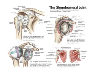
Anterior view of the right glenohumeral joint Posterior band ligament
showing the following external features of the
joint capsule: the capsular, coracohumeral, and Lateral aspect of the right glenohumeral
coracoacromial ligaments. joint showing the internal surface of the
joint. e humerus has been removed to
Cor expose the glenoid fossa.
on ac
omi ligaoacr
Acr me om
nt ia
Subacromial l
space Coracoacromial Short head of biceps (cut)
ligament Supraspinatus
Coracoid Coracobrachialis (cut)
process Supraspinatus
Infraspinatus
Biceps brachii tendon Subscapularis
(long head)
Teres minor
Long head of
Glenoid labrum biceps tendon (cut)
Teres major
Coracobrachialis (cut) Triceps
Inferior Side view of right glenohumeral joint with the
capsule Anterior Posterior
joint opened up to expose the articular surfaces.
e longitudinal diameter is depicted in the frontal Anterior and posterior views of the right shoulder
plane (purple) and the transverse diameter is showing the rotator cu muscles blending into and
depicted in the horizontal plane (blue-green). reinforcing the glenohumeral joint caspule.