2. RG • Volume 36 Number 7 Histed et al 2237
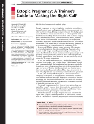
Figure 2. Heterotopic pregnancy. Longitudinal trans-
vaginal gray-scale US image demonstrates two em-
bryonic poles (arrows), with one intrauterine embryo
within the uterine fundus (left arrow), and the second
extrauterine embryo within the cervix (right arrow).
Both embryos had positive fetal cardiac activity.
Figure 1. Tubal ectopic pregnancy. Transverse transvaginal gray-scale (a) and transvaginal Doppler (b) US images
demonstrate an extrauterine pregnancy in the left adnexa with a yolk sac, an embryonic pole, and the ring-of-fire sign.
The ampulla accounts for more than 70% of all tubal ectopic pregnancies.
Treatment selection is dependent on clinical acuity,
desired future fertility, and medical comorbidities.
This online presentation reviews ectopic preg-
nancy in a case-based, multiple-choice question
format. After viewing the presentation, radiolo-
gists and radiology trainees should be able to
identify the expected US findings in a normal
IUP, recognize the variety of locations where an
ectopic pregnancy may be found, and discuss ap-
propriate treatment options.
Disclosures of Conflicts of Interest.—C.M.J. Activities related to
the present article: disclosed no relevant relationships. Activities
not related to the present article: disclosed no relevant relation-
ships. Other activities: author for UpToDate.
Suggested Readings
Ankum WM, Mol BW, van der Veen F, Bossuyt PM. Risk
factors for ectopic pregnancy: a meta-analysis. Fertil Steril
1996;65(6):1093–1099.
BarashJH,BuchananEM,HillsonC.Diagnosisandmanagement
ofectopicpregnancy.AmFamPhysician2014;90(1):34–40.
Bouyer J, Coste J, Shojaei T, et al. Risk factors for ectopic
pregnancy: a comprehensive analysis based on a large case-
control, population-based study in France. Am J Epidemiol
2003;157(3):185–194.
Chukus A, Tirada N, Restrepo R, Reddy NI. Uncommon im-
plantation sites of ectopic pregnancy: thinking beyond the
complexadnexalmass.RadioGraphics2015;35(3):946–959.
Dibble EH, Lourenco AP. Imaging unusual pregnancy im-
plantations: rare ectopic pregnancies and more. AJR Am J
Roentgenol 2016;30:1–13.
Doubilet PM, Benson CB, Bourne T, et al. Diagnostic criteria
for nonviable pregnancy early in the first trimester. N Engl
J Med 2013;369(15):1443–1451.
Levine D. Ectopic pregnancy. Radiology 2007;245(2):385–397.
LinEP,BhattS,DograVS.Diagnosticcluestoectopicpregnancy.
RadioGraphics 2008;28(6):1661–1671.
Marion LL, Meeks GR. Ectopic pregnancy: history, incidence,
epidemiology, and risk factors. Clin Obstet Gynecol
2012;55(2):376–386.
ParkerRA3rd,YanoM,TaiAW,FriedmanM,NarraVR,Menias
CO. MR imaging findings of ectopic pregnancy: a pictorial
review. RadioGraphics 2012;32(5):1445–1460.
uterus.The less common ectopic locations com-
pose up to 5% of ectopic pregnancies but are
diagnostically more challenging. A cervical ecto-
pic pregnancy can be mistaken for an abortion
in progress. Interstitial and cesarean section scar
ectopic pregnancies may be mistaken for normal
IUPs; due to inadequate surrounding myome-
trium and proximity to vascular structures, these
are at risk for devastating hemorrhage or uterine
rupture. Heterotopic pregnancy, a normal IUP
coexisting with an ectopic pregnancy, is uniquely
challenging as these patients are frequently un-
dergoing assisted reproductive techniques where
preservation of the viable pregnancy is para-
mount (Fig 2).
Treatment options include systemic metho-
trexate, local injection of potassium chloride or
methotrexate into the ectopic pregnancy, surgery, or
expectant management in limited clinical scenarios.