Litotrissia percutanea laparoscopica nel rene pelvico casi clinici
•
1 j'aime•1,218 vues

Litotrissia percutanea laparoscopica nel rene pelvico: casi clinici con un approccio diverso

Recommandé
Recommandé
Contenu connexe
Tendances
Tendances (20)
En vedette
En vedette (14)
Similaire à Litotrissia percutanea laparoscopica nel rene pelvico casi clinici
Similaire à Litotrissia percutanea laparoscopica nel rene pelvico casi clinici (20)
Plus de Merqurio
Plus de Merqurio (20)
Dernier
Dernier (20)
Litotrissia percutanea laparoscopica nel rene pelvico casi clinici
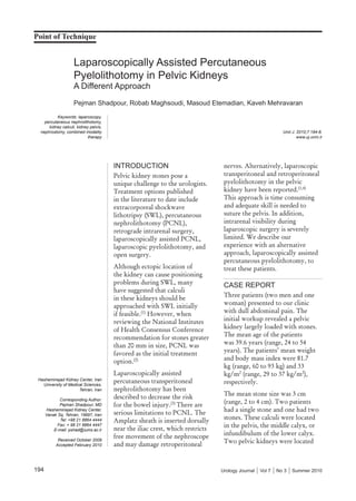
- 1. Point of Technique 194 Urology Journal Vol 7 No 3 Summer 2010 Laparoscopically Assisted Percutaneous Pyelolithotomy in Pelvic Kidneys A Different Approach Pejman Shadpour, Robab Maghsoudi, Masoud Etemadian, Kaveh Mehravaran Urol J. 2010;7:194-8. www.uj.unrc.ir Keywords: laparoscopy, percutaneous nephrolithotomy, kidney calculi, kidney pelvis, nephrostomy, combined modality therapy Hasheminejad Kidney Center, Iran University of Medical Sciences, Tehran, Iran Corresponding Author: Pejman Shadpour, MD Hasheminejad Kidney Center, Vanak Sq, Tehran, 19697, Iran Tel: +98 21 8864 4444 Fax: + 98 21 8864 4447 E-mail: pshad@iums.ac.ir Received October 2009 Accepted February 2010 INTRODUCTION Pelvic kidney stones pose a unique challenge to the urologists. Treatment options published in the literature to date include extracorporeal shockwave lithotripsy (SWL), percutaneous nephrolithotomy (PCNL), retrograde intrarenal surgery, laparoscopically assisted PCNL, laparoscopic pyelolithotomy, and open surgery. Although ectopic location of the kidney can cause positioning problems during SWL, many have suggested that calculi in these kidneys should be approached with SWL initially if feasible.(1) However, when reviewing the National Institutes of Health Consensus Conference recommendation for stones greater than 20 mm in size, PCNL was favored as the initial treatment option.(2) Laparoscopically assisted percutaneous transperitoneal nephrolithotomy has been described to decrease the risk for the bowel injury.(3) There are serious limitations to PCNL. The Amplatz sheath is inserted dorsally near the iliac crest, which restricts free movement of the nephroscope and may damage retroperitoneal nerves. Alternatively, laparoscopic transperitoneal and retroperitoneal pyelolithotomy in the pelvic kidney have been reported.(1,4) This approach is time consuming and adequate skill is needed to suture the pelvis. In addition, intrarenal visibility during laparoscopic surgery is severely limited. We describe our experience with an alternative approach, laparoscopically assisted percutaneous pyelolithotomy, to treat these patients. CASE REPORT Three patients (two men and one woman) presented to our clinic with dull abdominal pain. The initial workup revealed a pelvic kidney largely loaded with stones. The mean age of the patients was 39.6 years (range, 24 to 54 years). The patients’ mean weight and body mass index were 81.7 kg (range, 60 to 93 kg) and 33 kg/m2 (range, 29 to 37 kg/m2 ), respectively. The mean stone size was 3 cm (range, 2 to 4 cm). Two patients had a single stone and one had two stones. These calculi were located in the pelvis, the middle calyx, or infundibulum of the lower calyx. Two pelvic kidneys were located
- 2. Percutaneous Pyelolithotomy in Pelvic Kidneys—Shadpour et al 195Urology Journal Vol 7 No 3 Summer 2010 on the left and one on the right side (Figures 1 and 2). In addition to obvious obesity and short stature, one patient had history of previous open extraperitoneal pyelolithotomy of the affected kidney and another one had undergone abdominoplasty with tubal ligation. All had previous failed SWL of the pelvic kidney stone. Intervening bowel and/or extremely thick fat Figure 2. Pre-operative plain film and intravenous urographic image of one of the patients show left pelvic renal stones. Figure 1. Plain film and retrograde pyelography show right pelvic kidney stone in one patient.
- 3. Percutaneous Pyelolithotomy in Pelvic Kidneys—Shadpour et al 196 Urology Journal Vol 7 No 3 Summer 2010 layer precluded access to the kidney through the lumbodorsal approach in all three patients. Given these limitations, all patients consented to undergo laparoscopically assisted percutaneous pyelolithotomy. TECHNIQUE Pre-operative laboratory examination was within the normal limits. Intravenous urography and non contrast spiral computed tomography scan of the abdomen and the pelvis were performed pre- operatively for all three patients. On the day of surgery, after induction of general anesthesia and placing a ureteral catheter by cystoscopy, patients were prepped and draped in supine position. Laparoscopy was performed using two to three 5-mm ports in the midline. After exploration of the peritoneal cavity, the pelvic kidney was found and posterior peritoneum was incised to swiftly expose the renal pelvis by blunt dissection. The third trocar was needed only where dissection of previous adhesions became necessary. With adequate visualization of the typically abnormal renal vasculature, the safest path to enter the congenitally anterior renal pelvis was readily determined. An 18-gauge needle was inserted from the anterior abdominal wall into the renal pelvis. After full distention of the pelvis via the ureteral catheter, the tract was dilated in single pass by an Amplatz clad cone dilator. A 24-F rigid nephroscope was used to perform pyeloscopy. The stones were either removed intact or fragmented by Swiss Lithoclast Master Lithotripter (EMS, Bern, Switzerland). A 14-F closed tube drain was inserted through the lowermost laparoscopic port. The renal pelvis was not repaired after removal of the scope, but the ureteral catheter was left in place attached to a 14-F Foley catheter. Complete blood count, blood urea nitrogen, creatinine, and plain radiography were repeated the day after surgery. Both catheters were removed 24 hours postoperatively and the drain was removed on the second postoperative day. All patients were discharged from hospital 48 hours after the procedure. Kidney, ureter, bladder x-ray and renal ultrasonography were also carried out at weeks 2 and 12 postoperatively. RESULTS Mean operative time was 71 minutes (range, 48 to 113 minutes). Estimated blood loss was 26 mL (range, 8 to 50 mL). No patient received blood transfusion during or after the procedure. All three patients were rendered stone-free based on final intra-operative nephroscopic and fluoroscopic inspection of the pyelocalyceal system, which was confirmed by postoperative plain film and ultrasonography. Patients were discharged from the hospital after urine leakage discontinuation and documentation of stone-free state. The postoperative period remained uneventful with no clinically detectable leakage. DISCUSSION Although ectopic location of the kidney can cause problems during SWL due to intervening bowel anteriorly and bones posteriorly, calculi in these kidneys should be approached by this method initially if at all possible.(1) In the case of SWL failure, alternative modalities may be used. Our patients were not appropriate candidates for classic prone dorsal PCNL because of their obesity, short stature, and intervening bowels. Eshghi and colleagues described laparoscopically assisted PCNL in 1985 to deal with the frequent problems encountered by retrorenal intestines.(3) Holman and Toth reported good results and no major complications in 15 patients treated by laparoscopically assisted PCNL.(5) El-Kappany and associates presented the combination of laparoscopy and nephroscopy for treatment of ectopic pelvic kidney stones in 11 patients and concluded that this combination is feasible, safe, and effective for treatment of such stones.(6) Aron and coworkers reported laparoscopically assisted percutaneous nephrolithotomy in a patient with history of previous open pyelolithotomy.(7) In PCNL, the tract traverses renal parenchyma, and obtaining a suboptimal non trans-papillary route is common. The latter plus exaggerated angulation required to access middle and lower calyces in
- 4. Percutaneous Pyelolithotomy in Pelvic Kidneys—Shadpour et al 197Urology Journal Vol 7 No 3 Summer 2010 many pelvic kidneys can be associated with high probability of bleeding from infundibular or more extensive parenchymal laceration. This explains the frequent occurrence of bleeding that sometimes requires blood transfusion. Kramer and colleagues performed laparoscopic pyelolithotomy in three patients with a horseshoe kidney. There were no minor or major complications, and the estimated blood loss was <50 mL. The pelvis was incised and required suturing leading to mean operative time of 123 minutes (range, 74 to 150 minutes). They concluded that laparoscopic pyelolithotomy can be done safely, effectively, and efficiently with proper patient selection and adherence to standard laparoscopic surgical principles.(8) Alternatively, laparoscopic retroperitoneal pyelolithotomy was performed in a pelvic kidney by Harmon and associates.(1) Collins and coworkers reported the combination of laparoscopic pyelolithotomy and ultrasonic lithotripsy.(9) Their patient underwent uncomplicated laparoscopic pyelolithotomy. The stone has been located into an entrapment sack. The open end of the Endocatch sack was brought through a trocar site and a nephroscope and ultrasonic lithotripter were deployed. The stone was fragmented and aspirated in the standard manner, thereby, avoiding the need to extend the 12-mm trocar incision for stone extraction. The patient was stone-free and discharged in the morning of the first postoperative day without any complication.(9) In this study, we presented an entirely different approach for percutaneous pyelolithotomy. We used laparoscopic view to guide our nephroscope directly into the renal pelvis through a dilated needle puncture. By logical deduction based on the common feature of the anteriorly placed pelvis and posteriorly located renal parenchyma peculiar to this patient group, it is easy to see why accessing the renal parenchyma to perform laparoscopically assisted PCNL is conceivably more difficult than the method hereby described from the anatomical standpoint. Our patients were not amenable to the dorsal approach to the renal cortex even with laparoscopic dissection, for reasons mentioned above. Another well-known characteristic of pelvic kidneys is the presence of vascular variations, in which direct visualization afforded by laparoscopy can result in reduced associated risks. At the same time, percutaneous radially dilated access to the pelvis brings a novel minimally invasive method for access to the kidney without requiring incision and/or subsequent reclosure by suturing the urinary tract, thereby, saving time without causing any clinically significant leakage or risking vascular injury. Also of note is the much more adequate visualization of the pyelocalyceal interior in the submerged irrigant medium of the closed pyelocalyceal system through a nephroscope, instead of the encumbered tangential air or water view through a pyelotomy incision during classic laparoscopic pyelolithotomy of a pelvic kidney. It is also more efficient than putting the nephroscope into a gaping wide gas filled pyelotomy incision as described earlier.(8) There is a case report by Figge which although claimed to represent percutaneous transperitoneal nephrolithotomy, had actually involved a transpelvic route as described in our series. That case report, however, did not include any attempt at intracorporeal stone fragmentation, and did not require exploration of the entire calyceal system.(10) The fact that our patients had no postoperative leakage is particularly concerting. We believe this owes, at least in part, to deliberately leaving the ureter without an indwelling stent once stone- free state was ascertained postoperatively. In addition to allowing unimpeded reflux to exert high physiologic voiding pressures directly onto the pyelotomy site, a double J stent could often be blocked by clots and debris. Interestingly, the only instance of prolonged urinary drainage in a study by Holman and Toth was reported in a stented patient due to instrument malfunction.(5) Unstented subjects in Kramer and colleagues’ report had uneventful recovery.(8) Similarly, totally tubeless PCNL is now widely perceived to lessen urinary leakage significantly.
- 5. Percutaneous Pyelolithotomy in Pelvic Kidneys—Shadpour et al 198 Urology Journal Vol 7 No 3 Summer 2010 CONFLICT OF INTEREST None declared. REFERENCES 1. Harmon WJ, Kleer E, Segura JW. Laparoscopic pyelolithotomy for calculus removal in a pelvic kidney. J Urol. 1996;155:2019-20. 2. Segura JW, Preminger GM, Assimos DG, et al. Nephrolithiasis Clinical Guidelines Panel summary report on the management of staghorn calculi. The American Urological Association Nephrolithiasis Clinical Guidelines Panel. J Urol. 1994;151:1648-51. 3. Eshghi AM, Roth JS, Smith AD. Percutaneous transperitoneal approach to a pelvic kidney for endourological removal of staghorn calculus. J Urol. 1985;134:525-7. 4. Meria P, Milcent S, Desgrandchamps F, Mongiat-Artus P, Duclos JM, Teillac P. Management of pelvic stones larger than 20 mm: laparoscopic transperitoneal pyelolithotomy or percutaneous nephrolithotomy? Urol Int. 2005;75:322-6. 5. Holman E, Toth C. Laparoscopically assisted percutaneous transperitoneal nephrolithotomy in pelvic dystopic kidneys: experience in 15 successful cases. J Laparoendosc Adv Surg Tech A. 1998;8:431-5. 6. El-Kappany HA, El-Nahas AR, Shoma AM, El- Tabey NA, Eraky I, El-Kenawy MR. Combination of laparoscopy and nephroscopy for treatment of stones in pelvic ectopic kidneys. J Endourol. 2007;21:1131-6. 7. Aron M, Gupta NP, Goel R, Ansari MS. Laparoscopy- assisted percutaneous nephrolithotomy (PCNL) in previously operated ectopic pelvic kidney. Surg Laparosc Endosc Percutan Tech. 2005;15:41-3. 8. Kramer BA, Hammond L, Schwartz BF. Laparoscopic pyelolithotomy: indications and technique. J Endourol. 2007;21:860-1. 9. Collins S, Marruffo F, Durak E, et al. Laparoscopic pyelolithotomy with intraperitoneal ultrasonic lithotripsy: report of a novel minimally invasive technique for intracorporeal stone ablation. Surg Laparosc Endosc Percutan Tech. 2006;16:435-6. 10. Figge M. Percutaneous transperitoneal nephrolithotomy. Eur Urol. 1988;14:414-6.