Adipokines in Insulin Resistance Current Updates.pptx
•Télécharger en tant que PPTX, PDF•
1 j'aime•563 vues

Adipokines in Insulin Resistance Current Updates.pptx

Recommandé
Contenu connexe
Tendances
Tendances (20)
Similaire à Adipokines in Insulin Resistance Current Updates.pptx
Similaire à Adipokines in Insulin Resistance Current Updates.pptx (20)
Plus de Moustafa Rezk
Plus de Moustafa Rezk (20)
Dernier
Dernier (20)
Adipokines in Insulin Resistance Current Updates.pptx
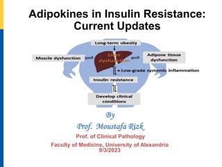
- 1. Adipokines in Insulin Resistance: Current Updates By Prof. Moustafa Rizk Prof. of Clinical Pathology Faculty of Medicine, University of Alexandria 9/3/2023
- 2. Agenda Adipose tissue as an endocrine organ Pro-inflammatory adipokines versus protective adipokines Pathophysiology of adipokines in the aetiology of insulin resistance
- 3. Fat Cell Development After fat cells have enlarged and energy intake continues to exceed expenditure, fat cells increase in number again. During growth, fat cells increase in number. When energy intake exceeds expenditure, fat cells increase in size. With fat loss, the size of the fat cells shrinks, but not the number.
- 4. Adipocytes
- 5. (WAT) depots in humans are shown in orange. Major subcutaneous WAT includes superficial and deep abdominal depots and gluteal-femoral depot. Major visceral WAT includes epicardial, omental and mesenteric.
- 6. Adipose tissue as an endocrine organ • Adipose tissue secretes a variety of bioactive peptides (adipokines). • Adipokines may locally regulate fat mass by modulating adipocyte size/number or angiogenesis and inversely increased fat mass leads to dysregulation of adipocyte functions.
- 7. Adipokines Adipokines not only regulate lipid metabolism but also function in a wide array of physiological or pathological processes as diverse as Host defense, Inflammation, Apoptosis, Autoimmunity, Cell differentiation, Organogenesis The complex role of adipokines in obesity, inflammation, and autoimmunity . Erin B.Taylor.March 2021.Clinical Science 135(6):731-752
- 8. “Adipokines” Forms : 1- Proteins 2- Hormones 3- Factors 4- Cytokines Act as 1- Endocrine 2- Paracrine 3- Autocrine Deposition of excess energy causes adipose tissue dysfunction low-grade chronic inflammation This chronic inflammation insulin resistance by modulating various metabolic pathways
- 9. Pathophysiology of adipokines in the aetiology of insulin resistance
- 10. 1- Adiponectin • Adiponectin consists of 244 amino acids and is located on chromosome 3q27 , a region associated with type 2 diabetes and CVD . • In healthy individuals, circulating adiponectin is normally between 5–30 u g/ml and is lower in individuals with adiposity, insulin resistance and type 2 diabetes • Numerous epidemiological studies suggest that adiponectin deficiency is associated with coronary artery disease and hypertension.
- 11. Hypoadiponectinemia - an additional CVD risk factor in T2D Hypoadiponectinemia is associated with endothelial dysfunction, greater carotid intima-media thickness(IMT), and coronary artery disease In vascular levels, actions of the adiponectin comprise reduction in expression of intercellular adhesion molecule-1(ICAM-1), vascular cell adhesion molecule1(VCAM-1) and E-selectin Inhibit the transformation of macrophages into foam cells and the proliferation and migration of smooth muscle cells which have a protective effect on atherosclerosis
- 12. • Thus, low serum adiponectin level is now considered a CVD risk factor, type 2 diabetic patients with low concentrations of this protein would have increased risk of developing premature arteriosclerosis. • In other words, lower levels of serum adiponectin is an additional CVD risk factor for the patients with type 2 diabetes
- 14. 2- Omentin Circulating omentin levels are decreased in : 1- Obesity, 2- Metabolic syndrome, 3- Type 2 diabetes (T2D) 4- Patients with coronary artery 5- Established carotid atherosclerosis Inversely correlated with insulin resistance,
- 15. 3-Visfatin Visfatin is an inflammatory mediator, based on its localization in macrophages within atherosclerotic lesion and its ability to induce matrix metalloproteinase (MMP)-9 in monocytes Elevated in 1- Obesity, 2- Insulin resistance, 3- Type 2 diabetes mellitus, 4- Pro-inflammatory states . Visfatin is involved in endothelial dysfunction (ED), which causes progression of atherosclerosis and therefore plays an important role in different forms of cardiovascular diseases.
- 16. 4- Leptin Leptin has emerged as a major inflammatory agent responsible for vascular inflammation, increased oxidative stress, endothelial dysfunction, and proliferation of vascular smooth muscle cells (VSMC), resulting in intimate hyperplasia. Patients with T2DM may be more risky for vascular inflammation mediated by leptin/adiponectin axis and subsequent damage leading to microvascular complications
- 17. 5-Resistin • Resistin is a member of a cysteine-rich protein termed as resistin like molecule (RELM) and circulates as a hexamer and trimer. • Hexameric form of this adipokine is more abundant, while trimeric form induces severe insulin resistance • The mechanism by which resistin causes the insulin resistance includes the activation of suppressor of cytokine signalling-3 (SOCS-3), which attenuates insulin-arbitrate signalling in adipocytes • In association with the toll-like receptor (TLR- 4), resistin stimulates insulin resistance in different cells. Adipose tissue-specific secretory factor (ADSF)
- 18. Resistin in metabolism, inflammation, and disease The FEBS Journal, Volume: 287, Issue: 15, Pages: 3141-3149, First published: 07 April 2020, DOI: (10.1111/febs.15322)
- 19. 6- Lipocalin-2 (Lcn2) Neutrophil gelatinase associated lipocalin (NGAL) • Lcn2 is highly expressed in adipocytes, liver, kidney and on macrophages and regulates apoptosis and innate immunity • The primary mechanism underlines the effect of Lcn2 on insulin resistance include the modulation of 12- lipoxygenase activity and TNF-á levels in adipose tissue • Study on LCN2 showed increased hepatic gluconeogenesis, debilitate lipid metabolism, impaired oxidation capacity of mitochondria, elevated inflammation favouring dyslipidemia due to diet-induced obesity, fatty liver disorders and insulin resistance. • Overexpression of this adipokine in adipose tissue has been reported for elevated fat mass, glucose intolerance and insulin resistance only in females via mitochondrial dysregulation Chella Krishnan K, Sabir S, Shum M, et al. Sex-specific metabolic functions of adipose Lipocalin-2. Mol Metab. 2019; 30:30-47.
- 20. Lipocalin 2 (LCN2) in organ damage Lipocalin 2 (LCN2) Expression in Hepatic Malfunction and Therapy. September 2016.Frontiers in Physiology 7(33)
- 21. 7- Retinol Binding Protein-4 (RBP-4) Higher expression of this adipokine in the adipocyte is inversely associated with the GLUT-4 expression in the adipocyte Thus, decreased GLUT-4 in adipocytes promotes higher expression of RBP-4, which inhibits insulin-mediated insulin receptor substrate-1 (IRS-1) phosphorylation that can contribute to insulin resistance During obesity, RBP-4 is preferentially produced by visceral fat depot than subcutaneous fat depot, suggesting the role of intra-abdominal adipose tissue in insulin resistance .
- 22. Retinol binding protein-4 links insulin resistance and heart failure The Associations between Retinol Binding Protein-4 and Cardiometabolic Profile: Intertwined-Intricate Relationship June 2020 Biomedical and Biotechnology Research Journal (BBRJ) 4(2)
- 23. 8-Chemerin Chemerin is a pro-inflammatory adipokine predominantly produced by white adipose tissue (WAT) that regulates the immune system (adaptive and innate), adipogenesis and metabolic homeostasis Overexpression of chemerin in adipose tissue causes insulin resistance in human skeletal muscles by modulating IRS-1, glucose uptake, glycogen synthase kinase 3 phosphorylation (GSK3P) Non-alcoholic fatty liver disease (NAFLD) is a common phenomenon in obesity, which is closely associated with chemerin induced increased insulin resistance
- 24. Christa Buechler et al . Chemerin Isoforms and Activity in Obesity. Int. J. Mol. Sci. 2019, 20(5) Effect of chemerin on the metabolic status of different organs
- 25. 9-TNF-á TNF-á adipocytokine induces insulin resistance via decreasing the tyrosine kinase activity of the insulin receptor This causes altered signalling pathways that can induce insulin resistance and related diseases
- 26. Tumor Necrosis Factor‐Alpha: Role in Development of Insulin Resistance and Pathogenesis of Type 2 Diabetes Mellitus J of Cellular Biochemistry, Volume: 119, Issue: 1, Pages: 105-110, First published: 01 June 2017, DOI: (10.1002/jcb.26174) c-Jun N-terminal kinase IκB kinase Insulin receptor substrate-1
- 27. an HY, Tan SL, et al . 2020. Development of a novel in vitro insulin resistance model in primary human tenocytes for diabetic tendinopathy esearch. Tumor Necrosis Factor‐Alpha: Role in Development of Insulin Resistance
- 28. 10- IL-6 Interleukin-6 is a pro-inflammatory cytokine secreted by many different cell types and tissues, including adipose tissue, and plays a significant role in the immune response. This adipocytokine contributes to low-grade chronic inflammatory state responsible for adipose tissue dysfunction . The mechanism of action by which this cytokine imparts its role in insulin resistance involves inhibitory effects on the gene transcription of PPAR gamma, GLUT-4 and IRS-1.
- 29. The balance between pro-inflammatory adipokines and protective adipokines is disturbed in type 2 diabetes Adipose tissue dysfunction
- 30. Schematic diagram representing the role of adipokines in insulin resistance and related diseases
- 31. CONCLUSION Dysfunctional adipose tissue secretes altered levels of adipokines that are associated with many health problems, including insulin resistance. Recent findings exhibit the role of adipokine induced insulin resistance as a major risk factor for the development of chronic diseases like neurodegenerative diseases, non-alcoholic fatty liver disease, chronic kidney diseases, cardiovascular diseases etc. However, determining the role of adipokines in the aetiology of insulin resistance may provide new opportunities for developing novel therapeutics for obesity arbitrates insulin resistance.
- 32. Questions or Comments Prof. Moustafa Rizk
Notes de l'éditeur
- Adipocytes are the major energy storage sites in the body, and they also have critical endocrine functions. There are two general classes of adipocytes; white adipocytes - which store energy as a single large lipid droplet and have important endocrine functions, and brown adipocytes - which store energy in multiple small lipid droplets but specifically for use as fuel to generate body heat (i.e. thermogenesis). Heat production by brown adipocytes is made possible by their unique expression of mitochondrial localized uncoupling protein 1 (Ucp1). However, these classifications are oversimplified because some white adipocytes can adopt brown adipocyte characteristics (termed brite or beige adipocytes) and vice versa depending on the temperature and diet. Adipose tissues are classified as brown adipose tissue (BAT) and white adipose tissue (WAT), originated from mesoderm and the mesenchymal stem cells during embryogenesis. BAT is rich in mitochondria; hence appear brown and predominantly involved in thermogenesis (heat production) via uncoupling proteins . Conversely, WAT is organ-specific and is further divided into visceral (mesenteric, retroperitoneal, omental and pericardial) and subcutaneous (beneath the skin) adipose depots; thus obesity-related consequences are primarily regulated by WAT
- Monocyte chemotactic protein-1 (MCP1) is a potent adipokine.
- The meaning of PARACRINE is of, relating to, promoted by, or being a substance secreted by a cell and acting on adjacent cells , Autocrine signaling is a form of cell communication in which a signal is released by a cell and then acts on that SAME cell, causing some alteration or effect.
- Resistin and human macrophages. Human resistin participates in inflammation, oxidative stress, and insulin resistance leading to T2DM. Deciphering the molecular crosstalk among them still needs further investigation. IF THIS IMAGE HAS BEEN PROVIDED BY OR IS OWNED BY A THIRD PARTY, AS INDICATED IN THE CAPTION LINE, THEN FURTHER PERMISSION MAY BE NEEDED BEFORE ANY FURTHER USE. PLEASE CONTACT WILEY'S PERMISSIONS DEPARTMENT ON PERMISSIONS@WILEY.COM OR USE THE RIGHTSLINK SERVICE BY CLICKING ON THE 'REQUEST PERMISSIONS' LINK ACCOMPANYING THIS ARTICLE. WILEY OR AUTHOR OWNED IMAGES MAY BE USED FOR NON-COMMERCIAL PURPOSES, SUBJECT TO PROPER CITATION OF THE ARTICLE, AUTHOR, AND PUBLISHER.
- The glucose transporter GLUT4 mediates insulin-stimulated glucose uptake in adipocytes and muscle by rapidly moving from intracellular storage sites to the plasma membrane. In insulin-resistant states such as obesity and type 2 diabetes, GLUT4 expression is decreased in adipose tissue but preserved in muscle.
- Effect of chemerin on the metabolic status of different organs (inconclusive results indicated by reverse arrows). Data published so far mostly agree that chemerin impairs skeletal muscle insulin response. This was not observed in the liver, here gluconeogenesis was enhanced in chemerin deficient mice. The function of chemerin on blood pressure was modified by gender. Chemerin further stimulated angiogenesis and vascular inflammation. Adipose tissue weight was not changed by chemerin. This adipokine may even improve insulin response of fat tissue although the number of adipose tissue resident macrophages was increased. Stimulatory and inhibitory effects of chemerin on glucose-induced release of insulin by pancreatic beta-cells was reported. Inconclusive findings may be partly explained by the different models studied.
- TNF-α plays a critical role in the development of insulin resistance in such a way that it reduces the expression of glucose transporter type 4 (GLUT4) which is an insulin-regulated glucose transporter and mainly located in adipocytes, skeletal, and cardiac muscles [Huang and Czech, 2007; Guilherme et al., 2008; Olson, 2012]. Serine phosphorylation of insulin receptor substrate-1 (IRS-1) induced by the activation of TNF-α, also works as an inhibitor of insulin receptor and down streams the signaling of phosphatidylinositol-3 kinase activation [Fasshauer and Paschke, 2003].
- n brief, the hypothetical pathomechanism starts with the binding of TNF-α to the TNF-α receptor on hTeno which initiates the phosphorylation of TNF receptor-associated factor 2 (TRAF2) and subsequently promotes activation of both c-Jun N-terminal kinase (JNK) pathway and IκB kinase (IKK). In particular, JNK and IKKs are both serine/threonine-specific protein kinase that catalyzes the phosphorylation of serine or threonine residues on target proteins. Activation of JNK is proposed to trigger serine phosphorylation of insulin receptor substrate-1 (IRS-1) instead of tyrosine phosphorylation, thus diminished the downstream pathways, i.e.: inhibits the phosphoinositide 3-kinases (PI3K) pathway and prohibits the translocation of GLUT4 intracellular vesicles to the transmembrane region, and eventually no glucose uptake by GLUT4. The glucose uptake in the cells is barely shuttled by GLUT1 via passive diffusion.
- Peroxisome proliferator-activated receptor gamma is a master regulator of adipogenesis in mammals,
- MCP-1(Monocyte Chemotactic Protein 1)