Intelligent and Automatic In vivo Detection and Quantification of Transplanted Cells in MRI
•Download as PPTX, PDF•
1 like•324 views

Muhammad Jamal Afridi, Arun Ross, and Erik M. Shapiro
Recommended

Recommended
More Related Content
What's hot
What's hot (20)
Similar to Intelligent and Automatic In vivo Detection and Quantification of Transplanted Cells in MRI
Similar to Intelligent and Automatic In vivo Detection and Quantification of Transplanted Cells in MRI (20)
More from Michigan State University Research
More from Michigan State University Research (20)
Recently uploaded
Recently uploaded (20)
Intelligent and Automatic In vivo Detection and Quantification of Transplanted Cells in MRI
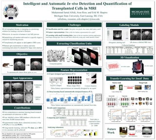
- 1. Challenges Intelligent and Automatic In vivo Detection and Quantification of Transplanted Cells in MRI Muhammad Jamal Afridi, Arun Ross, and Erik M. Shapiro Michigan State University, East Lansing, MI, USA Objective Extracting Classification Units Feature Representation Labeling ModuleMotivation Cell transplant therapy is emerging as a promising solution for treating a myriad of diseases. However, its success in humans is not fully proven. Quantifying cell number delivered to a specific organ is crucial for monitoring success. Transplanted cells appear as dark spots in MRI scans. Manual spot enumeration is tedious and time consuming and cannot be adopted for large scale analysis Transfer Learning for Small Data Classification unit for a spot: One pixel, two pixels, how many shall make a unit? Feature representation: What is the best feature representation for a spot? Learning with small training data: State-of-the-art machine learning approaches need large training data. How to train these effectively using only small amount of data? (1) Expert designed spot features (P-1) (2) Deep learning based automatically designed features (P-2) Contributions First comprehensive machine learning based research on automated cell detection in MRI. First, labelled cellular MRI database collection with more than 20,000 manual labels. Experimental results show a detection accuracy of up to 97.3% in vivo and 99.8%. Using transfer learning, with only 5% data, up to 88% in vivo accuracy can be achieved. Collected database Extracted patches Top PCA shapes Binary shape filters Context features Results and Comparison To automatically detect these spots in vivo. These feature representations are manually designed by an expert These feature representations are automatically learned from data in vitro in vivo LR Comparing spot numbers: Each tube is expected to have a spot number of ~2400 (B) shows a labeled MRI sliceDesigned Labeling tool in vitro scan in vivo scan {afridimu, rossarun, erik.shapiro}@msu.edu Spot Appearance Labeled MSCs Unlabeled MSCs Analyzing robustness: Future Work 3D Visualization We are thankful to the support provided by NIH grants DP2OD004362 (EMS), R21 CA185163(EMS), R01 DK107697(EMS), R21 AG041266 (EMS).