SlideShare a Scribd company logo
1 of 17
CHAPTER 64. Hyperbilirubinemia
INTRODUCTION
Hyperbilirubinemia is a common transitional finding occurring in 60-70% of term newborns and
almost all premature infants. An elevation of serum bilirubin concentration >2 mg/dL is found in
virtually all newborns in the first several days of life. Jaundice becomes clinically apparent at
serum bilirubin concentration of >5 mg/dL.
Bilirubin is the end product of the catabolism of heme and is produced primarily by the breakdown
of red blood cell hemoglobin. Other sources of heme include heme-containing proteins such as
myoglobin, cytochromes, and nitric oxide synthase. Bilirubin exists in several forms in the blood
but is predominantly bound to serum albumin. Free unconjugated bilirubin, and possibly other
forms, can enter the central nervous system (CNS) and become toxic to cells if concentration is
great enough. The precise mechanism is unknown.
Inside liver cells, unconjugated bilirubin is bound to ligandin, Z protein, and other binding proteins;
it is conjugated by uridine diphosphoglucuronyl transferase (UDPGT). Conjugated bilirubin is
water- soluble and can be excreted in the urine, but most of it is rapidly excreted as bile into the
intestine. Conjugated bilirubin is further metabolized by bacteria in the intestine and excreted in
the feces.
Hyperbilirubinemia presents in the neonate as either unconjugated hyperbilirubinemia or
conjugated hyperbilirubinemia. The two forms involve different pathophysiologic causes with
distinct potential complications.
UNCONJUGATED (INDIRECT) HYPERBILIRUBINEMIA
I. Definition. When the rate of bilirubin production exceeds the rate of bilirubin elimination, the
end result is an increase in the total serum bilirubin (TSB) concentration, resulting in the clinical
condition of hyperbilirubinemia, often called jaundice.
II. Classification and pathophysiology. The causes of unconjugated hyperbilirubinemia are listed in
Table 64-1.
A. Physiologic jaundice. In almost every newborn infant, particularly premature infants, a
physiologic elevation of serum unconjugated bilirubin develops during the first week of life,
usually in the second or third day, and resolves spontaneously. Jaundice that develops in the first
24 h of life is to be considered pathologic until proven otherwise.
1. Exclusion criteria
a. Unconjugated bilirubin level >12.9 mg/dL in the term infant.
b. Unconjugated bilirubin level >15 mg/dL in the preterm infant. (Threshold level may vary
depending on the gestational age and birth weight of infant)
c. Bilirubin level increasing at a rate >5 mg/dL/day.
d. Jaundice in the first 24 h of life.
e. Conjugated bilirubin level >2 mg/dL.
f. Clinical jaundice persisting >1 week in full-term infants or >2 weeks in preterm infants.
2. Physiology
a. Full-term infant. Serum unconjugated bilirubin progressively rises to a mean peak of 5-6
mg/dL by the third day of life in both white and black infants and a peak of 10 mg/dL at 3-4 days in
Asian babies. Figure 64-1 shows hour-specific nomogram with predictive value for subsequent
hyperbilirubinemia.
b. Preterm neonates. Liver function is less mature, and jaundice is more frequent and
pronounced. A peak concentration of 10-12 mg/dL is reached by the fifth day of life.
3. Mechanisms. A number of mechanisms have been suggested.
a. Increased bilirubin load because of the larger red blood cell volume, the shorter life span
of red blood cells, and increased enterohepatic recirculation of bilirubin in newborn infants.
b. Defective uptake by the liver as a result of decreased concentration of bilirubin binding
proteins, such as ligandin.
c. Defective conjugation because of reduced glucuronyl transferase activity in the term
newborn; even more pronounced in premature infants.
d. Impaired excretion into bile.
e. Overall impairment/immaturity of liver function.
B. Hemolytic anemia
1. Red blood cell defects. Hemolytic anemia may result from a congenital red blood cell defect
such as hereditary spherocytosis, infantile pyknocytosis, pyruvate kinase deficiency, glucose-6phosphate dehydrogenase (G6PD) deficiency, thalassemia, or vitamin K-induced hemolysis.
2. Acquired hemolytic anemia may be seen in blood group incompatibilities, such as ABO or
Rh incompatibility between infant and mother. It may also be associated with the use of certain
drugs (eg, sulfonamides) or with infection.
C. Polycythemia. The liver may not have the capacity to metabolize the increased bilirubin load
presented by the increased blood volume.
D. Blood extravasation. Sequestration of blood within body cavities can result in increased
bilirubin and thus overload the bilirubin degradation pathway. It may be seen with
cephalhematoma, intracranial and pulmonary hemorrhage, subcapsular hematoma of the liver,
excessive ecchymoses or petechiae, occult gastrointestinal hemorrhage, and large hemangiomas
(ie, Kasabach-Merritt syndrome).
TABLE 64-1. CAUSES OF UNCONJUGATED HYPERBILIRUBINEMIA
Physiologic jaundice Hemolytic anemia Congenital: red blood cell defects (hereditary
spherocytosis, infantile pyknocytosis, pyruvate kinase deficiency, G6PD deficiency, and
thalassemia) Acquired: blood group incompatibilities (ABO or Rh incompatibility), infection, and
drug-induced hemolysis Polycythemia Blood extravasation Defects of conjugation Congenital:
Crigler-Najjar syndrome (types I and II), Gilbert syndrome Acquired: Lucey-Driscoll syndrome
Breast-feeding and breast milk jaundice Metabolic disorders: galactosemia, hypothyroidism
Increased enterohepatic circulaton of bilirubin Substances/ disorders affecting binding of bilirubin
to albumin: drugs, fatty acids in nutritional products, asphyxia, acidosis, sepsis, hypothermia,
hyperosmolality, hypoglycemia
G6PD, glucose-6-phosphate dehydrogenase.
FIGURE 64-1. Hour-specific bilirubin nomogram with the predictive ability of the predischarge
bilirubin value for subsequent severe hyperbilirubinemia, >95th percentile tract. Reproduced with
permission from Bhutani VK, Johnson LH. Jaundice technologies; prediction of hyperbilirubinemia
in term and near term newborns. J Perinatol 2001;21:576
E. Defects of conjugation
1. Congenital deficiency of glucuronyl transferase
a. Crigler-Najjar syndrome type I. A severe form of uridine diphosphate (UPD) glucuronyl
transferase deficiency that is inherited as an autosomal recessive disorder. It is unresponsive to
phenobarbital therapy and has a poor prognosis.
b. Crigler-Najjar syndrome type II. Moderate deficiency of UDP-glucuronyl transferase. It is
responsive to phenobarbital therapy.
c. Gilbert syndrome is a mild form of UDP-glucuronyl transferase deficiency and is inherited
as an autosomal dominant condition, is benign, and is relatively common. It is responsive to
phenobarbital, although no specific therapy is necessary.
2. Glucuronyl transferase inhibition
a. Drugs such as novobiocin.
b. Lucey-Driscoll syndrome. An unspecified maternal gestational hormone, found in the
infant's serum interferes with the conjugation of bilirubin. This problem appears to resolve
spontaneously; however, in extreme cases, exchange transfusions may be required to avoid
kernicterus.
F. Breast milk jaundice (late onset) is a result of the prolongation of increased enterohepatic
recirculation of bilirubin because of a factor in human milk that promotes intestinal absorption. It
is characterized by a higher peak (10- 30 mg/dL, peaking by days 10-15 of life) and a slower decline
in the serum bilirubin concentration that can last for several weeks. It rarely appears before the
end of the first week of life for term or preterm infants. Interruption of breast-feeding for 24-48 h
at unacceptable bilirubin levels results in a rapid decline. Resumption of breast-feeding increases
bilirubin levels slightly but usually below previous levels. Breast milk jaundice may recur in 70% of
future pregnancies.
G. Metabolic disorders. Galactosemia, hypothyroidism, and maternal diabetes may be
associated with unconjugated hyperbilirubinemia.
H. Increased enterohepatic recirculation of unconjugated bilirubin as a result of pathologic
conditions such as cystic fibrosis, gastrointestinal obstruction (ie, pyloric stenosis, duodenal
atresia, annular pancreas), and ileus may result in exaggerated jaundice. Blood swallowed during
delivery and decreased caloric intake may also be contributing factors.
I. Substances and disorders affecting binding of bilirubin to albumin. Certain drugs occupy
bilirubin-binding sites on albumin and increase the amount of free unconjugated bilirubin that can
cross the blood-brain barrier. Drugs in which this effect may be significant include aspirin and
sulfonamides. Chloral hydrate is shown to compete for hepatic glucuronidation with bilirubin and
thus increase serum unconjugated bilirubin. Common drugs used in the neonates such as penicillin
and gentamicin have also been shown to compete with bilirubin for albumin binding sites. Fatty
acids
in nutritional products (eg, Intralipid) may also influence bilirubin binding to albumin, as may
asphyxia, acidosis, sepsis, hypothermia, hyperosmolality, and hypoglycemia.
III. Morbidity
A. General. Unconjugated bilirubin in high concentration can cross the blood-brain barrier,
penetrating the brain cells. This has been associated with neuronal dysfunction and death.
Bilirubin is toxic to the brain in several ways. It has been shown to depress O2 consumption and at
high concentration uncouples oxidative phosphorylation. Severe hyperbilirubinemia can cause
long-term neurologic sequelae; however, the safety of moderate degree of hyperbilirubinemia has
been debated. One study has shown that moderate hyperbilirubinemia (13.6-26.0 mg/dL) may be
associated with an increase in minor neurologic dysfunction.
B. Encephalopathy
1. Transient. Early bilirubin-induced neurologic dysfunction is transient and reversible.
Auditory brainstem evoked responses (ABRs) show significant prolongation of latency of specific
wavelengths. These changes may be reversed with either exchange transfusions or with
spontaneous decrease in bilirubin levels. More recent data suggest that with marked
hyperbilirubinemia, complete recovery of the ABR may be delayed. The auditory system serves as
an objective window in looking at the CNS in cases of severe hyperbilirubinemia, which can be
used as an early predictor of bilirubin encephalopathy.
2. Bilirubin encephalopathy. This is a preventable lifelong neurologic sequelae of untreated
hyperbilirubinemia. It is a clinically worsening encephalopathy (over 24 h) caused by bilirubin
toxicity to the basal ganglia and various brainstem nuclei.
a. Acute phase. A severely jaundiced infant develops lethargy, hypotonia, and poor sucking
response. If left untreated, the infant becomes hypertonic and may develop fever and a highpitched cry. Decerebrate posturing can occur at the end stage of the disease.
b. Chronic phase. Survivors usually develop choreoathetoid cerebral palsy, hearing loss,
dental dysplasia and paralysis of upward gaze, and, less often, mental retardation.
C. Kernicterus. A postmortem diagnosis of pathologic findings of bilirubin toxicity in the brain.
1. Staining and necroses of neurons in the basal ganglia, hippocampal cortex, subthalamic
nuclei, and cerebellum. Cerebral cortex is usually spared.
2. Bilirubin toxicity to the brain may be reversible if bilirubin levels fall before saturation of the
CNS nuclei occurs.
3. Controversy exists over bilirubin levels considered at risk for kernicterus. In nonhemolytic
states, and otherwise healthy term infants, levels of >25 mg/dL are considered to place the patient
at risk.
4. Kernicterus registry. Kernicterus is not a reportable condition in the United States;
therefore, its prevalence is not known. A pilot registry at a Pennsylvania hospital documented 90
cases from January 1984-June 2001.
IV. Clinical presentation
A. History
1. Family history. Family history of jaundice, anemia, splenectomy, or metabolic disorders is
significant. A history of a sibling with jaundice may suggest blood group incompatibility, breast
milk jaundice, or G6PD deficiency. A familial tendency of neonatal hyperbilirubinemia is present
regardless of breast-feeding and other risk factors.
2. Maternal history. Neonatal jaundice is increased with a history of maternal diabetes or
infection. Use of oxytocin, sulfonamides, antimalarials, and nitrofurantoins by the mother may
initiate hemolysis in G6PD-deficient infants. Delivery trauma, asphyxia, delayed cord clamping, and
prematurity are associated with an increased risk of hyperbilirubinemia in the infant.
3. Infant's history.
a. Breast-feeding. Poor breast-feeding may result in poor caloric intake, leading to
"starvation jaundice." (See prior discussion of breast milk jaundice).
b. Factors affecting the gastrointestinal tract, such as obstruction, decreased motility (ileus),
and delayed passage of meconium
c. Check for clinical symptoms such as vomiting and lethargy. Metabolic disorders, infection,
and bowel obstruction can present in this manner.
B. Signs and symptoms. Clinical jaundice is visible when the serum bilirubin level approaches 5- 7
mg/dL. Jaundice is often apparent first in the face, especially the nose, and then descending to the
torso and lower extremities as the degree of jaundice increases. Jaundice can be demonstrated in
some infants by pressing lightly on the skin with a finger. The yellow color is seen more easily in
the "fingerprint" area than in the surrounding skin. These signs should not appear within the first
24 h after birth in otherwise healthy infants.
Besides confirming the presence of jaundice, physical examination may also be helpful in
determining the cause of hyperbilirubinemia. Areas of bleeding such as cephalhematoma,
petechiae, or ecchymoses indicate blood extravasation. Hepatosplenomegaly may signify
hemolytic disease, liver disease, or infection. Physical signs of prematurity, intrauterine growth
retardation, and postmaturity may be helpful in elucidating a cause for hyperbilirubinemia.
Plethora is seen with polycythemia, pallor with hemolytic disease, and large infants with maternal
diabetes, all of which are associated with hyperbilirubinemia. Omphalitis, chorioretinitis,
microcephaly, and petechial and purpuric lesions suggest infectious causes of increased serum
bilirubin.
C. Neurologic examination. The appearance of abnormal neurologic signs heralds the onset of
early bilirubin encephalopathy. Initial signs include lethargy, poor feeding, vomiting, and
hypotonia. The persistently hyperbilirubinemic infant may go on to experience seizures (see
section III,B above). The progression of neurologic changes parallels the stages of bilirubin
encephalopathy from acute to chronic and irreversible changes.
V. Diagnosis
A. Laboratory studies. Hyperbilirubinemia should be investigated whenever pathologic causes
are suspected. The presence of a serum bilirubin level of ≥13 mg/dL in any infant requires initial
evaluation.
1. Total and direct bilirubin
a. Direct bilirubin = conjugated fraction.
b. Indirect bilirubin = unconjugated fraction. Derived by subtracting direct fraction from
TSB.
2. Complete blood cell count and reticulocyte count. A low hemoglobin or hematocrit
associated with a high reticulocyte count and the presence of nucleated red blood cells is highly
suggestive of hemolytic anemia. Polycythemia is defined as a venous blood hematocrit >65%. (Use
of capillary blood samples may cause underestimation of venous bilirubin values when the
bilirubin value is > 10 mg/dL). Total white blood cell count with differential and platelet count may
help detect sepsis. The peripheral blood smear aids in the diagnosis of hereditary spherocytosis
and other red blood cell defects.
3. Blood type and Rh status in mother and infant. ABO and Rh incompatibility can be easily
diagnosed by comparing infant and maternal blood types.
4. Direct Coombs' test. The test is usually positive in infant's with isoimmunization disorders.
This test does not correlate with the severity of jaundice.
5. Measurement of serum albumin may help to assess total bilirubin binding sites available
and whether there is a need for an albumin infusion.
6. Other laboratory tests. The urine should be tested for reducing substances (to rule out
galactosemia if the infant is receiving a galactose-containing formula) and for infectious agents. If
hemolysis is present, in the absence of ABO or Rh incompatibility, further testing by hemoglobin
electrophoresis, G6PD screening, or osmotic fragility testing may be required to diagnose red
blood cell defects. Persistent jaundice (>2 weeks of life) may require these types of investigation
as well as additional tests for thyroid and liver function, blood and urine cultures, and metabolic
screening workup, such as plasma amino acid and urine organic acid measurements.
B. Radiologic studies for suspected intestinal obstruction or blood extravasation into internal
organs and ultrasonography of the head to document intracranial or subdural hemorrhage might
be indicated.
C. Transcutaneous bilirubinometry (TcB). Measures the degree of yellow color in the skin and
subcutaneous tissues by selective wavelength reflection. Data suggest that this methodology is an
effective adjunct to nursing assessment in the term infant. A TcB value of >13 mg/dL should be
correlated with TSB.
D. Expired carbon monoxide breath analyzer. End-tidal carbon monoxide (CO) corrected for
ambient CO (ETCO2). An equimolar amount of CO is produced for every molecule of bilirubin
formed from the degradation of heme; therefore, measurement of CO in end-tidal breath is an
index of total bilirubin production. The method can alert the attending physician to the presence
of hemolysis irrespective of the timing of jaundice.
VI. Management. Three methods of treatment are commonly used to decrease the level of
unconjugated bilirubin: exchange transfusion, phototherapy, and pharmacologic therapy. As
noted, however, controversy persists as to what levels of serum bilirubin warrant therapy,
especially in otherwise healthy full-term infants.
A. Phototherapy (see also Chapter 39)
1. Indication. Most infants with pathologic jaundice are treated with phototherapy when it is
believed that bilirubin levels could enter the toxic range.
a. Practice parameters. The American Academy of Pediatrics (AAP) is currently revising the
guidelines for management of hyperbilirubinemia. The concern for kernicterus is being
reemphasized by the Subcommittee on Hyperbilirubinemia pending the full assessment and
analysis of the new approaches to the jaundiced infant. It is important to remember that the
current AAP guidelines are meant for healthy term newborn infants with no evidence of
hemolysis. Although there is continuing uncertainty about which TSB level warrants exchange
transfusions, the authors suggest that in an otherwise healthy term newborn a TSB level of 20
mg/dL at 48 h of life may be treated initially with phototherapy. If the TSB decreases by 1-2 mg/dL
within 4-6 h of starting phototherapy, exchange transfusion may not be necessary.
b. In preterm and sick term infants, a more conservative approach is usually adopted.
2. Using phototherapy effectively.
a. Light source. The most effective light source currently available for phototherapy is that
provided by blue fluorescent tubes. Bilirubin absorbs light maximally in the blue range (420-500
nm). Blue lamps with narrow spectral range (420-480 nm) are most effective. The blue light
reflection does interfere with skin color assessment and has been reported to cause dizziness and
nausea in those caring for these patients. Addition of a daylight fluorescent tube tempers these
side effects. Energy output or irradiance is a second variable that influences the efficacy of
phototherapy. Radiometer measurements should be minimally 5 uW/cm2/nm, with saturation for
bilirubin degradation at ~11 uW/cm2/nm.
b. Distance from the light to the infant. Light intensity is a function of the distance from the
light source; therefore, light source should be as close to the infant as possible (12-16 inches).
c. Surface area. The larger the skin surface area exposed, the more effective is the
phototherapy. The availability of fiberoptic pads (bili-blankets) make it easy to increase exposed
skin area to the bili-lights. In preterm infants, this type of "double-phototherapy" (overhead light
plus bili- blanket) is almost twice as effective as single phototherapy. Fiberoptic phototherapy
blankets (bili- blankets) can be wrapped around or placed just beneath the infant.
3. Supportive management. The infant's eyes should be covered with opaque patches for
overhead lamp phototherapy. However, this is not necessary when using only fiberoptic light
source. Animal experiments have shown retinal damage occurring as a result of phototherapy. To
maximize exposure, infants should be naked in servocontrolled incubators. Phototherapy does
increase insensible fluid losses. For infants weighing <1500 g, increase fluids by 0.5 mL/kg/h; for
those weighing >1500 g, increase by 1 mL/kg/h. Overhydration does not increase bilirubin
elimination.
4. Termination of phototherapy. Phototherapy is stopped when the following criteria are met:
a. The bilirubin level is low enough to eliminate the risk of kernicterus.
b. The infant is old enough to handle the bilirubin load.
5. Complications. Phototherapy is simple, safe, and inexpensive. Complications are very rare
but should not be overlooked.
a. The retinal effects of phototherapy on the exposed infant's eyes are unknown. However,
animal studies suggest that retinal degeneration may occur. Thus, eye shields must be used.
b. Increased insensible fluid loss increases fluid requirements by 25%. In addition, stools
may be looser and more frequent. Use of fiberoptic phototherapy results in lower insensible loss.
c. Bronze baby syndrome. With conjugated hyperbilirubinemia, phototherapy causes
photodestruction of copper porphyrins, causing urine and skin to become bronze.
d. Congenital erythropoietic porphyria is a rare syndrome in which phototherapy is
contraindicated. Exposure to visible light of moderate to high intensity will produce severe bullous
lesions on exposed skin and may lead to death.
B. Exchange transfusion (see also Chapter 21). Exchange transfusion is used when the risk of
kernicterus for a particular infant is significant. A double-volume exchange replaces 85% of the
circulating red blood cells and decreases the bilirubin level to about half of the preexchange value.
Besides removing bilirubin, an exchange transfusion can be used to correct severe anemia.
1. General guidelines. It appears that no specific level of bilirubin can be considered safe or
dangerous for all infants because patient-to-patient variations exist for the permeability of the
blood- brain barrier. The practice parameters published by the AAP in October 1994 recommend
exchange transfusions in healthy term newborns at TSB 25-30 mg/dL. The debate for upper limits
of serum bilirubin is directed to full-term infants with no evidence for associated disease.
Low birth weight infants are excluded from these considerations. The authors suggest that
each institution and its practicing physicians must establish their criteria for phototherapy and
exchange transfusion by gestational age, weight groups, postnatal age, and infant's condition
consistent with current standard of pediatric practice.
2. Factors that may affect the decision to perform exchange transfusion include the infant's
maturity, birth weight, age, rate of rise in bilirubin levels (>0.5 mg/dL/h), and presence of hypoxia,
acidosis, sepsis, or hypoproteinemia.
3. Albumin transfusions may be useful if bilirubin levels are >20 mg/dL and serum albumin
levels are <3 g/dL. Infusion of 1 g of albumin 1 h before exchange transfusion may improve the
yield of bilirubin removal. Fluid volume and cardiovascular status must be carefully considered
before giving albumin.
C. Pharmacology
1. Phenobarbital therapy
a. Action. Phenobarbital affects the metabolism of bilirubin by increasing the concentration
of ligandin in liver cells, inducing production of glucuronyl transferase, and enhancing bilirubin
excretion. Because it takes 3-7 days to become effective, phenobarbital is usually not helpful in
treating unconjugated hyperbilirubinemia in the newborn infant. The administration of
phenobarbital to newborns at the time that jaundice is first noted or even at birth is less effective
than administration to mothers during pregnancy for ≥2 weeks before delivery.
b. Indications. It may be useful to give phenobarbital 1-2 weeks before delivery to a
pregnant mother whose fetus has documented hemolytic disease to aid in reducing bilirubin levels
in the affected neonate. Phenobarbital is also used to treat type II glucuronyl transferase
deficiency and Gilbert syndrome in the infant.
2. Metalloporphyrins. A synthetic heme analog, metalloporphyrins have been shown to inhibit
heme oxygenase (HO), the rate-limiting enzyme in the catabolism of heme. By acting as a
competitive inhibitor, the metalloporphyrins decrease the production of bilirubin. Tinmesoporphyrin (SnMP) is a potent HO inhibitor that has been extensively studied. It has been
reported to be effective as a single intramuscular injection (6 mmol/ kg) in two patients with
hemolytic disease, resulting in a significant drop in TSB concentration, thereby avoiding the need
for exchange transfusion. Another report has shown that a single dose of SnMP can prevent the
development of severe hyperbilirubinemia in newborn infants with G6PD deficiency. The only
untoward effect noted was a non-dose-dependent, mild, transient erythema when used in
conjunction with phototherapy in a preterm infant.
3. Supportive management. Most breast-fed infants do not develop bilirubin levels of ≥20 mg/
dL in the first 8 days of life. For infants 6-8 days old, breast-fed and otherwise well, consider
interrupting breast-feeding for 48 h and use phototherapy if the bilirubin is >18 mg/dL.
CONJUGATED (DIRECT) HYPERBILIRUBINEMIA
I. Definition. Conjugated hyperbilirubinemia is a sign of hepatobiliary dysfunction. It usually
appears in the newborn infant after the first week of life, when the indirect hyperbilirubinemia of
physiologic jaundice has receded. When the direct bilirubin level is >2.0 mg/dL or is >20% of the
TSB, it is clinically significant.
II. Classification. Conjugated hyperbilirubinemia is caused by a defect or insufficiency in bile
secretion, biliary flow, or both, resulting in an inability to remove conjugated bilirubin from the
body. It is always pathologic. The term cholestasis is used to describe the group of disorders
associated with bilirubin excretion and is associated with a rise in serum conjugated bilirubin levels
and usually bile salts and phospholipids. Causes of conjugated hyperbilirubinemia are listed in
Table 64-2.
A. Extrahepatic biliary disease
1. Biliary atresia is the single most common cause of neonatal cholestasis, with an estimated
incidence of 1:10,000 live births. It is the reason for 50-60% of liver transplantations in children
worldwide. Its cause is poorly understood. It can be associated with polysplenia, cardiovascular
defects, and situs inversus.
2. Choledochal cysts are dilatations of extrahepatic biliary tree. Diagnosis is by
ultrasonography or nuclear medicine scanning.
3. Other extrahepatic biliary diseases are bile duct stenosis, spontaneous perforation of bile
duct, cholelithiasis, and neoplasm. These are uncommon conditions in neonates. Cholelithiasis in
infancy is usually related to other underlying conditions such as hemolysis, anatomic
malformation, use of medications (furosemide), or prolonged use of parenteral nutrition.
Cholecystectomy is rarely indicated.
B. Intrahepatic biliary disease
1. Intrahepatic bile duct paucity. This condition may be syndromic or nonsyndromic. The
syndromic form is called Alagille's syndrome (arteriohepatic dysplasia). This is characterized by a
constellation of features such as cholestasis, cardiovascular findings (pulmonary artery stenosis),
butterfly vertebrae, ocular findings (posterior embryotoxon), and characteristic facies. Liver biopsy
shows a paucity of interlobular bile ducts. Transmitted as autosomal dominant, this disorder has
been assigned to chromosome 20p12, a region of the chromosome containing the JAGGED1 gene.
2. Progressive intrahepatic cholestasis. These patients have chronic intrahepatic cholestasis
but with different pathogenesis and prognosis. Clinical features include chronic persistent
hepatocellular cholestasis, exclusion of other identifiable disorders, occurrence consistent with
autosomal recessive inheritance, and a combination of clinical, biochemical, and histologic
features. (See Haber & Lake, 1990, for detailed classification and description.)
3. Inspissated bile. Infants with moderate to severe hemolytic disease can develop bilirubin
overload and resulting cholestasis. Clinically, hepatosplenomegaly is often prominent and liver
enzymes are normal or slightly elevated. Cholestasis may persist up to 4 weeks; however, if it
persists beyond 4 weeks, it should not be attributed to bilirubin overload alone.
C. Hepatocellular disease
TABLE 64-2. CAUSES OF CONJUGATED HYPERBILIRUBINEMIA
Extrahepatic biliary disease Biliary atresia Choledochal cyst Bile duct stenosis Spontaneous
perforation of the bile duct Cholelithiasis Neoplasms Intrahepatic biliary disease Intrahepatic bile
duct paucity (syndromic or nonsyndromic) Progressive intrahepatic cholestasis Inspissated bile
Hepatocellular disease Metabolic and genetic defects α1-antitrypsin deficiency, cystic fibrosis,
Zellweger's syndrome, Dubin-Johnson and Rotor's syndromes, galactosemia Infections Total
parenteral nutrition Idiopathic neonatal hepatitis Neonatal hemochromatosis Miscellaneous Shock
Hypoperfusion State, ECMO INTRAHEPATIC CHOLESTASIS WITH NORMAL BILE DUCTS Infection
Viral: hepatitis B virus; non-A, non-B hepatitis virus; cytomegalovirus; herpes simplex virus;
coxsackie virus; Epstein- Barr virus; adenovirus Bacterial: Treponema pallidum, Escherichia coli,
group B streptococcus, Staphylococcus aureus, Listeria; urinary tract infection caused by E. coli or
Proteus spp, pneumococcus Other: Toxoplasma gondii Genetic disorders and inborn errors of
metabolism: Dubin- Johnson syndrome, Rotor's syndrome, galactosemia, hereditary fructose
intolerance, tyrosinemia, α1-antitrypsin deficiency, Byler disease, recurrent cholestasis with
lymphedema, cerebrohepatorenal syndrome, congenital erythropoietic porphyria, Niemann-Pick
disease, Menkes' kinky hair syndrome Idiopathic neonatal hepatitis (giant cell hepatitis) Total
parenteral nutrition-induced cholestasis
1. Metabolic and genetic defects. A significant number of identifiable metabolic and genetic
abnormalities can present with hepatocellular dysfunction.
a. α1-Antitrypsin deficiency. This is an autosomal recessive condition characterized by
accumulation of α1-antitrypsin in the hepatocyte, resulting in subsequent hepatocellular necrosis.
There are several phenotypes; however, only the homozygous Pi (protease inhibitor) ZZ and,
rarely, MZ types have been associated with liver disease in infancy. Most patients experience
jaundice in the first 8 weeks of life. Fifty to 70% of patients will go into remission by 6 months.
Otherwise, the clinical course is highly variable; some patients develop progressive liver failure.
b. Cystic fibrosis. Cholestasis can be an initial presentation of cystic fibrosis in infancy. Most
of these infants will also have meconium ileus. Liver biopsy of these patients will show excessive
biliary mucus, inspissated bile, mild inflammatory changes, and fibrosis. Patients who had resolved
neonatal jaundice are not at greater risk for liver disease later in life over other cystic fibrosis
patients.
c. Zellweger's or cerebrohepatorenal syndrome. This is a peroxisomal disorder and is
characterized by the absence of peroxisomes and deranged mitochondria. It is inherited as an
autosomal recessive trait and presents in the neonatal period with cholestasis, hepatomegaly,
profound hypotonia, and dysmorphic features. Diagnosis is confirmed by the presence of
abnormal levels of very-long-chain fatty acid in the serum. Prognosis is very poor. Most infants die
within 1 year. Survivors beyond 1 year of age have severe mental retardation and seizures.
d. Dubin-Johnson and Rotor's syndromes. These syndromes are rarely diagnosed in the
neonatal period, although they can initially present with cholestasis during that period. DubinJohnson syndrome is a nonhemolytic conjugated hyperbilirubinemia caused by deficiency in the
canalicular secretion of conjugated bilirubin. Liver biopsy is normal except for the presence of
pigmented granules. Rotor's syndrome is believed to be the result of a disturbance in the hepatic
storage of anions, characterized by lifelong presence of mild conjugated hyperbilirubinemia. Liver
biopsy is normal, but unlike Dubin-Johnson no pigment accumulation is noted. Both conditions are
inherited as autosomal recessive and have an excellent prognosis.
e. Galactosemia. This is an autosomal recessive disorder resulting from a deficiency of
galactose-1-phosphate uridyltransferase activity. Incidence is about 1:100,000. It is characterized
by presence of cholestasis, hepatomegaly, hypoglycemia, cataracts, vomiting, and failure to thrive.
Escherichia coli sepsis is the most devastating complication in the newborn period. The two other
enzymes, namely galactokinase and UDP galactose-4-epimerase, that are involved in the galactose
metabolic pathway are less common causes of galactosemia. The pathologic consequences are
thought to be secondary to accumulation of toxic metabolite, galactose-1-phosphate.
Galactosemia must be rapidly excluded in infants with early-onset cholestasis, with associated
emesis, acidosis, and gram-negative sepsis (particularly E. coli). These infants, while on lactosecontaining formula, will have galactose in the urine, resulting in a positive reducing substance in
the urine (Clinitest) but negative urine test for glucose (glucose oxidase).
f. Other metabolic and genetic disorders. Tyrosinemia, fructosemia, Niemann-Pick disease,
Gaucher's disease, Wolman's disease, and glycogenosis type IV are other metabolic and genetic
disorders that may present with cholestasis as an additional finding in the neonatal period.
2. Infection.
a. Congenital infection. Congenitally acquired infections have a spectrum of manifestations
but are usually asymptomatic. Congenital infection usually presents with other stigmata of the
disease. Vertical transmission of hepatitis viruses (B and C) is generally asymptomatic, but clinical
hepatitis, including hepatic failure, may develop at about 2 months of age. Coxsackievirus, EpsteinBarr virus, and adenovirus are known causes of neonatal hepatitis with elevated conjugated
fractions.
b. Sepsis. Direct bacterial infection of the liver may occur with overwhelming sepsis. Toxic
cholestasis with no direct invasion of the liver by microorganisms may be seen with urinary tract
infection, particularly with E. coli urosepsis.
3. Total parenteral nutrition (TPN) cholestasis. The frequency, not necessarily the severity, of
cholestasis is partly a function of the degree of prematurity. Cholestasis develops in >50% of
infants with birth weight of <1000 g and <10% of term infants after prolonged hyperalimentation.
No precise cause has been found; however, the most significant contributing factor was thought to
be the lack of enteral feeding. The resumption of normal enteral feeds has been associated with
improvement of cholestasis in 1-3 months, with no or little residual fibrosis and normal hepatic
function.
4. Idiopathic neonatal hepatitis (Table 64-3). This diagnosis applies when known infectious,
metabolic, and genetic causes have been ruled out. Jaundice and hepatosplenomegaly are major
physical findings. Liver biopsy demonstrates giant cell transformation, increased extramedullary
hematopoiesis, and inflammation. Prognosis is fair.
5. Miscellaneous.
a. Neonatal hemochromatosis (NH). Also known as neonatal iron storage disease, this
condition results from liver disease of intrauterine onset. It is associated with extrahepatic
deposits of stainable iron. Patients present with hepatocellular synthetic insufficiency
(hypoalbuminemia, coagulopathy, and low fibrinogen) with end-stage liver disease. Prognosis is
very poor and is almost always fatal.
b. Shock or hypoperfusion. Multiorgan involvement is not uncommon in cases of perinatal
asphyxia or shock/hypoperfusion state.
c. Extracorporeal membrane oxygenation (ECMO). Infants on ECMO may show an increase
in the prevalence and severity of cholestasis.
III. Clinical presentation. The classic clinical manifestations of neonatal cholestasis include
jaundice, acholic stools, and dark urine. Other findings may include hepatomegaly, splenomegaly,
pruritus, failure to thrive, decreased feeding/ appetite, ascites, and portal hypertension.
Unfortunately, individual signs and symptoms cannot differentiate between intra- versus
extrahepatic disease. The combination of clinical and historical findings can lead to possible
causes. The presence of nonhepatic findings will provide helpful clues to specific diagnosis, such as
the following:
A. Alagille's syndrome. Cardiovascular findings (peripheral pulmonic stenosis), vertebral
anomalies, ocular findings (posterior embryotoxon), and peculiar facies.
B. Zellweger's syndrome or cerebrohepatorenal syndrome. Profound hypotonia, seizures, and
dysmorphic features.
C. Congenital infections. Microcephaly, intracranial calcifications, intrauterine growth
retardation.
D. Galactosemia. Failure to thrive, vomiting, cataracts, and Gram-negative bacterial sepsis (E.
coli).
E. TPN-induced cholestasis. History of prematurity and prolonged use of hyperalimentation.
IV. Diagnosis
A. Laboratory studies
1. Bilirubin levels (total and direct).
2. Liver function tests. Serum glutamic oxaloacetic transaminase (SGOT [aspartate
transaminase, or AST]), serum glutamic pyruvic transaminase (SGPT [alanine transaminase, or
ALT]), and alkaline phosphatase may be helpful in monitoring the course of the disease.
3. Prothrombin time and partial thromboplastin time may be reliable indicators of liver
function.
4. γ-Glutamyl transpeptidase, 5′ -nucleotidase, and serum bile acids are also usually elevated
in cholestasis. Once the diagnosis of cholestasis is made, measurement of these markers of
cholestasis may not add any further information.
5. A complete blood cell count and reticulocyte count may be helpful if hepatitis is a
possibility because hemolysis may be found.
6. Serum cholesterol, triglycerides, and albumin levels. Triglyceride and cholesterol levels may
aid in nutritional management and assessment of liver failure. Albumin is a long-term indicator of
hepatic function.
7. Ammonia levels should be checked if liver failure is suspected.
8. Serum glucose levels should be checked if the infant appears ill.
9. Urine testing for reducing substances is a simple screening test that should always be
performed to detect for metabolic disease.
10. TORCH (toxoplasmosis, rubella, cytomegalovirus [CMV], and herpes simplex virus) titers
and urine cultures for CMV. The use of TORCH titers should be guided by the clinical presentation,
keeping in mind that CMV infection may be asymptomatic and congenital syphilis is treatable.
CMV immunoglobulins M and G (IgM and IgG), Venereal Disease Research Laboratory (VDRL), and
IgM-specific titers for herpes simplex, rubella, and toxoplasmosis may be useful in some cases.
11. Alpha-fetoprotein (AFP).
12. Other tests. More specific tests are indicated in the investigation of the specific causes of
conjugated hyperbilirubinemia.
a. Hepatitis. The maternal hepatitis B surface antigen (HBsAg) status should be known, and
the infant should also be tested.
b. Sepsis. If bacterial sepsis or urinary tract infection is suspected, appropriate cultures
should be obtained.
c. Metabolic disorders
i. Galactosemia and hereditary fructose intolerance. The urine should be tested for nonglucose-reducing substances. Enzymes involved in these disorders can also be assayed in the
blood.
ii. Tyrosinemia. High concentrations of tyrosine and methionine, and their metabolic
derivatives, will be seen in the urine.
iii. α1-Antitrypsin deficiency. Decreased serum α1-antitrypsin concentration and liver
biopsy showing periodic acid-Schiff (PAS)-positive cytoplasmic granules with variable degrees of
hepatic necrosis and fibrosis.
iv. Cystic fibrosis. A sweat test may diagnose cystic fibrosis.
B. Radiologic studies. Diagnosis of biliary atresia and other forms of extrahepatic biliary
obstruction by 8 weeks of age is required to prevent progression of the disease.
1. Ultrasonography. This is used to view the liver parenchyma and to diagnose dilatation of
the biliary tree; in extrahepatic obstruction, half of the infants will have dilated proximal ducts
within the liver. Choledochal cysts are seen. An inability to identify the gallbladder on
ultrasonography may result from obliteration of its lumen in biliary atresia. However, the presence
of a gallbladder does not rule out biliary atresia. Because this test is simple and noninvasive and
because of the reported coexistence of certain cholestatic diseases, we highly recommend that all
infants with cholestasis have this diagnostic test done.
2. Hepatobiliary imaging. Contrast agents are taken up by the liver and excreted into the bile.
HIDA (hepatobiliary iminodiacetic acid), EHIDA (ethyl hepatobiliary iminodiacetic acid), and PIPIDA
(p-isopropylacetanilide-iminodiacetic acid) are technetium labeled and provide a clear image of
the biliary tree after intravenous injection. Neonatal hepatitis, hyperalimentation, and septo- optic
dysplasia are reported causes of absent gastrointestinal contrast excretion and must be
considered in the diagnosis of biliary atresia. Administration of phenobarbital, 5 mg/kg/day for 5
days, in conjunction with hepatobiliary scanning may be helpful in distinguishing infants who do
not have biliary atresia. Excretion of the contrast medium may improve after phenobarbital
treatment.
C. Other studies
1. Percutaneous liver biopsy. This is a safe procedure in experienced hands. Biopsy findings
must be correlated with clinical and laboratory data.
2. Exploratory laparotomy with operative cholangiogram is sometimes indicated to correct
biliary atresia, especially if the just-mentioned tests are not diagnostic.
V. Management (see also Chapter 38)
A. Medical management
1. General plan. In cholestatic jaundice, promotion of bile flow and prevention of
malnutrition, vitamin deficiencies, and bleeding are goals of treatment.
2. Pharmacologic management. Phenobarbital and cholestyramine will promote bile flow and
decrease serum bilirubin and bile salt levels. Cholestyramine is a nonabsorbable anion exchange
resin that irreversibly binds salts in the intestine. This leads to increased fecal excretion of bile
salts and increased hepatic synthesis of bile salts from cholesterol, which may lower serum
cholesterol levels. Actigall (ursodeoxycholic acid) has also been successfully used in conjunction
with phenobarbital and cholestyramine.
B. Dietary management
1. Medium-chain triglycerides (MCT). Long-chain triglycerides are poorly absorbed in the
absence of sufficient bile salts. Therefore, infants with cholestasis often require a diet that
includes MCTs, which can be absorbed without the action of bile salts. Formulas containing MCTs
include Portagen and Pregestimil. Breast-fed cholestatic infants should be given supplemental
MCT.
2. Vitamin supplementation. Fat malabsorption will also interfere with maintenance of
adequate levels of fat-soluble vitamins in these infants. Supplementation of vitamins A, D, E, and K
is suggested. Extra vitamin K supplementation may be necessary if a bleeding tendency develops.
3. Dietary restrictions. Removal of galactose plus lactose and fructose plus sucrose may
prevent the development of cirrhosis and other manifestations of galactosemia and hereditary
fructose intolerance, respectively. Dietary restrictions may also be used to treat tyrosinemia but
usually are less successful. Most other metabolic causes of cholestatic jaundice have no specific
therapy.
C. Surgical management
1. Laparotomy with biopsy. If extrahepatic biliary obstruction is strongly suspected after
completion of an appropriate evaluation, exploratory laparotomy should be performed with
operative cholangiography and liver biopsy. Operative examination should be performed by
surgeons prepared to proceed with corrective procedures if necessary. Other causes of
extrahepatic biliary obstruction that may be diagnosed and treated during exploratory laparotomy
include choledochal cyst, spontaneous rupture of the bile duct, lymph node enlargement, tumors,
annular pancreas, pancreatic and hepatic cysts, and hemangioendothelioma of the pancreas or
liver. Inspissated bile syndrome caused by cystic fibrosis also requires surgical removal of
tenacious bile from the bile ducts.
2. Kasai procedure. Surgical procedure such as Kasai portoenterostomy should be done to
establish biliary drainage in patients diagnosed with biliary atresia. Optimal results are obtained if
the
procedure is done before 8 weeks of age. If the Kasai procedure is successful, most infants will
have deterioration of liver function over time and eventually need a liver transplant. The
procedure is used as a bridge to transplantation.
3. Liver transplantation. When end-stage liver disease is inevitable, liver transplantation is
considered. Biliary atresia is the most common indication for liver transplantation in the United
States. Overall, the success of liver transplantation has improved significantly, with reports of 5year survival rate of >80%. Some centers report a 1-year survival rate close to 90%.
D. Other treatments
1. Infectious diseases. Some of the infectious causes of hepatitis, such as hepatitis B virus,
herpes simplex virus, congenital syphilis, and bacterial infections, have specific therapeutic
regimens. Most other forms of infectious hepatitis resolve with no specific therapy.
2. TPN-induced conjugated hyperbilirubinemia will usually resolve once TPN is stopped. The
decision to continue TPN (with or without trace elements) in an infant with cholestasis must be
carefully considered. Resumption of normal enteral feeds is associated with clearing of cholestasis
in 4-12 weeks.
Ictericia fisiológica
Ictericia por leche materna
Hiperbilirrubinemia neonatal.
Conocer principios fisiológicos y fisiopatológicos de la ictericia. Criterios diagnósticos de
hiperbilirrubinemia del recién nacido exámenes de laboratorio, manejo y envío oportuno.
Diferenciar la ictericia fisiológica de la no fisiológica
Identificar datos de alarma neurológica y otras complicaciones

More Related Content

What's hot

Neonatal juindice
Neonatal juindiceNeonatal juindice
Neonatal juindiceEM OMSB
 
Neonatal hyperbilirubinemia management
Neonatal hyperbilirubinemia managementNeonatal hyperbilirubinemia management
Neonatal hyperbilirubinemia managementAnil Kumar KM
 
Neonatal jaundice
Neonatal jaundiceNeonatal jaundice
Neonatal jaundiceAzad Haleem
 
Neonatal jaundice (hyperbilirubinemia) by Rajiv Mavachi
Neonatal jaundice (hyperbilirubinemia) by Rajiv MavachiNeonatal jaundice (hyperbilirubinemia) by Rajiv Mavachi
Neonatal jaundice (hyperbilirubinemia) by Rajiv MavachiRajiv Mavachi
 
Neonatal jaundice
Neonatal jaundiceNeonatal jaundice
Neonatal jaundicebskanthb
 
Approach to a child with jaundice
Approach to a child with jaundice Approach to a child with jaundice
Approach to a child with jaundice Bala Sankar
 
Neonatal jaundice
Neonatal jaundiceNeonatal jaundice
Neonatal jaundiceakifab93
 
all you want to know about neonatal jaundice
all you want to know about neonatal jaundiceall you want to know about neonatal jaundice
all you want to know about neonatal jaundiceaws aliraqi
 
Tyrosinemia case presentation
Tyrosinemia  case presentationTyrosinemia  case presentation
Tyrosinemia case presentationSanjeev Kumar
 
Jaundice DR RAHUL PHYSIOLOGY SMS MC JAIPUR mobile no-8764324067
Jaundice DR RAHUL PHYSIOLOGY SMS MC JAIPUR mobile no-8764324067Jaundice DR RAHUL PHYSIOLOGY SMS MC JAIPUR mobile no-8764324067
Jaundice DR RAHUL PHYSIOLOGY SMS MC JAIPUR mobile no-8764324067Dr.Rahul ,Jaipur
 

What's hot (20)

Neonatal juindice
Neonatal juindiceNeonatal juindice
Neonatal juindice
 
Neonatal hyperbilirubinemia management
Neonatal hyperbilirubinemia managementNeonatal hyperbilirubinemia management
Neonatal hyperbilirubinemia management
 
Neonatal jaundice
Neonatal jaundiceNeonatal jaundice
Neonatal jaundice
 
Inborn Errors of Aminoacid Metabolism
Inborn Errors of Aminoacid MetabolismInborn Errors of Aminoacid Metabolism
Inborn Errors of Aminoacid Metabolism
 
Neonatal jaundice
Neonatal jaundiceNeonatal jaundice
Neonatal jaundice
 
Neonatal jaundice (hyperbilirubinemia) by Rajiv Mavachi
Neonatal jaundice (hyperbilirubinemia) by Rajiv MavachiNeonatal jaundice (hyperbilirubinemia) by Rajiv Mavachi
Neonatal jaundice (hyperbilirubinemia) by Rajiv Mavachi
 
Neonatal jaundice
Neonatal jaundiceNeonatal jaundice
Neonatal jaundice
 
Approach to a child with jaundice
Approach to a child with jaundice Approach to a child with jaundice
Approach to a child with jaundice
 
Hyperbilirubinemia
HyperbilirubinemiaHyperbilirubinemia
Hyperbilirubinemia
 
Neonatal jaundice
Neonatal jaundiceNeonatal jaundice
Neonatal jaundice
 
Hyperbilirubinemia
HyperbilirubinemiaHyperbilirubinemia
Hyperbilirubinemia
 
Metabolic cad
Metabolic cadMetabolic cad
Metabolic cad
 
Hyperbilirubinemia
HyperbilirubinemiaHyperbilirubinemia
Hyperbilirubinemia
 
Neonatal Jaundice
Neonatal JaundiceNeonatal Jaundice
Neonatal Jaundice
 
Neonatal jaundice
Neonatal jaundiceNeonatal jaundice
Neonatal jaundice
 
Gilbert syndrome
Gilbert syndromeGilbert syndrome
Gilbert syndrome
 
Hyperbilirubinemia
HyperbilirubinemiaHyperbilirubinemia
Hyperbilirubinemia
 
all you want to know about neonatal jaundice
all you want to know about neonatal jaundiceall you want to know about neonatal jaundice
all you want to know about neonatal jaundice
 
Tyrosinemia case presentation
Tyrosinemia  case presentationTyrosinemia  case presentation
Tyrosinemia case presentation
 
Jaundice DR RAHUL PHYSIOLOGY SMS MC JAIPUR mobile no-8764324067
Jaundice DR RAHUL PHYSIOLOGY SMS MC JAIPUR mobile no-8764324067Jaundice DR RAHUL PHYSIOLOGY SMS MC JAIPUR mobile no-8764324067
Jaundice DR RAHUL PHYSIOLOGY SMS MC JAIPUR mobile no-8764324067
 

Similar to Newborn Hyperbilirubinemia Guide

Approach to child with Neonatal Hyperbilirubinemia
Approach to child with Neonatal HyperbilirubinemiaApproach to child with Neonatal Hyperbilirubinemia
Approach to child with Neonatal HyperbilirubinemiaDr. Amit Anand
 
Neonatal jaundice
Neonatal jaundiceNeonatal jaundice
Neonatal jaundiceAlya Imad
 
Copy of 2,Neonatal_Jaundice.pptx
Copy of 2,Neonatal_Jaundice.pptxCopy of 2,Neonatal_Jaundice.pptx
Copy of 2,Neonatal_Jaundice.pptxNatanA7
 
2017 lecture 1 neonatal Jaundice..pptx
2017 lecture 1 neonatal Jaundice..pptx2017 lecture 1 neonatal Jaundice..pptx
2017 lecture 1 neonatal Jaundice..pptxاحمد فياض
 
Jaundice (pathophysiology)
Jaundice (pathophysiology)Jaundice (pathophysiology)
Jaundice (pathophysiology)Devlop Shrestha
 
Neonatal Jaundice.pptx
Neonatal Jaundice.pptxNeonatal Jaundice.pptx
Neonatal Jaundice.pptxYusra Khan
 
Hiperbilirrubinemia directa
Hiperbilirrubinemia directaHiperbilirrubinemia directa
Hiperbilirrubinemia directamanuel azabache
 
Neonatal jaundice
Neonatal jaundiceNeonatal jaundice
Neonatal jaundiceCSN Vittal
 
Neonatal icterus.pptx
Neonatal icterus.pptxNeonatal icterus.pptx
Neonatal icterus.pptxL Ngahneilam
 
Neonatal jaundice - 2017
Neonatal jaundice   - 2017Neonatal jaundice   - 2017
Neonatal jaundice - 2017Sayed Ahmed
 
Neonatal jaundice presentation
Neonatal jaundice presentationNeonatal jaundice presentation
Neonatal jaundice presentationSHRUTISHARMA569
 

Similar to Newborn Hyperbilirubinemia Guide (20)

Approach to child with Neonatal Hyperbilirubinemia
Approach to child with Neonatal HyperbilirubinemiaApproach to child with Neonatal Hyperbilirubinemia
Approach to child with Neonatal Hyperbilirubinemia
 
Neonatal jaundice
Neonatal jaundiceNeonatal jaundice
Neonatal jaundice
 
Neonatal jaundice
Neonatal jaundiceNeonatal jaundice
Neonatal jaundice
 
Copy of 2,Neonatal_Jaundice.pptx
Copy of 2,Neonatal_Jaundice.pptxCopy of 2,Neonatal_Jaundice.pptx
Copy of 2,Neonatal_Jaundice.pptx
 
2017 lecture 1 neonatal Jaundice..pptx
2017 lecture 1 neonatal Jaundice..pptx2017 lecture 1 neonatal Jaundice..pptx
2017 lecture 1 neonatal Jaundice..pptx
 
Neonatal Jaundice
Neonatal JaundiceNeonatal Jaundice
Neonatal Jaundice
 
Neonatal jaundice
Neonatal jaundiceNeonatal jaundice
Neonatal jaundice
 
Jaundice (pathophysiology)
Jaundice (pathophysiology)Jaundice (pathophysiology)
Jaundice (pathophysiology)
 
Neonatal Jaundice.pptx
Neonatal Jaundice.pptxNeonatal Jaundice.pptx
Neonatal Jaundice.pptx
 
Neonatal jaundice
Neonatal jaundiceNeonatal jaundice
Neonatal jaundice
 
Hiperbilirrubinemia directa
Hiperbilirrubinemia directaHiperbilirrubinemia directa
Hiperbilirrubinemia directa
 
Neonatal jaundice
Neonatal jaundiceNeonatal jaundice
Neonatal jaundice
 
Jaundice
JaundiceJaundice
Jaundice
 
Ictericia
IctericiaIctericia
Ictericia
 
Neonatal icterus.pptx
Neonatal icterus.pptxNeonatal icterus.pptx
Neonatal icterus.pptx
 
neontal joundice.pptx
neontal joundice.pptxneontal joundice.pptx
neontal joundice.pptx
 
Neonatal jaundice - 2017
Neonatal jaundice   - 2017Neonatal jaundice   - 2017
Neonatal jaundice - 2017
 
Neonatal jaundice presentation
Neonatal jaundice presentationNeonatal jaundice presentation
Neonatal jaundice presentation
 
Yusuf Transient & persistent hypoglycemia in neonates
Yusuf Transient & persistent hypoglycemia in neonatesYusuf Transient & persistent hypoglycemia in neonates
Yusuf Transient & persistent hypoglycemia in neonates
 
Jaundice
JaundiceJaundice
Jaundice
 

Recently uploaded

Night 7k to 12k Navi Mumbai Call Girl Photo 👉 BOOK NOW 9833363713 👈 ♀️ night ...
Night 7k to 12k Navi Mumbai Call Girl Photo 👉 BOOK NOW 9833363713 👈 ♀️ night ...Night 7k to 12k Navi Mumbai Call Girl Photo 👉 BOOK NOW 9833363713 👈 ♀️ night ...
Night 7k to 12k Navi Mumbai Call Girl Photo 👉 BOOK NOW 9833363713 👈 ♀️ night ...aartirawatdelhi
 
Call Girls Horamavu WhatsApp Number 7001035870 Meeting With Bangalore Escorts
Call Girls Horamavu WhatsApp Number 7001035870 Meeting With Bangalore EscortsCall Girls Horamavu WhatsApp Number 7001035870 Meeting With Bangalore Escorts
Call Girls Horamavu WhatsApp Number 7001035870 Meeting With Bangalore Escortsvidya singh
 
All Time Service Available Call Girls Marine Drive 📳 9820252231 For 18+ VIP C...
All Time Service Available Call Girls Marine Drive 📳 9820252231 For 18+ VIP C...All Time Service Available Call Girls Marine Drive 📳 9820252231 For 18+ VIP C...
All Time Service Available Call Girls Marine Drive 📳 9820252231 For 18+ VIP C...Arohi Goyal
 
Call Girls Dehradun Just Call 9907093804 Top Class Call Girl Service Available
Call Girls Dehradun Just Call 9907093804 Top Class Call Girl Service AvailableCall Girls Dehradun Just Call 9907093804 Top Class Call Girl Service Available
Call Girls Dehradun Just Call 9907093804 Top Class Call Girl Service AvailableDipal Arora
 
Call Girls Bareilly Just Call 9907093804 Top Class Call Girl Service Available
Call Girls Bareilly Just Call 9907093804 Top Class Call Girl Service AvailableCall Girls Bareilly Just Call 9907093804 Top Class Call Girl Service Available
Call Girls Bareilly Just Call 9907093804 Top Class Call Girl Service AvailableDipal Arora
 
Call Girls Aurangabad Just Call 9907093804 Top Class Call Girl Service Available
Call Girls Aurangabad Just Call 9907093804 Top Class Call Girl Service AvailableCall Girls Aurangabad Just Call 9907093804 Top Class Call Girl Service Available
Call Girls Aurangabad Just Call 9907093804 Top Class Call Girl Service AvailableDipal Arora
 
(Low Rate RASHMI ) Rate Of Call Girls Jaipur ❣ 8445551418 ❣ Elite Models & Ce...
(Low Rate RASHMI ) Rate Of Call Girls Jaipur ❣ 8445551418 ❣ Elite Models & Ce...(Low Rate RASHMI ) Rate Of Call Girls Jaipur ❣ 8445551418 ❣ Elite Models & Ce...
(Low Rate RASHMI ) Rate Of Call Girls Jaipur ❣ 8445551418 ❣ Elite Models & Ce...parulsinha
 
Premium Bangalore Call Girls Jigani Dail 6378878445 Escort Service For Hot Ma...
Premium Bangalore Call Girls Jigani Dail 6378878445 Escort Service For Hot Ma...Premium Bangalore Call Girls Jigani Dail 6378878445 Escort Service For Hot Ma...
Premium Bangalore Call Girls Jigani Dail 6378878445 Escort Service For Hot Ma...tanya dube
 
VIP Service Call Girls Sindhi Colony 📳 7877925207 For 18+ VIP Call Girl At Th...
VIP Service Call Girls Sindhi Colony 📳 7877925207 For 18+ VIP Call Girl At Th...VIP Service Call Girls Sindhi Colony 📳 7877925207 For 18+ VIP Call Girl At Th...
VIP Service Call Girls Sindhi Colony 📳 7877925207 For 18+ VIP Call Girl At Th...jageshsingh5554
 
Top Rated Hyderabad Call Girls Erragadda ⟟ 6297143586 ⟟ Call Me For Genuine ...
Top Rated  Hyderabad Call Girls Erragadda ⟟ 6297143586 ⟟ Call Me For Genuine ...Top Rated  Hyderabad Call Girls Erragadda ⟟ 6297143586 ⟟ Call Me For Genuine ...
Top Rated Hyderabad Call Girls Erragadda ⟟ 6297143586 ⟟ Call Me For Genuine ...chandars293
 
Call Girls Bhubaneswar Just Call 9907093804 Top Class Call Girl Service Avail...
Call Girls Bhubaneswar Just Call 9907093804 Top Class Call Girl Service Avail...Call Girls Bhubaneswar Just Call 9907093804 Top Class Call Girl Service Avail...
Call Girls Bhubaneswar Just Call 9907093804 Top Class Call Girl Service Avail...Dipal Arora
 
Night 7k to 12k Chennai City Center Call Girls 👉👉 7427069034⭐⭐ 100% Genuine E...
Night 7k to 12k Chennai City Center Call Girls 👉👉 7427069034⭐⭐ 100% Genuine E...Night 7k to 12k Chennai City Center Call Girls 👉👉 7427069034⭐⭐ 100% Genuine E...
Night 7k to 12k Chennai City Center Call Girls 👉👉 7427069034⭐⭐ 100% Genuine E...hotbabesbook
 
Bangalore Call Girls Nelamangala Number 7001035870 Meetin With Bangalore Esc...
Bangalore Call Girls Nelamangala Number 7001035870  Meetin With Bangalore Esc...Bangalore Call Girls Nelamangala Number 7001035870  Meetin With Bangalore Esc...
Bangalore Call Girls Nelamangala Number 7001035870 Meetin With Bangalore Esc...narwatsonia7
 
Manyata Tech Park ( Call Girls ) Bangalore ✔ 6297143586 ✔ Hot Model With Sexy...
Manyata Tech Park ( Call Girls ) Bangalore ✔ 6297143586 ✔ Hot Model With Sexy...Manyata Tech Park ( Call Girls ) Bangalore ✔ 6297143586 ✔ Hot Model With Sexy...
Manyata Tech Park ( Call Girls ) Bangalore ✔ 6297143586 ✔ Hot Model With Sexy...vidya singh
 
The Most Attractive Hyderabad Call Girls Kothapet 𖠋 6297143586 𖠋 Will You Mis...
The Most Attractive Hyderabad Call Girls Kothapet 𖠋 6297143586 𖠋 Will You Mis...The Most Attractive Hyderabad Call Girls Kothapet 𖠋 6297143586 𖠋 Will You Mis...
The Most Attractive Hyderabad Call Girls Kothapet 𖠋 6297143586 𖠋 Will You Mis...chandars293
 
Call Girls Jabalpur Just Call 9907093804 Top Class Call Girl Service Available
Call Girls Jabalpur Just Call 9907093804 Top Class Call Girl Service AvailableCall Girls Jabalpur Just Call 9907093804 Top Class Call Girl Service Available
Call Girls Jabalpur Just Call 9907093804 Top Class Call Girl Service AvailableDipal Arora
 
Call Girls Gwalior Just Call 9907093804 Top Class Call Girl Service Available
Call Girls Gwalior Just Call 9907093804 Top Class Call Girl Service AvailableCall Girls Gwalior Just Call 9907093804 Top Class Call Girl Service Available
Call Girls Gwalior Just Call 9907093804 Top Class Call Girl Service AvailableDipal Arora
 
Call Girls Ooty Just Call 9907093804 Top Class Call Girl Service Available
Call Girls Ooty Just Call 9907093804 Top Class Call Girl Service AvailableCall Girls Ooty Just Call 9907093804 Top Class Call Girl Service Available
Call Girls Ooty Just Call 9907093804 Top Class Call Girl Service AvailableDipal Arora
 
Best Rate (Hyderabad) Call Girls Jahanuma ⟟ 8250192130 ⟟ High Class Call Girl...
Best Rate (Hyderabad) Call Girls Jahanuma ⟟ 8250192130 ⟟ High Class Call Girl...Best Rate (Hyderabad) Call Girls Jahanuma ⟟ 8250192130 ⟟ High Class Call Girl...
Best Rate (Hyderabad) Call Girls Jahanuma ⟟ 8250192130 ⟟ High Class Call Girl...astropune
 
Premium Call Girls Cottonpet Whatsapp 7001035870 Independent Escort Service
Premium Call Girls Cottonpet Whatsapp 7001035870 Independent Escort ServicePremium Call Girls Cottonpet Whatsapp 7001035870 Independent Escort Service
Premium Call Girls Cottonpet Whatsapp 7001035870 Independent Escort Servicevidya singh
 

Recently uploaded (20)

Night 7k to 12k Navi Mumbai Call Girl Photo 👉 BOOK NOW 9833363713 👈 ♀️ night ...
Night 7k to 12k Navi Mumbai Call Girl Photo 👉 BOOK NOW 9833363713 👈 ♀️ night ...Night 7k to 12k Navi Mumbai Call Girl Photo 👉 BOOK NOW 9833363713 👈 ♀️ night ...
Night 7k to 12k Navi Mumbai Call Girl Photo 👉 BOOK NOW 9833363713 👈 ♀️ night ...
 
Call Girls Horamavu WhatsApp Number 7001035870 Meeting With Bangalore Escorts
Call Girls Horamavu WhatsApp Number 7001035870 Meeting With Bangalore EscortsCall Girls Horamavu WhatsApp Number 7001035870 Meeting With Bangalore Escorts
Call Girls Horamavu WhatsApp Number 7001035870 Meeting With Bangalore Escorts
 
All Time Service Available Call Girls Marine Drive 📳 9820252231 For 18+ VIP C...
All Time Service Available Call Girls Marine Drive 📳 9820252231 For 18+ VIP C...All Time Service Available Call Girls Marine Drive 📳 9820252231 For 18+ VIP C...
All Time Service Available Call Girls Marine Drive 📳 9820252231 For 18+ VIP C...
 
Call Girls Dehradun Just Call 9907093804 Top Class Call Girl Service Available
Call Girls Dehradun Just Call 9907093804 Top Class Call Girl Service AvailableCall Girls Dehradun Just Call 9907093804 Top Class Call Girl Service Available
Call Girls Dehradun Just Call 9907093804 Top Class Call Girl Service Available
 
Call Girls Bareilly Just Call 9907093804 Top Class Call Girl Service Available
Call Girls Bareilly Just Call 9907093804 Top Class Call Girl Service AvailableCall Girls Bareilly Just Call 9907093804 Top Class Call Girl Service Available
Call Girls Bareilly Just Call 9907093804 Top Class Call Girl Service Available
 
Call Girls Aurangabad Just Call 9907093804 Top Class Call Girl Service Available
Call Girls Aurangabad Just Call 9907093804 Top Class Call Girl Service AvailableCall Girls Aurangabad Just Call 9907093804 Top Class Call Girl Service Available
Call Girls Aurangabad Just Call 9907093804 Top Class Call Girl Service Available
 
(Low Rate RASHMI ) Rate Of Call Girls Jaipur ❣ 8445551418 ❣ Elite Models & Ce...
(Low Rate RASHMI ) Rate Of Call Girls Jaipur ❣ 8445551418 ❣ Elite Models & Ce...(Low Rate RASHMI ) Rate Of Call Girls Jaipur ❣ 8445551418 ❣ Elite Models & Ce...
(Low Rate RASHMI ) Rate Of Call Girls Jaipur ❣ 8445551418 ❣ Elite Models & Ce...
 
Premium Bangalore Call Girls Jigani Dail 6378878445 Escort Service For Hot Ma...
Premium Bangalore Call Girls Jigani Dail 6378878445 Escort Service For Hot Ma...Premium Bangalore Call Girls Jigani Dail 6378878445 Escort Service For Hot Ma...
Premium Bangalore Call Girls Jigani Dail 6378878445 Escort Service For Hot Ma...
 
VIP Service Call Girls Sindhi Colony 📳 7877925207 For 18+ VIP Call Girl At Th...
VIP Service Call Girls Sindhi Colony 📳 7877925207 For 18+ VIP Call Girl At Th...VIP Service Call Girls Sindhi Colony 📳 7877925207 For 18+ VIP Call Girl At Th...
VIP Service Call Girls Sindhi Colony 📳 7877925207 For 18+ VIP Call Girl At Th...
 
Top Rated Hyderabad Call Girls Erragadda ⟟ 6297143586 ⟟ Call Me For Genuine ...
Top Rated  Hyderabad Call Girls Erragadda ⟟ 6297143586 ⟟ Call Me For Genuine ...Top Rated  Hyderabad Call Girls Erragadda ⟟ 6297143586 ⟟ Call Me For Genuine ...
Top Rated Hyderabad Call Girls Erragadda ⟟ 6297143586 ⟟ Call Me For Genuine ...
 
Call Girls Bhubaneswar Just Call 9907093804 Top Class Call Girl Service Avail...
Call Girls Bhubaneswar Just Call 9907093804 Top Class Call Girl Service Avail...Call Girls Bhubaneswar Just Call 9907093804 Top Class Call Girl Service Avail...
Call Girls Bhubaneswar Just Call 9907093804 Top Class Call Girl Service Avail...
 
Night 7k to 12k Chennai City Center Call Girls 👉👉 7427069034⭐⭐ 100% Genuine E...
Night 7k to 12k Chennai City Center Call Girls 👉👉 7427069034⭐⭐ 100% Genuine E...Night 7k to 12k Chennai City Center Call Girls 👉👉 7427069034⭐⭐ 100% Genuine E...
Night 7k to 12k Chennai City Center Call Girls 👉👉 7427069034⭐⭐ 100% Genuine E...
 
Bangalore Call Girls Nelamangala Number 7001035870 Meetin With Bangalore Esc...
Bangalore Call Girls Nelamangala Number 7001035870  Meetin With Bangalore Esc...Bangalore Call Girls Nelamangala Number 7001035870  Meetin With Bangalore Esc...
Bangalore Call Girls Nelamangala Number 7001035870 Meetin With Bangalore Esc...
 
Manyata Tech Park ( Call Girls ) Bangalore ✔ 6297143586 ✔ Hot Model With Sexy...
Manyata Tech Park ( Call Girls ) Bangalore ✔ 6297143586 ✔ Hot Model With Sexy...Manyata Tech Park ( Call Girls ) Bangalore ✔ 6297143586 ✔ Hot Model With Sexy...
Manyata Tech Park ( Call Girls ) Bangalore ✔ 6297143586 ✔ Hot Model With Sexy...
 
The Most Attractive Hyderabad Call Girls Kothapet 𖠋 6297143586 𖠋 Will You Mis...
The Most Attractive Hyderabad Call Girls Kothapet 𖠋 6297143586 𖠋 Will You Mis...The Most Attractive Hyderabad Call Girls Kothapet 𖠋 6297143586 𖠋 Will You Mis...
The Most Attractive Hyderabad Call Girls Kothapet 𖠋 6297143586 𖠋 Will You Mis...
 
Call Girls Jabalpur Just Call 9907093804 Top Class Call Girl Service Available
Call Girls Jabalpur Just Call 9907093804 Top Class Call Girl Service AvailableCall Girls Jabalpur Just Call 9907093804 Top Class Call Girl Service Available
Call Girls Jabalpur Just Call 9907093804 Top Class Call Girl Service Available
 
Call Girls Gwalior Just Call 9907093804 Top Class Call Girl Service Available
Call Girls Gwalior Just Call 9907093804 Top Class Call Girl Service AvailableCall Girls Gwalior Just Call 9907093804 Top Class Call Girl Service Available
Call Girls Gwalior Just Call 9907093804 Top Class Call Girl Service Available
 
Call Girls Ooty Just Call 9907093804 Top Class Call Girl Service Available
Call Girls Ooty Just Call 9907093804 Top Class Call Girl Service AvailableCall Girls Ooty Just Call 9907093804 Top Class Call Girl Service Available
Call Girls Ooty Just Call 9907093804 Top Class Call Girl Service Available
 
Best Rate (Hyderabad) Call Girls Jahanuma ⟟ 8250192130 ⟟ High Class Call Girl...
Best Rate (Hyderabad) Call Girls Jahanuma ⟟ 8250192130 ⟟ High Class Call Girl...Best Rate (Hyderabad) Call Girls Jahanuma ⟟ 8250192130 ⟟ High Class Call Girl...
Best Rate (Hyderabad) Call Girls Jahanuma ⟟ 8250192130 ⟟ High Class Call Girl...
 
Premium Call Girls Cottonpet Whatsapp 7001035870 Independent Escort Service
Premium Call Girls Cottonpet Whatsapp 7001035870 Independent Escort ServicePremium Call Girls Cottonpet Whatsapp 7001035870 Independent Escort Service
Premium Call Girls Cottonpet Whatsapp 7001035870 Independent Escort Service
 

Newborn Hyperbilirubinemia Guide

  • 1. CHAPTER 64. Hyperbilirubinemia INTRODUCTION Hyperbilirubinemia is a common transitional finding occurring in 60-70% of term newborns and almost all premature infants. An elevation of serum bilirubin concentration >2 mg/dL is found in virtually all newborns in the first several days of life. Jaundice becomes clinically apparent at serum bilirubin concentration of >5 mg/dL. Bilirubin is the end product of the catabolism of heme and is produced primarily by the breakdown of red blood cell hemoglobin. Other sources of heme include heme-containing proteins such as myoglobin, cytochromes, and nitric oxide synthase. Bilirubin exists in several forms in the blood but is predominantly bound to serum albumin. Free unconjugated bilirubin, and possibly other forms, can enter the central nervous system (CNS) and become toxic to cells if concentration is great enough. The precise mechanism is unknown. Inside liver cells, unconjugated bilirubin is bound to ligandin, Z protein, and other binding proteins; it is conjugated by uridine diphosphoglucuronyl transferase (UDPGT). Conjugated bilirubin is water- soluble and can be excreted in the urine, but most of it is rapidly excreted as bile into the intestine. Conjugated bilirubin is further metabolized by bacteria in the intestine and excreted in the feces. Hyperbilirubinemia presents in the neonate as either unconjugated hyperbilirubinemia or conjugated hyperbilirubinemia. The two forms involve different pathophysiologic causes with distinct potential complications. UNCONJUGATED (INDIRECT) HYPERBILIRUBINEMIA I. Definition. When the rate of bilirubin production exceeds the rate of bilirubin elimination, the end result is an increase in the total serum bilirubin (TSB) concentration, resulting in the clinical condition of hyperbilirubinemia, often called jaundice. II. Classification and pathophysiology. The causes of unconjugated hyperbilirubinemia are listed in Table 64-1. A. Physiologic jaundice. In almost every newborn infant, particularly premature infants, a physiologic elevation of serum unconjugated bilirubin develops during the first week of life, usually in the second or third day, and resolves spontaneously. Jaundice that develops in the first 24 h of life is to be considered pathologic until proven otherwise. 1. Exclusion criteria a. Unconjugated bilirubin level >12.9 mg/dL in the term infant. b. Unconjugated bilirubin level >15 mg/dL in the preterm infant. (Threshold level may vary depending on the gestational age and birth weight of infant) c. Bilirubin level increasing at a rate >5 mg/dL/day.
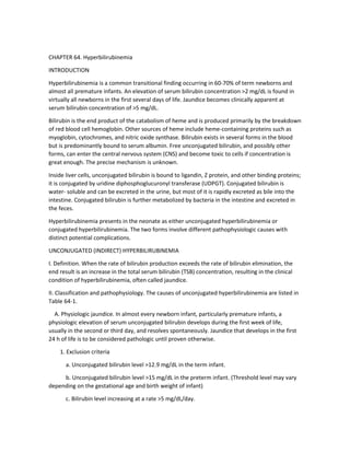
  • 2. d. Jaundice in the first 24 h of life. e. Conjugated bilirubin level >2 mg/dL. f. Clinical jaundice persisting >1 week in full-term infants or >2 weeks in preterm infants. 2. Physiology a. Full-term infant. Serum unconjugated bilirubin progressively rises to a mean peak of 5-6 mg/dL by the third day of life in both white and black infants and a peak of 10 mg/dL at 3-4 days in Asian babies. Figure 64-1 shows hour-specific nomogram with predictive value for subsequent hyperbilirubinemia. b. Preterm neonates. Liver function is less mature, and jaundice is more frequent and pronounced. A peak concentration of 10-12 mg/dL is reached by the fifth day of life. 3. Mechanisms. A number of mechanisms have been suggested. a. Increased bilirubin load because of the larger red blood cell volume, the shorter life span of red blood cells, and increased enterohepatic recirculation of bilirubin in newborn infants. b. Defective uptake by the liver as a result of decreased concentration of bilirubin binding proteins, such as ligandin. c. Defective conjugation because of reduced glucuronyl transferase activity in the term newborn; even more pronounced in premature infants. d. Impaired excretion into bile. e. Overall impairment/immaturity of liver function. B. Hemolytic anemia 1. Red blood cell defects. Hemolytic anemia may result from a congenital red blood cell defect such as hereditary spherocytosis, infantile pyknocytosis, pyruvate kinase deficiency, glucose-6phosphate dehydrogenase (G6PD) deficiency, thalassemia, or vitamin K-induced hemolysis. 2. Acquired hemolytic anemia may be seen in blood group incompatibilities, such as ABO or Rh incompatibility between infant and mother. It may also be associated with the use of certain drugs (eg, sulfonamides) or with infection. C. Polycythemia. The liver may not have the capacity to metabolize the increased bilirubin load presented by the increased blood volume. D. Blood extravasation. Sequestration of blood within body cavities can result in increased bilirubin and thus overload the bilirubin degradation pathway. It may be seen with cephalhematoma, intracranial and pulmonary hemorrhage, subcapsular hematoma of the liver, excessive ecchymoses or petechiae, occult gastrointestinal hemorrhage, and large hemangiomas (ie, Kasabach-Merritt syndrome). TABLE 64-1. CAUSES OF UNCONJUGATED HYPERBILIRUBINEMIA
  • 3. Physiologic jaundice Hemolytic anemia Congenital: red blood cell defects (hereditary spherocytosis, infantile pyknocytosis, pyruvate kinase deficiency, G6PD deficiency, and thalassemia) Acquired: blood group incompatibilities (ABO or Rh incompatibility), infection, and drug-induced hemolysis Polycythemia Blood extravasation Defects of conjugation Congenital: Crigler-Najjar syndrome (types I and II), Gilbert syndrome Acquired: Lucey-Driscoll syndrome Breast-feeding and breast milk jaundice Metabolic disorders: galactosemia, hypothyroidism Increased enterohepatic circulaton of bilirubin Substances/ disorders affecting binding of bilirubin to albumin: drugs, fatty acids in nutritional products, asphyxia, acidosis, sepsis, hypothermia, hyperosmolality, hypoglycemia G6PD, glucose-6-phosphate dehydrogenase. FIGURE 64-1. Hour-specific bilirubin nomogram with the predictive ability of the predischarge bilirubin value for subsequent severe hyperbilirubinemia, >95th percentile tract. Reproduced with permission from Bhutani VK, Johnson LH. Jaundice technologies; prediction of hyperbilirubinemia in term and near term newborns. J Perinatol 2001;21:576 E. Defects of conjugation 1. Congenital deficiency of glucuronyl transferase a. Crigler-Najjar syndrome type I. A severe form of uridine diphosphate (UPD) glucuronyl transferase deficiency that is inherited as an autosomal recessive disorder. It is unresponsive to phenobarbital therapy and has a poor prognosis. b. Crigler-Najjar syndrome type II. Moderate deficiency of UDP-glucuronyl transferase. It is responsive to phenobarbital therapy. c. Gilbert syndrome is a mild form of UDP-glucuronyl transferase deficiency and is inherited as an autosomal dominant condition, is benign, and is relatively common. It is responsive to phenobarbital, although no specific therapy is necessary. 2. Glucuronyl transferase inhibition a. Drugs such as novobiocin. b. Lucey-Driscoll syndrome. An unspecified maternal gestational hormone, found in the infant's serum interferes with the conjugation of bilirubin. This problem appears to resolve spontaneously; however, in extreme cases, exchange transfusions may be required to avoid kernicterus. F. Breast milk jaundice (late onset) is a result of the prolongation of increased enterohepatic recirculation of bilirubin because of a factor in human milk that promotes intestinal absorption. It is characterized by a higher peak (10- 30 mg/dL, peaking by days 10-15 of life) and a slower decline in the serum bilirubin concentration that can last for several weeks. It rarely appears before the end of the first week of life for term or preterm infants. Interruption of breast-feeding for 24-48 h at unacceptable bilirubin levels results in a rapid decline. Resumption of breast-feeding increases bilirubin levels slightly but usually below previous levels. Breast milk jaundice may recur in 70% of future pregnancies.
  • 4. G. Metabolic disorders. Galactosemia, hypothyroidism, and maternal diabetes may be associated with unconjugated hyperbilirubinemia. H. Increased enterohepatic recirculation of unconjugated bilirubin as a result of pathologic conditions such as cystic fibrosis, gastrointestinal obstruction (ie, pyloric stenosis, duodenal atresia, annular pancreas), and ileus may result in exaggerated jaundice. Blood swallowed during delivery and decreased caloric intake may also be contributing factors. I. Substances and disorders affecting binding of bilirubin to albumin. Certain drugs occupy bilirubin-binding sites on albumin and increase the amount of free unconjugated bilirubin that can cross the blood-brain barrier. Drugs in which this effect may be significant include aspirin and sulfonamides. Chloral hydrate is shown to compete for hepatic glucuronidation with bilirubin and thus increase serum unconjugated bilirubin. Common drugs used in the neonates such as penicillin and gentamicin have also been shown to compete with bilirubin for albumin binding sites. Fatty acids in nutritional products (eg, Intralipid) may also influence bilirubin binding to albumin, as may asphyxia, acidosis, sepsis, hypothermia, hyperosmolality, and hypoglycemia. III. Morbidity A. General. Unconjugated bilirubin in high concentration can cross the blood-brain barrier, penetrating the brain cells. This has been associated with neuronal dysfunction and death. Bilirubin is toxic to the brain in several ways. It has been shown to depress O2 consumption and at high concentration uncouples oxidative phosphorylation. Severe hyperbilirubinemia can cause long-term neurologic sequelae; however, the safety of moderate degree of hyperbilirubinemia has been debated. One study has shown that moderate hyperbilirubinemia (13.6-26.0 mg/dL) may be associated with an increase in minor neurologic dysfunction. B. Encephalopathy 1. Transient. Early bilirubin-induced neurologic dysfunction is transient and reversible. Auditory brainstem evoked responses (ABRs) show significant prolongation of latency of specific wavelengths. These changes may be reversed with either exchange transfusions or with spontaneous decrease in bilirubin levels. More recent data suggest that with marked hyperbilirubinemia, complete recovery of the ABR may be delayed. The auditory system serves as an objective window in looking at the CNS in cases of severe hyperbilirubinemia, which can be used as an early predictor of bilirubin encephalopathy. 2. Bilirubin encephalopathy. This is a preventable lifelong neurologic sequelae of untreated hyperbilirubinemia. It is a clinically worsening encephalopathy (over 24 h) caused by bilirubin toxicity to the basal ganglia and various brainstem nuclei. a. Acute phase. A severely jaundiced infant develops lethargy, hypotonia, and poor sucking response. If left untreated, the infant becomes hypertonic and may develop fever and a highpitched cry. Decerebrate posturing can occur at the end stage of the disease. b. Chronic phase. Survivors usually develop choreoathetoid cerebral palsy, hearing loss, dental dysplasia and paralysis of upward gaze, and, less often, mental retardation.
  • 5. C. Kernicterus. A postmortem diagnosis of pathologic findings of bilirubin toxicity in the brain. 1. Staining and necroses of neurons in the basal ganglia, hippocampal cortex, subthalamic nuclei, and cerebellum. Cerebral cortex is usually spared. 2. Bilirubin toxicity to the brain may be reversible if bilirubin levels fall before saturation of the CNS nuclei occurs. 3. Controversy exists over bilirubin levels considered at risk for kernicterus. In nonhemolytic states, and otherwise healthy term infants, levels of >25 mg/dL are considered to place the patient at risk. 4. Kernicterus registry. Kernicterus is not a reportable condition in the United States; therefore, its prevalence is not known. A pilot registry at a Pennsylvania hospital documented 90 cases from January 1984-June 2001. IV. Clinical presentation A. History 1. Family history. Family history of jaundice, anemia, splenectomy, or metabolic disorders is significant. A history of a sibling with jaundice may suggest blood group incompatibility, breast milk jaundice, or G6PD deficiency. A familial tendency of neonatal hyperbilirubinemia is present regardless of breast-feeding and other risk factors. 2. Maternal history. Neonatal jaundice is increased with a history of maternal diabetes or infection. Use of oxytocin, sulfonamides, antimalarials, and nitrofurantoins by the mother may initiate hemolysis in G6PD-deficient infants. Delivery trauma, asphyxia, delayed cord clamping, and prematurity are associated with an increased risk of hyperbilirubinemia in the infant. 3. Infant's history. a. Breast-feeding. Poor breast-feeding may result in poor caloric intake, leading to "starvation jaundice." (See prior discussion of breast milk jaundice). b. Factors affecting the gastrointestinal tract, such as obstruction, decreased motility (ileus), and delayed passage of meconium c. Check for clinical symptoms such as vomiting and lethargy. Metabolic disorders, infection, and bowel obstruction can present in this manner. B. Signs and symptoms. Clinical jaundice is visible when the serum bilirubin level approaches 5- 7 mg/dL. Jaundice is often apparent first in the face, especially the nose, and then descending to the torso and lower extremities as the degree of jaundice increases. Jaundice can be demonstrated in some infants by pressing lightly on the skin with a finger. The yellow color is seen more easily in the "fingerprint" area than in the surrounding skin. These signs should not appear within the first 24 h after birth in otherwise healthy infants. Besides confirming the presence of jaundice, physical examination may also be helpful in determining the cause of hyperbilirubinemia. Areas of bleeding such as cephalhematoma,
  • 6. petechiae, or ecchymoses indicate blood extravasation. Hepatosplenomegaly may signify hemolytic disease, liver disease, or infection. Physical signs of prematurity, intrauterine growth retardation, and postmaturity may be helpful in elucidating a cause for hyperbilirubinemia. Plethora is seen with polycythemia, pallor with hemolytic disease, and large infants with maternal diabetes, all of which are associated with hyperbilirubinemia. Omphalitis, chorioretinitis, microcephaly, and petechial and purpuric lesions suggest infectious causes of increased serum bilirubin. C. Neurologic examination. The appearance of abnormal neurologic signs heralds the onset of early bilirubin encephalopathy. Initial signs include lethargy, poor feeding, vomiting, and hypotonia. The persistently hyperbilirubinemic infant may go on to experience seizures (see section III,B above). The progression of neurologic changes parallels the stages of bilirubin encephalopathy from acute to chronic and irreversible changes. V. Diagnosis A. Laboratory studies. Hyperbilirubinemia should be investigated whenever pathologic causes are suspected. The presence of a serum bilirubin level of ≥13 mg/dL in any infant requires initial evaluation. 1. Total and direct bilirubin a. Direct bilirubin = conjugated fraction. b. Indirect bilirubin = unconjugated fraction. Derived by subtracting direct fraction from TSB. 2. Complete blood cell count and reticulocyte count. A low hemoglobin or hematocrit associated with a high reticulocyte count and the presence of nucleated red blood cells is highly suggestive of hemolytic anemia. Polycythemia is defined as a venous blood hematocrit >65%. (Use of capillary blood samples may cause underestimation of venous bilirubin values when the bilirubin value is > 10 mg/dL). Total white blood cell count with differential and platelet count may help detect sepsis. The peripheral blood smear aids in the diagnosis of hereditary spherocytosis and other red blood cell defects. 3. Blood type and Rh status in mother and infant. ABO and Rh incompatibility can be easily diagnosed by comparing infant and maternal blood types. 4. Direct Coombs' test. The test is usually positive in infant's with isoimmunization disorders. This test does not correlate with the severity of jaundice. 5. Measurement of serum albumin may help to assess total bilirubin binding sites available and whether there is a need for an albumin infusion. 6. Other laboratory tests. The urine should be tested for reducing substances (to rule out galactosemia if the infant is receiving a galactose-containing formula) and for infectious agents. If hemolysis is present, in the absence of ABO or Rh incompatibility, further testing by hemoglobin electrophoresis, G6PD screening, or osmotic fragility testing may be required to diagnose red blood cell defects. Persistent jaundice (>2 weeks of life) may require these types of investigation
  • 7. as well as additional tests for thyroid and liver function, blood and urine cultures, and metabolic screening workup, such as plasma amino acid and urine organic acid measurements. B. Radiologic studies for suspected intestinal obstruction or blood extravasation into internal organs and ultrasonography of the head to document intracranial or subdural hemorrhage might be indicated. C. Transcutaneous bilirubinometry (TcB). Measures the degree of yellow color in the skin and subcutaneous tissues by selective wavelength reflection. Data suggest that this methodology is an effective adjunct to nursing assessment in the term infant. A TcB value of >13 mg/dL should be correlated with TSB. D. Expired carbon monoxide breath analyzer. End-tidal carbon monoxide (CO) corrected for ambient CO (ETCO2). An equimolar amount of CO is produced for every molecule of bilirubin formed from the degradation of heme; therefore, measurement of CO in end-tidal breath is an index of total bilirubin production. The method can alert the attending physician to the presence of hemolysis irrespective of the timing of jaundice. VI. Management. Three methods of treatment are commonly used to decrease the level of unconjugated bilirubin: exchange transfusion, phototherapy, and pharmacologic therapy. As noted, however, controversy persists as to what levels of serum bilirubin warrant therapy, especially in otherwise healthy full-term infants. A. Phototherapy (see also Chapter 39) 1. Indication. Most infants with pathologic jaundice are treated with phototherapy when it is believed that bilirubin levels could enter the toxic range. a. Practice parameters. The American Academy of Pediatrics (AAP) is currently revising the guidelines for management of hyperbilirubinemia. The concern for kernicterus is being reemphasized by the Subcommittee on Hyperbilirubinemia pending the full assessment and analysis of the new approaches to the jaundiced infant. It is important to remember that the current AAP guidelines are meant for healthy term newborn infants with no evidence of hemolysis. Although there is continuing uncertainty about which TSB level warrants exchange transfusions, the authors suggest that in an otherwise healthy term newborn a TSB level of 20 mg/dL at 48 h of life may be treated initially with phototherapy. If the TSB decreases by 1-2 mg/dL within 4-6 h of starting phototherapy, exchange transfusion may not be necessary. b. In preterm and sick term infants, a more conservative approach is usually adopted. 2. Using phototherapy effectively. a. Light source. The most effective light source currently available for phototherapy is that provided by blue fluorescent tubes. Bilirubin absorbs light maximally in the blue range (420-500 nm). Blue lamps with narrow spectral range (420-480 nm) are most effective. The blue light reflection does interfere with skin color assessment and has been reported to cause dizziness and nausea in those caring for these patients. Addition of a daylight fluorescent tube tempers these side effects. Energy output or irradiance is a second variable that influences the efficacy of
  • 8. phototherapy. Radiometer measurements should be minimally 5 uW/cm2/nm, with saturation for bilirubin degradation at ~11 uW/cm2/nm. b. Distance from the light to the infant. Light intensity is a function of the distance from the light source; therefore, light source should be as close to the infant as possible (12-16 inches). c. Surface area. The larger the skin surface area exposed, the more effective is the phototherapy. The availability of fiberoptic pads (bili-blankets) make it easy to increase exposed skin area to the bili-lights. In preterm infants, this type of "double-phototherapy" (overhead light plus bili- blanket) is almost twice as effective as single phototherapy. Fiberoptic phototherapy blankets (bili- blankets) can be wrapped around or placed just beneath the infant. 3. Supportive management. The infant's eyes should be covered with opaque patches for overhead lamp phototherapy. However, this is not necessary when using only fiberoptic light source. Animal experiments have shown retinal damage occurring as a result of phototherapy. To maximize exposure, infants should be naked in servocontrolled incubators. Phototherapy does increase insensible fluid losses. For infants weighing <1500 g, increase fluids by 0.5 mL/kg/h; for those weighing >1500 g, increase by 1 mL/kg/h. Overhydration does not increase bilirubin elimination. 4. Termination of phototherapy. Phototherapy is stopped when the following criteria are met: a. The bilirubin level is low enough to eliminate the risk of kernicterus. b. The infant is old enough to handle the bilirubin load. 5. Complications. Phototherapy is simple, safe, and inexpensive. Complications are very rare but should not be overlooked. a. The retinal effects of phototherapy on the exposed infant's eyes are unknown. However, animal studies suggest that retinal degeneration may occur. Thus, eye shields must be used. b. Increased insensible fluid loss increases fluid requirements by 25%. In addition, stools may be looser and more frequent. Use of fiberoptic phototherapy results in lower insensible loss. c. Bronze baby syndrome. With conjugated hyperbilirubinemia, phototherapy causes photodestruction of copper porphyrins, causing urine and skin to become bronze. d. Congenital erythropoietic porphyria is a rare syndrome in which phototherapy is contraindicated. Exposure to visible light of moderate to high intensity will produce severe bullous lesions on exposed skin and may lead to death. B. Exchange transfusion (see also Chapter 21). Exchange transfusion is used when the risk of kernicterus for a particular infant is significant. A double-volume exchange replaces 85% of the circulating red blood cells and decreases the bilirubin level to about half of the preexchange value. Besides removing bilirubin, an exchange transfusion can be used to correct severe anemia. 1. General guidelines. It appears that no specific level of bilirubin can be considered safe or dangerous for all infants because patient-to-patient variations exist for the permeability of the blood- brain barrier. The practice parameters published by the AAP in October 1994 recommend
  • 9. exchange transfusions in healthy term newborns at TSB 25-30 mg/dL. The debate for upper limits of serum bilirubin is directed to full-term infants with no evidence for associated disease. Low birth weight infants are excluded from these considerations. The authors suggest that each institution and its practicing physicians must establish their criteria for phototherapy and exchange transfusion by gestational age, weight groups, postnatal age, and infant's condition consistent with current standard of pediatric practice. 2. Factors that may affect the decision to perform exchange transfusion include the infant's maturity, birth weight, age, rate of rise in bilirubin levels (>0.5 mg/dL/h), and presence of hypoxia, acidosis, sepsis, or hypoproteinemia. 3. Albumin transfusions may be useful if bilirubin levels are >20 mg/dL and serum albumin levels are <3 g/dL. Infusion of 1 g of albumin 1 h before exchange transfusion may improve the yield of bilirubin removal. Fluid volume and cardiovascular status must be carefully considered before giving albumin. C. Pharmacology 1. Phenobarbital therapy a. Action. Phenobarbital affects the metabolism of bilirubin by increasing the concentration of ligandin in liver cells, inducing production of glucuronyl transferase, and enhancing bilirubin excretion. Because it takes 3-7 days to become effective, phenobarbital is usually not helpful in treating unconjugated hyperbilirubinemia in the newborn infant. The administration of phenobarbital to newborns at the time that jaundice is first noted or even at birth is less effective than administration to mothers during pregnancy for ≥2 weeks before delivery. b. Indications. It may be useful to give phenobarbital 1-2 weeks before delivery to a pregnant mother whose fetus has documented hemolytic disease to aid in reducing bilirubin levels in the affected neonate. Phenobarbital is also used to treat type II glucuronyl transferase deficiency and Gilbert syndrome in the infant. 2. Metalloporphyrins. A synthetic heme analog, metalloporphyrins have been shown to inhibit heme oxygenase (HO), the rate-limiting enzyme in the catabolism of heme. By acting as a competitive inhibitor, the metalloporphyrins decrease the production of bilirubin. Tinmesoporphyrin (SnMP) is a potent HO inhibitor that has been extensively studied. It has been reported to be effective as a single intramuscular injection (6 mmol/ kg) in two patients with hemolytic disease, resulting in a significant drop in TSB concentration, thereby avoiding the need for exchange transfusion. Another report has shown that a single dose of SnMP can prevent the development of severe hyperbilirubinemia in newborn infants with G6PD deficiency. The only untoward effect noted was a non-dose-dependent, mild, transient erythema when used in conjunction with phototherapy in a preterm infant. 3. Supportive management. Most breast-fed infants do not develop bilirubin levels of ≥20 mg/ dL in the first 8 days of life. For infants 6-8 days old, breast-fed and otherwise well, consider interrupting breast-feeding for 48 h and use phototherapy if the bilirubin is >18 mg/dL.
  • 10. CONJUGATED (DIRECT) HYPERBILIRUBINEMIA I. Definition. Conjugated hyperbilirubinemia is a sign of hepatobiliary dysfunction. It usually appears in the newborn infant after the first week of life, when the indirect hyperbilirubinemia of physiologic jaundice has receded. When the direct bilirubin level is >2.0 mg/dL or is >20% of the TSB, it is clinically significant. II. Classification. Conjugated hyperbilirubinemia is caused by a defect or insufficiency in bile secretion, biliary flow, or both, resulting in an inability to remove conjugated bilirubin from the body. It is always pathologic. The term cholestasis is used to describe the group of disorders associated with bilirubin excretion and is associated with a rise in serum conjugated bilirubin levels and usually bile salts and phospholipids. Causes of conjugated hyperbilirubinemia are listed in Table 64-2. A. Extrahepatic biliary disease 1. Biliary atresia is the single most common cause of neonatal cholestasis, with an estimated incidence of 1:10,000 live births. It is the reason for 50-60% of liver transplantations in children worldwide. Its cause is poorly understood. It can be associated with polysplenia, cardiovascular defects, and situs inversus. 2. Choledochal cysts are dilatations of extrahepatic biliary tree. Diagnosis is by ultrasonography or nuclear medicine scanning. 3. Other extrahepatic biliary diseases are bile duct stenosis, spontaneous perforation of bile duct, cholelithiasis, and neoplasm. These are uncommon conditions in neonates. Cholelithiasis in infancy is usually related to other underlying conditions such as hemolysis, anatomic malformation, use of medications (furosemide), or prolonged use of parenteral nutrition. Cholecystectomy is rarely indicated. B. Intrahepatic biliary disease 1. Intrahepatic bile duct paucity. This condition may be syndromic or nonsyndromic. The syndromic form is called Alagille's syndrome (arteriohepatic dysplasia). This is characterized by a constellation of features such as cholestasis, cardiovascular findings (pulmonary artery stenosis), butterfly vertebrae, ocular findings (posterior embryotoxon), and characteristic facies. Liver biopsy shows a paucity of interlobular bile ducts. Transmitted as autosomal dominant, this disorder has been assigned to chromosome 20p12, a region of the chromosome containing the JAGGED1 gene. 2. Progressive intrahepatic cholestasis. These patients have chronic intrahepatic cholestasis but with different pathogenesis and prognosis. Clinical features include chronic persistent hepatocellular cholestasis, exclusion of other identifiable disorders, occurrence consistent with autosomal recessive inheritance, and a combination of clinical, biochemical, and histologic features. (See Haber & Lake, 1990, for detailed classification and description.) 3. Inspissated bile. Infants with moderate to severe hemolytic disease can develop bilirubin overload and resulting cholestasis. Clinically, hepatosplenomegaly is often prominent and liver enzymes are normal or slightly elevated. Cholestasis may persist up to 4 weeks; however, if it persists beyond 4 weeks, it should not be attributed to bilirubin overload alone.
  • 11. C. Hepatocellular disease TABLE 64-2. CAUSES OF CONJUGATED HYPERBILIRUBINEMIA Extrahepatic biliary disease Biliary atresia Choledochal cyst Bile duct stenosis Spontaneous perforation of the bile duct Cholelithiasis Neoplasms Intrahepatic biliary disease Intrahepatic bile duct paucity (syndromic or nonsyndromic) Progressive intrahepatic cholestasis Inspissated bile Hepatocellular disease Metabolic and genetic defects α1-antitrypsin deficiency, cystic fibrosis, Zellweger's syndrome, Dubin-Johnson and Rotor's syndromes, galactosemia Infections Total parenteral nutrition Idiopathic neonatal hepatitis Neonatal hemochromatosis Miscellaneous Shock Hypoperfusion State, ECMO INTRAHEPATIC CHOLESTASIS WITH NORMAL BILE DUCTS Infection Viral: hepatitis B virus; non-A, non-B hepatitis virus; cytomegalovirus; herpes simplex virus; coxsackie virus; Epstein- Barr virus; adenovirus Bacterial: Treponema pallidum, Escherichia coli, group B streptococcus, Staphylococcus aureus, Listeria; urinary tract infection caused by E. coli or Proteus spp, pneumococcus Other: Toxoplasma gondii Genetic disorders and inborn errors of metabolism: Dubin- Johnson syndrome, Rotor's syndrome, galactosemia, hereditary fructose intolerance, tyrosinemia, α1-antitrypsin deficiency, Byler disease, recurrent cholestasis with lymphedema, cerebrohepatorenal syndrome, congenital erythropoietic porphyria, Niemann-Pick disease, Menkes' kinky hair syndrome Idiopathic neonatal hepatitis (giant cell hepatitis) Total parenteral nutrition-induced cholestasis 1. Metabolic and genetic defects. A significant number of identifiable metabolic and genetic abnormalities can present with hepatocellular dysfunction. a. α1-Antitrypsin deficiency. This is an autosomal recessive condition characterized by accumulation of α1-antitrypsin in the hepatocyte, resulting in subsequent hepatocellular necrosis. There are several phenotypes; however, only the homozygous Pi (protease inhibitor) ZZ and, rarely, MZ types have been associated with liver disease in infancy. Most patients experience jaundice in the first 8 weeks of life. Fifty to 70% of patients will go into remission by 6 months. Otherwise, the clinical course is highly variable; some patients develop progressive liver failure. b. Cystic fibrosis. Cholestasis can be an initial presentation of cystic fibrosis in infancy. Most of these infants will also have meconium ileus. Liver biopsy of these patients will show excessive biliary mucus, inspissated bile, mild inflammatory changes, and fibrosis. Patients who had resolved neonatal jaundice are not at greater risk for liver disease later in life over other cystic fibrosis patients. c. Zellweger's or cerebrohepatorenal syndrome. This is a peroxisomal disorder and is characterized by the absence of peroxisomes and deranged mitochondria. It is inherited as an autosomal recessive trait and presents in the neonatal period with cholestasis, hepatomegaly, profound hypotonia, and dysmorphic features. Diagnosis is confirmed by the presence of abnormal levels of very-long-chain fatty acid in the serum. Prognosis is very poor. Most infants die within 1 year. Survivors beyond 1 year of age have severe mental retardation and seizures. d. Dubin-Johnson and Rotor's syndromes. These syndromes are rarely diagnosed in the neonatal period, although they can initially present with cholestasis during that period. DubinJohnson syndrome is a nonhemolytic conjugated hyperbilirubinemia caused by deficiency in the canalicular secretion of conjugated bilirubin. Liver biopsy is normal except for the presence of
  • 12. pigmented granules. Rotor's syndrome is believed to be the result of a disturbance in the hepatic storage of anions, characterized by lifelong presence of mild conjugated hyperbilirubinemia. Liver biopsy is normal, but unlike Dubin-Johnson no pigment accumulation is noted. Both conditions are inherited as autosomal recessive and have an excellent prognosis. e. Galactosemia. This is an autosomal recessive disorder resulting from a deficiency of galactose-1-phosphate uridyltransferase activity. Incidence is about 1:100,000. It is characterized by presence of cholestasis, hepatomegaly, hypoglycemia, cataracts, vomiting, and failure to thrive. Escherichia coli sepsis is the most devastating complication in the newborn period. The two other enzymes, namely galactokinase and UDP galactose-4-epimerase, that are involved in the galactose metabolic pathway are less common causes of galactosemia. The pathologic consequences are thought to be secondary to accumulation of toxic metabolite, galactose-1-phosphate. Galactosemia must be rapidly excluded in infants with early-onset cholestasis, with associated emesis, acidosis, and gram-negative sepsis (particularly E. coli). These infants, while on lactosecontaining formula, will have galactose in the urine, resulting in a positive reducing substance in the urine (Clinitest) but negative urine test for glucose (glucose oxidase). f. Other metabolic and genetic disorders. Tyrosinemia, fructosemia, Niemann-Pick disease, Gaucher's disease, Wolman's disease, and glycogenosis type IV are other metabolic and genetic disorders that may present with cholestasis as an additional finding in the neonatal period. 2. Infection. a. Congenital infection. Congenitally acquired infections have a spectrum of manifestations but are usually asymptomatic. Congenital infection usually presents with other stigmata of the disease. Vertical transmission of hepatitis viruses (B and C) is generally asymptomatic, but clinical hepatitis, including hepatic failure, may develop at about 2 months of age. Coxsackievirus, EpsteinBarr virus, and adenovirus are known causes of neonatal hepatitis with elevated conjugated fractions. b. Sepsis. Direct bacterial infection of the liver may occur with overwhelming sepsis. Toxic cholestasis with no direct invasion of the liver by microorganisms may be seen with urinary tract infection, particularly with E. coli urosepsis. 3. Total parenteral nutrition (TPN) cholestasis. The frequency, not necessarily the severity, of cholestasis is partly a function of the degree of prematurity. Cholestasis develops in >50% of infants with birth weight of <1000 g and <10% of term infants after prolonged hyperalimentation. No precise cause has been found; however, the most significant contributing factor was thought to be the lack of enteral feeding. The resumption of normal enteral feeds has been associated with improvement of cholestasis in 1-3 months, with no or little residual fibrosis and normal hepatic function. 4. Idiopathic neonatal hepatitis (Table 64-3). This diagnosis applies when known infectious, metabolic, and genetic causes have been ruled out. Jaundice and hepatosplenomegaly are major physical findings. Liver biopsy demonstrates giant cell transformation, increased extramedullary hematopoiesis, and inflammation. Prognosis is fair. 5. Miscellaneous.
  • 13. a. Neonatal hemochromatosis (NH). Also known as neonatal iron storage disease, this condition results from liver disease of intrauterine onset. It is associated with extrahepatic deposits of stainable iron. Patients present with hepatocellular synthetic insufficiency (hypoalbuminemia, coagulopathy, and low fibrinogen) with end-stage liver disease. Prognosis is very poor and is almost always fatal. b. Shock or hypoperfusion. Multiorgan involvement is not uncommon in cases of perinatal asphyxia or shock/hypoperfusion state. c. Extracorporeal membrane oxygenation (ECMO). Infants on ECMO may show an increase in the prevalence and severity of cholestasis. III. Clinical presentation. The classic clinical manifestations of neonatal cholestasis include jaundice, acholic stools, and dark urine. Other findings may include hepatomegaly, splenomegaly, pruritus, failure to thrive, decreased feeding/ appetite, ascites, and portal hypertension. Unfortunately, individual signs and symptoms cannot differentiate between intra- versus extrahepatic disease. The combination of clinical and historical findings can lead to possible causes. The presence of nonhepatic findings will provide helpful clues to specific diagnosis, such as the following: A. Alagille's syndrome. Cardiovascular findings (peripheral pulmonic stenosis), vertebral anomalies, ocular findings (posterior embryotoxon), and peculiar facies. B. Zellweger's syndrome or cerebrohepatorenal syndrome. Profound hypotonia, seizures, and dysmorphic features. C. Congenital infections. Microcephaly, intracranial calcifications, intrauterine growth retardation. D. Galactosemia. Failure to thrive, vomiting, cataracts, and Gram-negative bacterial sepsis (E. coli). E. TPN-induced cholestasis. History of prematurity and prolonged use of hyperalimentation. IV. Diagnosis A. Laboratory studies 1. Bilirubin levels (total and direct). 2. Liver function tests. Serum glutamic oxaloacetic transaminase (SGOT [aspartate transaminase, or AST]), serum glutamic pyruvic transaminase (SGPT [alanine transaminase, or ALT]), and alkaline phosphatase may be helpful in monitoring the course of the disease. 3. Prothrombin time and partial thromboplastin time may be reliable indicators of liver function. 4. γ-Glutamyl transpeptidase, 5′ -nucleotidase, and serum bile acids are also usually elevated in cholestasis. Once the diagnosis of cholestasis is made, measurement of these markers of cholestasis may not add any further information.
  • 14. 5. A complete blood cell count and reticulocyte count may be helpful if hepatitis is a possibility because hemolysis may be found. 6. Serum cholesterol, triglycerides, and albumin levels. Triglyceride and cholesterol levels may aid in nutritional management and assessment of liver failure. Albumin is a long-term indicator of hepatic function. 7. Ammonia levels should be checked if liver failure is suspected. 8. Serum glucose levels should be checked if the infant appears ill. 9. Urine testing for reducing substances is a simple screening test that should always be performed to detect for metabolic disease. 10. TORCH (toxoplasmosis, rubella, cytomegalovirus [CMV], and herpes simplex virus) titers and urine cultures for CMV. The use of TORCH titers should be guided by the clinical presentation, keeping in mind that CMV infection may be asymptomatic and congenital syphilis is treatable. CMV immunoglobulins M and G (IgM and IgG), Venereal Disease Research Laboratory (VDRL), and IgM-specific titers for herpes simplex, rubella, and toxoplasmosis may be useful in some cases. 11. Alpha-fetoprotein (AFP). 12. Other tests. More specific tests are indicated in the investigation of the specific causes of conjugated hyperbilirubinemia. a. Hepatitis. The maternal hepatitis B surface antigen (HBsAg) status should be known, and the infant should also be tested. b. Sepsis. If bacterial sepsis or urinary tract infection is suspected, appropriate cultures should be obtained. c. Metabolic disorders i. Galactosemia and hereditary fructose intolerance. The urine should be tested for nonglucose-reducing substances. Enzymes involved in these disorders can also be assayed in the blood. ii. Tyrosinemia. High concentrations of tyrosine and methionine, and their metabolic derivatives, will be seen in the urine. iii. α1-Antitrypsin deficiency. Decreased serum α1-antitrypsin concentration and liver biopsy showing periodic acid-Schiff (PAS)-positive cytoplasmic granules with variable degrees of hepatic necrosis and fibrosis. iv. Cystic fibrosis. A sweat test may diagnose cystic fibrosis. B. Radiologic studies. Diagnosis of biliary atresia and other forms of extrahepatic biliary obstruction by 8 weeks of age is required to prevent progression of the disease. 1. Ultrasonography. This is used to view the liver parenchyma and to diagnose dilatation of the biliary tree; in extrahepatic obstruction, half of the infants will have dilated proximal ducts within the liver. Choledochal cysts are seen. An inability to identify the gallbladder on
  • 15. ultrasonography may result from obliteration of its lumen in biliary atresia. However, the presence of a gallbladder does not rule out biliary atresia. Because this test is simple and noninvasive and because of the reported coexistence of certain cholestatic diseases, we highly recommend that all infants with cholestasis have this diagnostic test done. 2. Hepatobiliary imaging. Contrast agents are taken up by the liver and excreted into the bile. HIDA (hepatobiliary iminodiacetic acid), EHIDA (ethyl hepatobiliary iminodiacetic acid), and PIPIDA (p-isopropylacetanilide-iminodiacetic acid) are technetium labeled and provide a clear image of the biliary tree after intravenous injection. Neonatal hepatitis, hyperalimentation, and septo- optic dysplasia are reported causes of absent gastrointestinal contrast excretion and must be considered in the diagnosis of biliary atresia. Administration of phenobarbital, 5 mg/kg/day for 5 days, in conjunction with hepatobiliary scanning may be helpful in distinguishing infants who do not have biliary atresia. Excretion of the contrast medium may improve after phenobarbital treatment. C. Other studies 1. Percutaneous liver biopsy. This is a safe procedure in experienced hands. Biopsy findings must be correlated with clinical and laboratory data. 2. Exploratory laparotomy with operative cholangiogram is sometimes indicated to correct biliary atresia, especially if the just-mentioned tests are not diagnostic. V. Management (see also Chapter 38) A. Medical management 1. General plan. In cholestatic jaundice, promotion of bile flow and prevention of malnutrition, vitamin deficiencies, and bleeding are goals of treatment. 2. Pharmacologic management. Phenobarbital and cholestyramine will promote bile flow and decrease serum bilirubin and bile salt levels. Cholestyramine is a nonabsorbable anion exchange resin that irreversibly binds salts in the intestine. This leads to increased fecal excretion of bile salts and increased hepatic synthesis of bile salts from cholesterol, which may lower serum cholesterol levels. Actigall (ursodeoxycholic acid) has also been successfully used in conjunction with phenobarbital and cholestyramine. B. Dietary management 1. Medium-chain triglycerides (MCT). Long-chain triglycerides are poorly absorbed in the absence of sufficient bile salts. Therefore, infants with cholestasis often require a diet that includes MCTs, which can be absorbed without the action of bile salts. Formulas containing MCTs include Portagen and Pregestimil. Breast-fed cholestatic infants should be given supplemental MCT. 2. Vitamin supplementation. Fat malabsorption will also interfere with maintenance of adequate levels of fat-soluble vitamins in these infants. Supplementation of vitamins A, D, E, and K is suggested. Extra vitamin K supplementation may be necessary if a bleeding tendency develops.
  • 16. 3. Dietary restrictions. Removal of galactose plus lactose and fructose plus sucrose may prevent the development of cirrhosis and other manifestations of galactosemia and hereditary fructose intolerance, respectively. Dietary restrictions may also be used to treat tyrosinemia but usually are less successful. Most other metabolic causes of cholestatic jaundice have no specific therapy. C. Surgical management 1. Laparotomy with biopsy. If extrahepatic biliary obstruction is strongly suspected after completion of an appropriate evaluation, exploratory laparotomy should be performed with operative cholangiography and liver biopsy. Operative examination should be performed by surgeons prepared to proceed with corrective procedures if necessary. Other causes of extrahepatic biliary obstruction that may be diagnosed and treated during exploratory laparotomy include choledochal cyst, spontaneous rupture of the bile duct, lymph node enlargement, tumors, annular pancreas, pancreatic and hepatic cysts, and hemangioendothelioma of the pancreas or liver. Inspissated bile syndrome caused by cystic fibrosis also requires surgical removal of tenacious bile from the bile ducts. 2. Kasai procedure. Surgical procedure such as Kasai portoenterostomy should be done to establish biliary drainage in patients diagnosed with biliary atresia. Optimal results are obtained if the procedure is done before 8 weeks of age. If the Kasai procedure is successful, most infants will have deterioration of liver function over time and eventually need a liver transplant. The procedure is used as a bridge to transplantation. 3. Liver transplantation. When end-stage liver disease is inevitable, liver transplantation is considered. Biliary atresia is the most common indication for liver transplantation in the United States. Overall, the success of liver transplantation has improved significantly, with reports of 5year survival rate of >80%. Some centers report a 1-year survival rate close to 90%. D. Other treatments 1. Infectious diseases. Some of the infectious causes of hepatitis, such as hepatitis B virus, herpes simplex virus, congenital syphilis, and bacterial infections, have specific therapeutic regimens. Most other forms of infectious hepatitis resolve with no specific therapy. 2. TPN-induced conjugated hyperbilirubinemia will usually resolve once TPN is stopped. The decision to continue TPN (with or without trace elements) in an infant with cholestasis must be carefully considered. Resumption of normal enteral feeds is associated with clearing of cholestasis in 4-12 weeks.
  • 17. Ictericia fisiológica Ictericia por leche materna Hiperbilirrubinemia neonatal. Conocer principios fisiológicos y fisiopatológicos de la ictericia. Criterios diagnósticos de hiperbilirrubinemia del recién nacido exámenes de laboratorio, manejo y envío oportuno. Diferenciar la ictericia fisiológica de la no fisiológica Identificar datos de alarma neurológica y otras complicaciones