SlideShare une entreprise Scribd logo
1  sur  62
Télécharger pour lire hors ligne
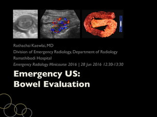
Emergency US:
Bowel Evaluation
Rathachai Kaewlai,MD
Division of Emergency Radiology, Department of Radiology
Ramathibodi Hospital
Emergency Radiology Minicourse 2016 | 28 Jun 2016 12:30-13:30
Disclosure of Commercial Interest
I have no relationships
with commercial interests to disclose
This talk is adapted from an award-winning presentation at
the annual scientific meeting of the American Society
of Emergency Radiology (ASER) in Boston (2013)
Key Points
Transabdominal US has potential to help diagnose and
manage acute GI disorders and mimics
US is most effective when prevalence of the suspected
conditions is high
Outline
Introduction
US technique
Normal bowel wall “gut signature”
US of abnormal bowel
Pathologies
Introduction
Availability, affordability and speed of US imaging
High level of operator skills and experience
Image fromAmberUSA.com
Principles
Most bowel pathologic findings displace bowel gas and
feces, making them stand out against normal bowel
segments
Image fromweb.duke.edu
Selecting the Probes
Curvilinear, low freq (3-5 MHz)
Linear, high freq (10+ MHz)
3D curvilinear probe
US Technique
Start with curvilinear probe to examine
Area of pain: solid organs, peritoneal fluid
Layout of large bowel
Distribution and motility of small bowel
US Technique
Focal bowel masses, segments of wall
thickening and dilated loops may be apparent
on curvilinear probe but require high-
frequency linear probes to identify and
characterize changes in bowel wall layers
Graded Compression US
Puylaert originally described this technique in 1986
“Gradual progressive increase in pressure the operator
applies to the probe while making gentle sweeping
movements”
Radiology 1986; 158:355-360
Graded Compression US:
Suggested Techniques
Step Probe Area(s) of Scanning Organs Visualized
0 C All 4 quadrants with
curvilinear probe
Any free fluid
1 L LLQ for calibrating scan
parameters
Sigmoid colon crossing psoas and
anterior to iliac vessels
2 L RLQ Find ascending colon
Find IC valve
Find terminal ileum
3 L If found pathology,
specifically scanning this area
Bowel of interest (point of
tenderness or abnormality suspected)
4 L ”Mowing the lawn” Check entire colon
Check entire small bowel
5 L Additional views Bowel of interest
First, Adjusting Scan Parameters
Focused examination linear
high-frequency probe may start
in left iliac fossa
Find sigmoid colon
Easily identified
Constant anatomy crossing left psoas and iliac vessels
Adjust scan parameters (depth, gain, focus, etc)
Image fromRadiologyAssistant.nl
Scanning Right Iliac Fossa
Find ascending colon
Find terminal ileum (in
continuity with IC valve)
Scanning Right Iliac Fossa
If suspicious bowel segment seen, specifically examine
Bowel thickening
Alteration of individual bowel layers
Vascularity
Extraintestinal abnormalities such as thickened
mesenteric fat, interloop fluid, lymph node enlargement
Mowing the Lawn
Systematic technique to survey
entire intestine in abd
Overlapping vertical sweeps of
high-frequency probe up and
down the abdomen (manner of
lawnmower)
Additional Views
Posterior manual compression
Left lateral decubitus (assess retrocecal area)
Transvaginal
Images from Janitz E et al. J Am Osteo Coll Radiol 2016;5:5
Normal Bowel Wall:
Mucus Pattern
Collapsed bowel
containing highly reflective
mucus in its center
Target appearance
Normal Bowel Wall:
Gas Pattern
Gas-filled bowel causing
echogenic band with
artifacts underneath
Only anterior part of
bowel wall visualized
Normal Bowel Wall:
Fluid Pattern
Fluid/feces-filled bowel
loops
Normal Bowel Wall
“Gut Signature”
High frequency probes (15+ MHz)
Five alternating bands of high and low echogenicity
Most easily seen when fluid-filled bowel or ascites
Image fromRadiologyAssistant.nl
Normal Bowel Wall
“Gut Signature”
Layer 1 Superficial mucosa Fine bright line
Layer 2 Deep mucosa Gray
Layer 3 Submucosa Bright
Layer 4 Muscularis propria Dark
Layer 5 Serosa Fine bright line
Image fromRadiologyAssistant.nl
Normal Bowel Wall
“Gut Signature”
At least two most prominent layers are evident due to
relative thickness and high contrast
Bright submucosa (Layer 3)
Dark muscularis propria (Layer 4)
(Layer 2 is lost among luminal contents)
Image fromRadiologyAssistant.nl
Normal Bowel: In General
Up to 2-3 mm thick varying on contraction, relaxation
No Doppler signal in normal bowel wall
Healthy bowel can be compressed and shifted by probe
pressure (limited compressibility in obese individuals)
Kralik R et al.Gastroenterol Res Pract 2013, article ID896704
Terminal ileum without and with compression
Normal Bowel: SB vs. Colon
Features Small Bowel Colon
Location Central Peripheral
(picture frame)
Appearance Folds (lesser distally) Haustra
Contents Fluid or dry
Minimal air
Feces
Air
Spontaneous peristalsis Should be seen in
healthy segments
Rarely
Easy to compress and
displace by US probe
Yes, easily Yes for mesenteric
segments
US FINDINGS OF
ABNORMAL BOWEL
Wall thickening (m/c, most striking)
Altered gut signature
Narrow or dilated lumen
Plasticity, mobility, peristalsis
Altered blood flow
Extramural changes
Mesenteric lymph node
Bowel Wall Thickening
Focal, segmental or diffuse
Circumferential or partially circumferential
Target sign, ring sign, pseudokidney sign
Edema, hemorrhage, inflammation, tumor or other
infiltrations
Wall Thickening:
Tumors vs. Infection/inflammation
US Features Tumors Infection/inflammati
on
Length of involvement Short Long
Thickness More (usually >1 cm)
Irregular
Less except ischemia
Smooth
Circumference Yes,asymmetric Yes,symmetric
Gut signature Lost Preserved (not always)
If absent,suggest ischemia
Vascular flow Increased,high RI Increased,low RI
If absent,suggest ischemia
Altered Gut Signature
In diseased segment, bowel layer pattern may be
Preserved
Exaggerated
Distorted
Diminished or obliterated
Bowel Lumen: Dilatation
Aneurysmal dilatation (mostly seen in lymphoma)
Dilatation proximal to obstructing lesion: initially with
increased peristalsis, later without peristalsis (DDx
paralytic ileus)
Images from UltrasoundCases.info
Bowel Lumen: Narrowing
Narrow lumen 2/2 thickening or stricture
Bowel Plasticity, Mobility and
Peristalsis
Most diseases stiffen bowel segments
Rigid
Less compressible
Less easily displaced
Reduced or absent peristalsis
Tethering, architectural distortion with reduced
peristalsis suggest more chronic or aggressive process
such as transmural inflammation or malignancy
Bowel Non-compressibility
Lack of compressibility may be
due to appendicitis,
intussusception, bowel
malignancy or luminal distension
from obstruction
Cystic or hypoechoic mass DDx
Altered Blood Flow
Normal bowel wall perfusion
cannot be demonstrated by
color or power Doppler
Presence of flow =
pathologic perfusion (eg,
hyperemia in actively
inflamed segments)Appendicitis
Superb Microvascular Imaging
(SMI)
Algorithm based on Doppler signals
Separate flow signals from overlaying tissue motion
artifacts by removing global motion signals
Images from Sara O hara, Toshiba Inc
Extraluminal Changes
Bowel wall disease may involve adjacent loops or solid
organ, or result from external disease
Peri-intestinal fluid, collections, abscesses, fistulas
Altered mesenteric fat
Extraluminal Changes: Fluid
Septated fluid collection 2/2 malignant peritoneal disease
Extraluminal Changes: Free Air
Thin echogenic line with
posterior reverberation
between abdominal wall and
anterior hepatic surface
Left lateral decubitus
Shifting in real time
Fat Stranding
Increased fat echo
In RLQ,this is 73%
sensitive and 98% specific
for inflammatory disease
Compare with
contralateral abdominal
fat echo
Mesenteric Lymph Node
Size
Shape (oval or round)
Echotexture (hyperechoic or hypoechoic,
heterogeneous)
Smooth or irregular surface
Conglomeration or matting
Images from UltrasoundCases.info, case 496
Valsalva Maneuver
Help detect hernias of bowel, mesentery and omentum
Intermittent hernia – show reducibility
Contiguity of mass with intraperitoneal space
Better depiction of hernia sac or abdominal wall defect
Transvaginal Imaging
Deep position of appendix
Terminal ileitis
Sigmoid/rectal inflammation
Pelvic masses or abscesses
PATHOLOGIES
Appendicitis
Diverticulitis and epiploic appendagitis
Obstruction and hernia
Appendicitis
Most common emergency surgical condition
Luminal obstruction usually by fecalith or appendicolith
Luminal distension, mucosal ischemia and necrosis
Bacterial invasion, transmural inflammation, full-
thickness infarction and perforation
Image from PathologyOutlines.com
US of the Appendix
Thin, blind-ending tube with typical gut signature
Continued with cecal pole
Rising 1-2 cm below ileocecal valve
Variable length (average 8 cm, range 1-24 cm)
Location (m/c pelvic/descending and retrocecal)
Acute Appendicitis: US
Non-compressible
Maximum outer diameter >6 mm
Use of threshold alone cautioned -
not always true (normal appendix
can be >6 mm, filled c feces and air)
Sensitivity 100% but specificity 68%
Appendicolith
Appendicolith not reliable of
inflammation
Strategic Approach in Imaging
Appendicitis
Prospective comparative studies of CT and US in acute
appendicitis favor CT
Graded compression US SE 78, SP 83
CT SE 91, SP 90
US-first strategy issues with unavailability of skilled
operators within acceptable timeframe. However, US
does not increase perforation rates or delay surgery
Issues Ahead
Paradigm shift to ATBs Rx of
uncomplicated appendicitis
would shift burden of
imaging to identifying those
cases with complications
WebMD
Mimics of Acute Appendicitis
Gynecologic causes
Acute diverticulitis
Epiploic appendagitis
Mesenteric adenitis
Ovarian or paraovarian cyst 17
Gastroenteritis 14
Cystitis 9
Hemorrhagic ovarian cyst 6
Inflammatory bowel disease 5
Pyelonephritis 4
Nephrolithiasis 3
Obstructing ureteric calculus 3
Ruptured ovarian follicle 3
Colitis 3
Intussusception,hydrosalpinx,hip
effusion,SBO, cholelithiasis
1
each
Trout	AT	et	al.	Pediatr Radiol 2012
US of Acute Appendicitis:
Ramathibodi Experience
N=238, 72% positivity rate
50% likely appendicitis at US. Of
these, 91% went to OR wo CT
41% of US – non-visualized
appendix. Of these, 51% had
appendicitis
7% of US – alternative dx
Kaewlai R, LertlumsakulsubW,Srichareon P. Ultrasound Med Biol
2015;41:1605
US of Acute Appendicitis:
Ramathibodi Experience
For ”non-visualized” appendix at US,Alvarado score
most helpful factor to determine risk of appendicitis
If Alvarado >/=7, almost all would have appendicitis
If Alvarado 4-6, 2/3 of cases would have appendicitis
Our CT performance (Trimankha P et al.unpublished data)
SE 89, SP 90, PPV 87, NPV 91, accuracy 89
Colonic Diverticulosis
Most colonic diverticula “false diverticula”
Prevalence increases with age (>50% of >70yo)
M/C sigmoid colon
Vary in size from tiny intramural, transient to
permanent protrusion up to 2-3 cm
Acute Diverticulitis
Retention of fecal matter within diverticulum causing
abrasion, infection/inflammation of wall, abscess,
perforation
Incidence of diverticulitis increases with duration of
diverticulosis
Image frommedlibes.com
US of Diverticulosis
Bright “ears” out with the bowel wall
and acoustic shadowing
Thin diverticular wall with reduced
gut signature
Echogenic band traversing hypoechoic
thickened muscularis propria
Images from UltrasoundCases.info
US of Diverticulitis
Protrusion from colon wall
Ill-defined margin
Surrounding echogenic
noncompressible fat
Loss of wall signature
Dilated, Fluid-filled Loops
Identification
Which loops are dilated?
Only small bowel dilatation or large bowel as well?
If small bowel dilates, is it localized or diffuse?
Peristalsis: increased (early obstruction) or lacking (ileus
or late-staged obstruction)
Images from UltrasoundCases.info
Small bowel ileus
Take Home Messages
Steps in performing graded compression US (0 to 5)
Use high-frequency linear probe for thin patients
Gradual compression!
Look at bowel in all quadrants, not just the site of pain
Special Thanks
Drs Orachart Udompanich and Kamonporn Limchavalit,
Bumrungrad International Hospital for providing several
cases
Dr Sirote Wongwaisayawan for preparing the detailed
information of diseases
Dr Ruedeekorn Suwannanon for nice graphics
THANK YOU FOR
YOUR ATTENTION

Contenu connexe

Tendances

Ultrasound of the abdominal wall hernias
Ultrasound of the abdominal wall herniasUltrasound of the abdominal wall hernias
Ultrasound of the abdominal wall herniasSamir Haffar
 
small intestine imaging
small intestine imagingsmall intestine imaging
small intestine imagingSumer Yadav
 
Presentation1, radiological imaging of barium studies.
Presentation1, radiological imaging of barium studies.Presentation1, radiological imaging of barium studies.
Presentation1, radiological imaging of barium studies.Abdellah Nazeer
 
Imaging of Bowel Obstruction
Imaging of Bowel ObstructionImaging of Bowel Obstruction
Imaging of Bowel ObstructionRathachai Kaewlai
 
Presentation1.pptx, ultrasound examination of the liver and gall bladder.
Presentation1.pptx, ultrasound examination of the liver and gall bladder.Presentation1.pptx, ultrasound examination of the liver and gall bladder.
Presentation1.pptx, ultrasound examination of the liver and gall bladder.Abdellah Nazeer
 
Radiological approach to gastric ulcer disease
Radiological approach to gastric ulcer diseaseRadiological approach to gastric ulcer disease
Radiological approach to gastric ulcer diseaseNavneet Ranjan
 
Liver Ultrasound
Liver UltrasoundLiver Ultrasound
Liver UltrasoundSafi. Khan
 
Imaging in acute abdominal pain
Imaging in acute abdominal painImaging in acute abdominal pain
Imaging in acute abdominal painSCGH ED CME
 
Doppler ultrasound of the portal system - Normal findings
Doppler ultrasound of the portal system - Normal findingsDoppler ultrasound of the portal system - Normal findings
Doppler ultrasound of the portal system - Normal findingsSamir Haffar
 
Ultrasound renal stone differential diagnosis .
Ultrasound renal stone differential diagnosis .Ultrasound renal stone differential diagnosis .
Ultrasound renal stone differential diagnosis .AHMED ESAWY
 
Ultrasound of groin & anterior abdominal wall hernias
Ultrasound of groin & anterior abdominal wall herniasUltrasound of groin & anterior abdominal wall hernias
Ultrasound of groin & anterior abdominal wall herniasSamir Haffar
 
Breast ultrasound
Breast ultrasoundBreast ultrasound
Breast ultrasoundairwave12
 
Sonographic evaluation of breast Dr. Muhammad Bin Zulfiqar
Sonographic evaluation of breast Dr. Muhammad Bin ZulfiqarSonographic evaluation of breast Dr. Muhammad Bin Zulfiqar
Sonographic evaluation of breast Dr. Muhammad Bin ZulfiqarDr. Muhammad Bin Zulfiqar
 
Presentation2.pptx, radiological imaging of gastric lesions.
Presentation2.pptx, radiological imaging of gastric lesions.Presentation2.pptx, radiological imaging of gastric lesions.
Presentation2.pptx, radiological imaging of gastric lesions.Abdellah Nazeer
 
Presentation1, interpretation of x ray of the abdomen.
Presentation1, interpretation of x ray of the abdomen.Presentation1, interpretation of x ray of the abdomen.
Presentation1, interpretation of x ray of the abdomen.Abdellah Nazeer
 

Tendances (20)

Ultrasoud hernia
Ultrasoud herniaUltrasoud hernia
Ultrasoud hernia
 
Ultrasound of the abdominal wall hernias
Ultrasound of the abdominal wall herniasUltrasound of the abdominal wall hernias
Ultrasound of the abdominal wall hernias
 
small intestine imaging
small intestine imagingsmall intestine imaging
small intestine imaging
 
Presentation1, radiological imaging of barium studies.
Presentation1, radiological imaging of barium studies.Presentation1, radiological imaging of barium studies.
Presentation1, radiological imaging of barium studies.
 
Imaging of Bowel Obstruction
Imaging of Bowel ObstructionImaging of Bowel Obstruction
Imaging of Bowel Obstruction
 
Presentation1.pptx, ultrasound examination of the liver and gall bladder.
Presentation1.pptx, ultrasound examination of the liver and gall bladder.Presentation1.pptx, ultrasound examination of the liver and gall bladder.
Presentation1.pptx, ultrasound examination of the liver and gall bladder.
 
Breast ultrasound techniques
Breast ultrasound techniquesBreast ultrasound techniques
Breast ultrasound techniques
 
Radiological approach to gastric ulcer disease
Radiological approach to gastric ulcer diseaseRadiological approach to gastric ulcer disease
Radiological approach to gastric ulcer disease
 
Liver Ultrasound
Liver UltrasoundLiver Ultrasound
Liver Ultrasound
 
Imaging in Appendicitis
Imaging in AppendicitisImaging in Appendicitis
Imaging in Appendicitis
 
Imaging in acute abdominal pain
Imaging in acute abdominal painImaging in acute abdominal pain
Imaging in acute abdominal pain
 
Doppler ultrasound of the portal system - Normal findings
Doppler ultrasound of the portal system - Normal findingsDoppler ultrasound of the portal system - Normal findings
Doppler ultrasound of the portal system - Normal findings
 
Ultrasound renal stone differential diagnosis .
Ultrasound renal stone differential diagnosis .Ultrasound renal stone differential diagnosis .
Ultrasound renal stone differential diagnosis .
 
Ultrasound of groin & anterior abdominal wall hernias
Ultrasound of groin & anterior abdominal wall herniasUltrasound of groin & anterior abdominal wall hernias
Ultrasound of groin & anterior abdominal wall hernias
 
Level II usg
Level II usgLevel II usg
Level II usg
 
Breast ultrasound
Breast ultrasoundBreast ultrasound
Breast ultrasound
 
Sonographic evaluation of breast Dr. Muhammad Bin Zulfiqar
Sonographic evaluation of breast Dr. Muhammad Bin ZulfiqarSonographic evaluation of breast Dr. Muhammad Bin Zulfiqar
Sonographic evaluation of breast Dr. Muhammad Bin Zulfiqar
 
Acute Abdomen Imaging
Acute Abdomen ImagingAcute Abdomen Imaging
Acute Abdomen Imaging
 
Presentation2.pptx, radiological imaging of gastric lesions.
Presentation2.pptx, radiological imaging of gastric lesions.Presentation2.pptx, radiological imaging of gastric lesions.
Presentation2.pptx, radiological imaging of gastric lesions.
 
Presentation1, interpretation of x ray of the abdomen.
Presentation1, interpretation of x ray of the abdomen.Presentation1, interpretation of x ray of the abdomen.
Presentation1, interpretation of x ray of the abdomen.
 

Similaire à Emergency Ultrasound: Bowel

Intestinal Obstruction .DR MAHIPAL., INDIA
Intestinal Obstruction .DR MAHIPAL., INDIAIntestinal Obstruction .DR MAHIPAL., INDIA
Intestinal Obstruction .DR MAHIPAL., INDIAmahipal33
 
Intestinal Obstruction
Intestinal ObstructionIntestinal Obstruction
Intestinal Obstructiondrcerof
 
Congenital gastrointestinal anomalies
Congenital gastrointestinal  anomaliesCongenital gastrointestinal  anomalies
Congenital gastrointestinal anomaliesDev Lakhera
 
congenitalgastrointestinalanomalies-161121183124.pdf
congenitalgastrointestinalanomalies-161121183124.pdfcongenitalgastrointestinalanomalies-161121183124.pdf
congenitalgastrointestinalanomalies-161121183124.pdfDeekshaPpt
 
Imaging of small bowel pathologies
Imaging of small bowel pathologiesImaging of small bowel pathologies
Imaging of small bowel pathologiesGirendra Shankar
 
Presentation1.pptx, radiological imaging of large bowel diseases
Presentation1.pptx, radiological imaging of large bowel diseasesPresentation1.pptx, radiological imaging of large bowel diseases
Presentation1.pptx, radiological imaging of large bowel diseasesAbdellah Nazeer
 
Diagnostic modalities for Gastric diseases.pptx
Diagnostic modalities for Gastric diseases.pptxDiagnostic modalities for Gastric diseases.pptx
Diagnostic modalities for Gastric diseases.pptxPushpa Lal Bhadel
 
Bowel obstruction colorectal ca
Bowel obstruction colorectal caBowel obstruction colorectal ca
Bowel obstruction colorectal caElstella Ehicheoya
 
Imaging in pain abdomen
Imaging in pain abdomenImaging in pain abdomen
Imaging in pain abdomenRunal Shah
 
quick abdomen radiology review .pptx
quick abdomen radiology review .pptxquick abdomen radiology review .pptx
quick abdomen radiology review .pptxMohammed Ali
 
intestinal obstruction.pptx
intestinal obstruction.pptxintestinal obstruction.pptx
intestinal obstruction.pptxselma446644
 
Aid of Imaging in diagnosis of Acute Abdomen
Aid of Imaging in diagnosis of Acute AbdomenAid of Imaging in diagnosis of Acute Abdomen
Aid of Imaging in diagnosis of Acute AbdomenDr. MD. Majedul Islam
 
Intussusception
IntussusceptionIntussusception
Intussusceptionairwave12
 
Presentation1, Ultrasound of the bowel loops and the lymph nodes..pptx
Presentation1, Ultrasound of the bowel loops and the lymph nodes..pptxPresentation1, Ultrasound of the bowel loops and the lymph nodes..pptx
Presentation1, Ultrasound of the bowel loops and the lymph nodes..pptxAbdellah Nazeer
 
Presentation1, ultrasound of the bowel loops and the lymph nodes.
Presentation1, ultrasound of the bowel loops and the lymph nodes.Presentation1, ultrasound of the bowel loops and the lymph nodes.
Presentation1, ultrasound of the bowel loops and the lymph nodes.AbdullahNazeerYassin
 
Radiological diagnostics of Gastro-Intestinal System
Radiological diagnostics of Gastro-Intestinal SystemRadiological diagnostics of Gastro-Intestinal System
Radiological diagnostics of Gastro-Intestinal SystemEneutron
 

Similaire à Emergency Ultrasound: Bowel (20)

Intestinal Obstruction .DR MAHIPAL., INDIA
Intestinal Obstruction .DR MAHIPAL., INDIAIntestinal Obstruction .DR MAHIPAL., INDIA
Intestinal Obstruction .DR MAHIPAL., INDIA
 
Intestinal Obstruction
Intestinal ObstructionIntestinal Obstruction
Intestinal Obstruction
 
Congenital gastrointestinal anomalies
Congenital gastrointestinal  anomaliesCongenital gastrointestinal  anomalies
Congenital gastrointestinal anomalies
 
congenitalgastrointestinalanomalies-161121183124.pdf
congenitalgastrointestinalanomalies-161121183124.pdfcongenitalgastrointestinalanomalies-161121183124.pdf
congenitalgastrointestinalanomalies-161121183124.pdf
 
Dr.raju sharma 2
Dr.raju sharma  2Dr.raju sharma  2
Dr.raju sharma 2
 
Imaging of small bowel pathologies
Imaging of small bowel pathologiesImaging of small bowel pathologies
Imaging of small bowel pathologies
 
Acute abdomen
Acute abdomenAcute abdomen
Acute abdomen
 
Presentation1.pptx, radiological imaging of large bowel diseases
Presentation1.pptx, radiological imaging of large bowel diseasesPresentation1.pptx, radiological imaging of large bowel diseases
Presentation1.pptx, radiological imaging of large bowel diseases
 
Diagnostic modalities for Gastric diseases.pptx
Diagnostic modalities for Gastric diseases.pptxDiagnostic modalities for Gastric diseases.pptx
Diagnostic modalities for Gastric diseases.pptx
 
Bowel obstruction colorectal ca
Bowel obstruction colorectal caBowel obstruction colorectal ca
Bowel obstruction colorectal ca
 
Imaging in pain abdomen
Imaging in pain abdomenImaging in pain abdomen
Imaging in pain abdomen
 
quick abdomen radiology review .pptx
quick abdomen radiology review .pptxquick abdomen radiology review .pptx
quick abdomen radiology review .pptx
 
intestinal obstruction.pptx
intestinal obstruction.pptxintestinal obstruction.pptx
intestinal obstruction.pptx
 
Aid of Imaging in diagnosis of Acute Abdomen
Aid of Imaging in diagnosis of Acute AbdomenAid of Imaging in diagnosis of Acute Abdomen
Aid of Imaging in diagnosis of Acute Abdomen
 
ACUTE ABDOMEN
ACUTE ABDOMENACUTE ABDOMEN
ACUTE ABDOMEN
 
Intussusception
IntussusceptionIntussusception
Intussusception
 
Presentation1, Ultrasound of the bowel loops and the lymph nodes..pptx
Presentation1, Ultrasound of the bowel loops and the lymph nodes..pptxPresentation1, Ultrasound of the bowel loops and the lymph nodes..pptx
Presentation1, Ultrasound of the bowel loops and the lymph nodes..pptx
 
Presentation1, ultrasound of the bowel loops and the lymph nodes.
Presentation1, ultrasound of the bowel loops and the lymph nodes.Presentation1, ultrasound of the bowel loops and the lymph nodes.
Presentation1, ultrasound of the bowel loops and the lymph nodes.
 
Radiological diagnostics of Gastro-Intestinal System
Radiological diagnostics of Gastro-Intestinal SystemRadiological diagnostics of Gastro-Intestinal System
Radiological diagnostics of Gastro-Intestinal System
 
Acute abdomen
Acute abdomenAcute abdomen
Acute abdomen
 

Plus de Rathachai Kaewlai

Ultrasound in Obstetric Emergencies by Dr Wannanee Meennuch
Ultrasound in Obstetric Emergencies by Dr Wannanee MeennuchUltrasound in Obstetric Emergencies by Dr Wannanee Meennuch
Ultrasound in Obstetric Emergencies by Dr Wannanee MeennuchRathachai Kaewlai
 
Stone protocol CT: Why, How and Pitfalls
Stone protocol CT: Why, How and PitfallsStone protocol CT: Why, How and Pitfalls
Stone protocol CT: Why, How and PitfallsRathachai Kaewlai
 
Dengue and Bedside Ultrasound
Dengue and Bedside UltrasoundDengue and Bedside Ultrasound
Dengue and Bedside UltrasoundRathachai Kaewlai
 
Body CT for Emergency Physicians
Body CT for Emergency PhysiciansBody CT for Emergency Physicians
Body CT for Emergency PhysiciansRathachai Kaewlai
 
Neuro-imaging in Emergency Conditions
Neuro-imaging in Emergency ConditionsNeuro-imaging in Emergency Conditions
Neuro-imaging in Emergency ConditionsRathachai Kaewlai
 
Trauma Imaging and Intervention: JCMS2015
Trauma Imaging and Intervention: JCMS2015Trauma Imaging and Intervention: JCMS2015
Trauma Imaging and Intervention: JCMS2015Rathachai Kaewlai
 
CT Radiation Management: Why and How
CT Radiation Management: Why and HowCT Radiation Management: Why and How
CT Radiation Management: Why and HowRathachai Kaewlai
 
Practical Points in Emergency CT for Emergency Physicians
Practical Points in Emergency CT for Emergency PhysiciansPractical Points in Emergency CT for Emergency Physicians
Practical Points in Emergency CT for Emergency PhysiciansRathachai Kaewlai
 
Traumatic Brain Injury Pearls and Pitfalls (2014)
Traumatic Brain Injury Pearls and Pitfalls (2014)Traumatic Brain Injury Pearls and Pitfalls (2014)
Traumatic Brain Injury Pearls and Pitfalls (2014)Rathachai Kaewlai
 
Imaging of Non-traumatic Intracranial Hemorrhage
Imaging of Non-traumatic Intracranial HemorrhageImaging of Non-traumatic Intracranial Hemorrhage
Imaging of Non-traumatic Intracranial HemorrhageRathachai Kaewlai
 
Imaging of Traumatic Brain Injury
Imaging of Traumatic Brain InjuryImaging of Traumatic Brain Injury
Imaging of Traumatic Brain InjuryRathachai Kaewlai
 
Imaging Acute Abdomen (Part 1)
Imaging Acute Abdomen (Part 1)Imaging Acute Abdomen (Part 1)
Imaging Acute Abdomen (Part 1)Rathachai Kaewlai
 
Imaging Of Facial Trauma Part 3 (2) 2
Imaging Of Facial Trauma Part 3 (2) 2Imaging Of Facial Trauma Part 3 (2) 2
Imaging Of Facial Trauma Part 3 (2) 2Rathachai Kaewlai
 
Imaging Of Facial Trauma Part 3 1
Imaging Of Facial Trauma Part 3 1Imaging Of Facial Trauma Part 3 1
Imaging Of Facial Trauma Part 3 1Rathachai Kaewlai
 

Plus de Rathachai Kaewlai (20)

Ultrasound in Obstetric Emergencies by Dr Wannanee Meennuch
Ultrasound in Obstetric Emergencies by Dr Wannanee MeennuchUltrasound in Obstetric Emergencies by Dr Wannanee Meennuch
Ultrasound in Obstetric Emergencies by Dr Wannanee Meennuch
 
Stone protocol CT: Why, How and Pitfalls
Stone protocol CT: Why, How and PitfallsStone protocol CT: Why, How and Pitfalls
Stone protocol CT: Why, How and Pitfalls
 
Dengue and Bedside Ultrasound
Dengue and Bedside UltrasoundDengue and Bedside Ultrasound
Dengue and Bedside Ultrasound
 
Body CT for Emergency Physicians
Body CT for Emergency PhysiciansBody CT for Emergency Physicians
Body CT for Emergency Physicians
 
Neuro-imaging in Emergency Conditions
Neuro-imaging in Emergency ConditionsNeuro-imaging in Emergency Conditions
Neuro-imaging in Emergency Conditions
 
Emergency CT: Updates
Emergency CT: UpdatesEmergency CT: Updates
Emergency CT: Updates
 
Postmortem CT (PMCT)
Postmortem CT (PMCT)Postmortem CT (PMCT)
Postmortem CT (PMCT)
 
Imaging 3.0
Imaging 3.0Imaging 3.0
Imaging 3.0
 
Trauma Imaging and Intervention: JCMS2015
Trauma Imaging and Intervention: JCMS2015Trauma Imaging and Intervention: JCMS2015
Trauma Imaging and Intervention: JCMS2015
 
CT Radiation Management: Why and How
CT Radiation Management: Why and HowCT Radiation Management: Why and How
CT Radiation Management: Why and How
 
Practical Points in Emergency CT for Emergency Physicians
Practical Points in Emergency CT for Emergency PhysiciansPractical Points in Emergency CT for Emergency Physicians
Practical Points in Emergency CT for Emergency Physicians
 
Traumatic Brain Injury Pearls and Pitfalls (2014)
Traumatic Brain Injury Pearls and Pitfalls (2014)Traumatic Brain Injury Pearls and Pitfalls (2014)
Traumatic Brain Injury Pearls and Pitfalls (2014)
 
Imaging of Facial Trauma
Imaging of Facial TraumaImaging of Facial Trauma
Imaging of Facial Trauma
 
Imaging of Non-traumatic Intracranial Hemorrhage
Imaging of Non-traumatic Intracranial HemorrhageImaging of Non-traumatic Intracranial Hemorrhage
Imaging of Non-traumatic Intracranial Hemorrhage
 
Imaging of Thoracic Trauma
Imaging of Thoracic TraumaImaging of Thoracic Trauma
Imaging of Thoracic Trauma
 
Imaging of Abdominal Trauma
Imaging of Abdominal TraumaImaging of Abdominal Trauma
Imaging of Abdominal Trauma
 
Imaging of Traumatic Brain Injury
Imaging of Traumatic Brain InjuryImaging of Traumatic Brain Injury
Imaging of Traumatic Brain Injury
 
Imaging Acute Abdomen (Part 1)
Imaging Acute Abdomen (Part 1)Imaging Acute Abdomen (Part 1)
Imaging Acute Abdomen (Part 1)
 
Imaging Of Facial Trauma Part 3 (2) 2
Imaging Of Facial Trauma Part 3 (2) 2Imaging Of Facial Trauma Part 3 (2) 2
Imaging Of Facial Trauma Part 3 (2) 2
 
Imaging Of Facial Trauma Part 3 1
Imaging Of Facial Trauma Part 3 1Imaging Of Facial Trauma Part 3 1
Imaging Of Facial Trauma Part 3 1
 

Dernier

Giftedness: Understanding Everyday Neurobiology for Self-Knowledge
Giftedness: Understanding Everyday Neurobiology for Self-KnowledgeGiftedness: Understanding Everyday Neurobiology for Self-Knowledge
Giftedness: Understanding Everyday Neurobiology for Self-Knowledgeassessoriafabianodea
 
METHODS OF ACQUIRING KNOWLEDGE IN NURSING.pptx by navdeep kaur
METHODS OF ACQUIRING KNOWLEDGE IN NURSING.pptx by navdeep kaurMETHODS OF ACQUIRING KNOWLEDGE IN NURSING.pptx by navdeep kaur
METHODS OF ACQUIRING KNOWLEDGE IN NURSING.pptx by navdeep kaurNavdeep Kaur
 
systemic bacteriology (7)............pptx
systemic bacteriology (7)............pptxsystemic bacteriology (7)............pptx
systemic bacteriology (7)............pptxEyobAlemu11
 
COVID-19 (NOVEL CORONA VIRUS DISEASE PANDEMIC ).pptx
COVID-19  (NOVEL CORONA  VIRUS DISEASE PANDEMIC ).pptxCOVID-19  (NOVEL CORONA  VIRUS DISEASE PANDEMIC ).pptx
COVID-19 (NOVEL CORONA VIRUS DISEASE PANDEMIC ).pptxBibekananda shah
 
Presentació "Real-Life VR Integration for Mild Cognitive Impairment Rehabilit...
Presentació "Real-Life VR Integration for Mild Cognitive Impairment Rehabilit...Presentació "Real-Life VR Integration for Mild Cognitive Impairment Rehabilit...
Presentació "Real-Life VR Integration for Mild Cognitive Impairment Rehabilit...Badalona Serveis Assistencials
 
SGK HÓA SINH NĂNG LƯỢNG SINH HỌC 2006.pdf
SGK HÓA SINH NĂNG LƯỢNG SINH HỌC 2006.pdfSGK HÓA SINH NĂNG LƯỢNG SINH HỌC 2006.pdf
SGK HÓA SINH NĂNG LƯỢNG SINH HỌC 2006.pdfHongBiThi1
 
Basic principles involved in the traditional systems of medicine PDF.pdf
Basic principles involved in the traditional systems of medicine PDF.pdfBasic principles involved in the traditional systems of medicine PDF.pdf
Basic principles involved in the traditional systems of medicine PDF.pdfDivya Kanojiya
 
World-Health-Day-2024-My-Health-My-Right.pptx
World-Health-Day-2024-My-Health-My-Right.pptxWorld-Health-Day-2024-My-Health-My-Right.pptx
World-Health-Day-2024-My-Health-My-Right.pptxEx WHO/USAID
 
Informed Consent Empowering Healthcare Decision-Making.pptx
Informed Consent Empowering Healthcare Decision-Making.pptxInformed Consent Empowering Healthcare Decision-Making.pptx
Informed Consent Empowering Healthcare Decision-Making.pptxSasikiranMarri
 
ANTI-DIABETICS DRUGS - PTEROCARPUS AND GYMNEMA
ANTI-DIABETICS DRUGS - PTEROCARPUS AND GYMNEMAANTI-DIABETICS DRUGS - PTEROCARPUS AND GYMNEMA
ANTI-DIABETICS DRUGS - PTEROCARPUS AND GYMNEMADivya Kanojiya
 
Introduction to Sports Injuries by- Dr. Anjali Rai
Introduction to Sports Injuries by- Dr. Anjali RaiIntroduction to Sports Injuries by- Dr. Anjali Rai
Introduction to Sports Injuries by- Dr. Anjali RaiGoogle
 
PHYSIOTHERAPY IN HEART TRANSPLANTATION..
PHYSIOTHERAPY IN HEART TRANSPLANTATION..PHYSIOTHERAPY IN HEART TRANSPLANTATION..
PHYSIOTHERAPY IN HEART TRANSPLANTATION..AneriPatwari
 
MedDRA-A-Comprehensive-Guide-to-Standardized-Medical-Terminology.pdf
MedDRA-A-Comprehensive-Guide-to-Standardized-Medical-Terminology.pdfMedDRA-A-Comprehensive-Guide-to-Standardized-Medical-Terminology.pdf
MedDRA-A-Comprehensive-Guide-to-Standardized-Medical-Terminology.pdfSasikiranMarri
 
SYNDESMOTIC INJURY- ANATOMICAL REPAIR.pptx
SYNDESMOTIC INJURY- ANATOMICAL REPAIR.pptxSYNDESMOTIC INJURY- ANATOMICAL REPAIR.pptx
SYNDESMOTIC INJURY- ANATOMICAL REPAIR.pptxdrashraf369
 
Statistical modeling in pharmaceutical research and development.
Statistical modeling in pharmaceutical research and development.Statistical modeling in pharmaceutical research and development.
Statistical modeling in pharmaceutical research and development.ANJALI
 
Presentation on Parasympathetic Nervous System
Presentation on Parasympathetic Nervous SystemPresentation on Parasympathetic Nervous System
Presentation on Parasympathetic Nervous SystemPrerana Jadhav
 
LESSON PLAN ON fever.pdf child health nursing
LESSON PLAN ON fever.pdf child health nursingLESSON PLAN ON fever.pdf child health nursing
LESSON PLAN ON fever.pdf child health nursingSakthi Kathiravan
 
Primary headache and facial pain. (2024)
Primary headache and facial pain. (2024)Primary headache and facial pain. (2024)
Primary headache and facial pain. (2024)Mohamed Rizk Khodair
 
L1.INTRODUCTION to ENDOCRINOLOGY MEDICINE.pptx
L1.INTRODUCTION to ENDOCRINOLOGY MEDICINE.pptxL1.INTRODUCTION to ENDOCRINOLOGY MEDICINE.pptx
L1.INTRODUCTION to ENDOCRINOLOGY MEDICINE.pptxDr Bilal Natiq
 
Clinical Pharmacotherapy of Scabies Disease
Clinical Pharmacotherapy of Scabies DiseaseClinical Pharmacotherapy of Scabies Disease
Clinical Pharmacotherapy of Scabies DiseaseSreenivasa Reddy Thalla
 

Dernier (20)

Giftedness: Understanding Everyday Neurobiology for Self-Knowledge
Giftedness: Understanding Everyday Neurobiology for Self-KnowledgeGiftedness: Understanding Everyday Neurobiology for Self-Knowledge
Giftedness: Understanding Everyday Neurobiology for Self-Knowledge
 
METHODS OF ACQUIRING KNOWLEDGE IN NURSING.pptx by navdeep kaur
METHODS OF ACQUIRING KNOWLEDGE IN NURSING.pptx by navdeep kaurMETHODS OF ACQUIRING KNOWLEDGE IN NURSING.pptx by navdeep kaur
METHODS OF ACQUIRING KNOWLEDGE IN NURSING.pptx by navdeep kaur
 
systemic bacteriology (7)............pptx
systemic bacteriology (7)............pptxsystemic bacteriology (7)............pptx
systemic bacteriology (7)............pptx
 
COVID-19 (NOVEL CORONA VIRUS DISEASE PANDEMIC ).pptx
COVID-19  (NOVEL CORONA  VIRUS DISEASE PANDEMIC ).pptxCOVID-19  (NOVEL CORONA  VIRUS DISEASE PANDEMIC ).pptx
COVID-19 (NOVEL CORONA VIRUS DISEASE PANDEMIC ).pptx
 
Presentació "Real-Life VR Integration for Mild Cognitive Impairment Rehabilit...
Presentació "Real-Life VR Integration for Mild Cognitive Impairment Rehabilit...Presentació "Real-Life VR Integration for Mild Cognitive Impairment Rehabilit...
Presentació "Real-Life VR Integration for Mild Cognitive Impairment Rehabilit...
 
SGK HÓA SINH NĂNG LƯỢNG SINH HỌC 2006.pdf
SGK HÓA SINH NĂNG LƯỢNG SINH HỌC 2006.pdfSGK HÓA SINH NĂNG LƯỢNG SINH HỌC 2006.pdf
SGK HÓA SINH NĂNG LƯỢNG SINH HỌC 2006.pdf
 
Basic principles involved in the traditional systems of medicine PDF.pdf
Basic principles involved in the traditional systems of medicine PDF.pdfBasic principles involved in the traditional systems of medicine PDF.pdf
Basic principles involved in the traditional systems of medicine PDF.pdf
 
World-Health-Day-2024-My-Health-My-Right.pptx
World-Health-Day-2024-My-Health-My-Right.pptxWorld-Health-Day-2024-My-Health-My-Right.pptx
World-Health-Day-2024-My-Health-My-Right.pptx
 
Informed Consent Empowering Healthcare Decision-Making.pptx
Informed Consent Empowering Healthcare Decision-Making.pptxInformed Consent Empowering Healthcare Decision-Making.pptx
Informed Consent Empowering Healthcare Decision-Making.pptx
 
ANTI-DIABETICS DRUGS - PTEROCARPUS AND GYMNEMA
ANTI-DIABETICS DRUGS - PTEROCARPUS AND GYMNEMAANTI-DIABETICS DRUGS - PTEROCARPUS AND GYMNEMA
ANTI-DIABETICS DRUGS - PTEROCARPUS AND GYMNEMA
 
Introduction to Sports Injuries by- Dr. Anjali Rai
Introduction to Sports Injuries by- Dr. Anjali RaiIntroduction to Sports Injuries by- Dr. Anjali Rai
Introduction to Sports Injuries by- Dr. Anjali Rai
 
PHYSIOTHERAPY IN HEART TRANSPLANTATION..
PHYSIOTHERAPY IN HEART TRANSPLANTATION..PHYSIOTHERAPY IN HEART TRANSPLANTATION..
PHYSIOTHERAPY IN HEART TRANSPLANTATION..
 
MedDRA-A-Comprehensive-Guide-to-Standardized-Medical-Terminology.pdf
MedDRA-A-Comprehensive-Guide-to-Standardized-Medical-Terminology.pdfMedDRA-A-Comprehensive-Guide-to-Standardized-Medical-Terminology.pdf
MedDRA-A-Comprehensive-Guide-to-Standardized-Medical-Terminology.pdf
 
SYNDESMOTIC INJURY- ANATOMICAL REPAIR.pptx
SYNDESMOTIC INJURY- ANATOMICAL REPAIR.pptxSYNDESMOTIC INJURY- ANATOMICAL REPAIR.pptx
SYNDESMOTIC INJURY- ANATOMICAL REPAIR.pptx
 
Statistical modeling in pharmaceutical research and development.
Statistical modeling in pharmaceutical research and development.Statistical modeling in pharmaceutical research and development.
Statistical modeling in pharmaceutical research and development.
 
Presentation on Parasympathetic Nervous System
Presentation on Parasympathetic Nervous SystemPresentation on Parasympathetic Nervous System
Presentation on Parasympathetic Nervous System
 
LESSON PLAN ON fever.pdf child health nursing
LESSON PLAN ON fever.pdf child health nursingLESSON PLAN ON fever.pdf child health nursing
LESSON PLAN ON fever.pdf child health nursing
 
Primary headache and facial pain. (2024)
Primary headache and facial pain. (2024)Primary headache and facial pain. (2024)
Primary headache and facial pain. (2024)
 
L1.INTRODUCTION to ENDOCRINOLOGY MEDICINE.pptx
L1.INTRODUCTION to ENDOCRINOLOGY MEDICINE.pptxL1.INTRODUCTION to ENDOCRINOLOGY MEDICINE.pptx
L1.INTRODUCTION to ENDOCRINOLOGY MEDICINE.pptx
 
Clinical Pharmacotherapy of Scabies Disease
Clinical Pharmacotherapy of Scabies DiseaseClinical Pharmacotherapy of Scabies Disease
Clinical Pharmacotherapy of Scabies Disease
 

Emergency Ultrasound: Bowel

  • 1. Emergency US: Bowel Evaluation Rathachai Kaewlai,MD Division of Emergency Radiology, Department of Radiology Ramathibodi Hospital Emergency Radiology Minicourse 2016 | 28 Jun 2016 12:30-13:30
  • 2. Disclosure of Commercial Interest I have no relationships with commercial interests to disclose
  • 3. This talk is adapted from an award-winning presentation at the annual scientific meeting of the American Society of Emergency Radiology (ASER) in Boston (2013)
  • 4. Key Points Transabdominal US has potential to help diagnose and manage acute GI disorders and mimics US is most effective when prevalence of the suspected conditions is high
  • 5. Outline Introduction US technique Normal bowel wall “gut signature” US of abnormal bowel Pathologies
  • 6. Introduction Availability, affordability and speed of US imaging High level of operator skills and experience Image fromAmberUSA.com
  • 7. Principles Most bowel pathologic findings displace bowel gas and feces, making them stand out against normal bowel segments Image fromweb.duke.edu
  • 8. Selecting the Probes Curvilinear, low freq (3-5 MHz) Linear, high freq (10+ MHz) 3D curvilinear probe
  • 9. US Technique Start with curvilinear probe to examine Area of pain: solid organs, peritoneal fluid Layout of large bowel Distribution and motility of small bowel
  • 10. US Technique Focal bowel masses, segments of wall thickening and dilated loops may be apparent on curvilinear probe but require high- frequency linear probes to identify and characterize changes in bowel wall layers
  • 11. Graded Compression US Puylaert originally described this technique in 1986 “Gradual progressive increase in pressure the operator applies to the probe while making gentle sweeping movements” Radiology 1986; 158:355-360
  • 12. Graded Compression US: Suggested Techniques Step Probe Area(s) of Scanning Organs Visualized 0 C All 4 quadrants with curvilinear probe Any free fluid 1 L LLQ for calibrating scan parameters Sigmoid colon crossing psoas and anterior to iliac vessels 2 L RLQ Find ascending colon Find IC valve Find terminal ileum 3 L If found pathology, specifically scanning this area Bowel of interest (point of tenderness or abnormality suspected) 4 L ”Mowing the lawn” Check entire colon Check entire small bowel 5 L Additional views Bowel of interest
  • 13. First, Adjusting Scan Parameters Focused examination linear high-frequency probe may start in left iliac fossa Find sigmoid colon Easily identified Constant anatomy crossing left psoas and iliac vessels Adjust scan parameters (depth, gain, focus, etc) Image fromRadiologyAssistant.nl
  • 14. Scanning Right Iliac Fossa Find ascending colon Find terminal ileum (in continuity with IC valve)
  • 15. Scanning Right Iliac Fossa If suspicious bowel segment seen, specifically examine Bowel thickening Alteration of individual bowel layers Vascularity Extraintestinal abnormalities such as thickened mesenteric fat, interloop fluid, lymph node enlargement
  • 16. Mowing the Lawn Systematic technique to survey entire intestine in abd Overlapping vertical sweeps of high-frequency probe up and down the abdomen (manner of lawnmower)
  • 17. Additional Views Posterior manual compression Left lateral decubitus (assess retrocecal area) Transvaginal Images from Janitz E et al. J Am Osteo Coll Radiol 2016;5:5
  • 18. Normal Bowel Wall: Mucus Pattern Collapsed bowel containing highly reflective mucus in its center Target appearance
  • 19. Normal Bowel Wall: Gas Pattern Gas-filled bowel causing echogenic band with artifacts underneath Only anterior part of bowel wall visualized
  • 20. Normal Bowel Wall: Fluid Pattern Fluid/feces-filled bowel loops
  • 21. Normal Bowel Wall “Gut Signature” High frequency probes (15+ MHz) Five alternating bands of high and low echogenicity Most easily seen when fluid-filled bowel or ascites Image fromRadiologyAssistant.nl
  • 22. Normal Bowel Wall “Gut Signature” Layer 1 Superficial mucosa Fine bright line Layer 2 Deep mucosa Gray Layer 3 Submucosa Bright Layer 4 Muscularis propria Dark Layer 5 Serosa Fine bright line Image fromRadiologyAssistant.nl
  • 23. Normal Bowel Wall “Gut Signature” At least two most prominent layers are evident due to relative thickness and high contrast Bright submucosa (Layer 3) Dark muscularis propria (Layer 4) (Layer 2 is lost among luminal contents) Image fromRadiologyAssistant.nl
  • 24. Normal Bowel: In General Up to 2-3 mm thick varying on contraction, relaxation No Doppler signal in normal bowel wall Healthy bowel can be compressed and shifted by probe pressure (limited compressibility in obese individuals) Kralik R et al.Gastroenterol Res Pract 2013, article ID896704 Terminal ileum without and with compression
  • 25. Normal Bowel: SB vs. Colon Features Small Bowel Colon Location Central Peripheral (picture frame) Appearance Folds (lesser distally) Haustra Contents Fluid or dry Minimal air Feces Air Spontaneous peristalsis Should be seen in healthy segments Rarely Easy to compress and displace by US probe Yes, easily Yes for mesenteric segments
  • 26. US FINDINGS OF ABNORMAL BOWEL Wall thickening (m/c, most striking) Altered gut signature Narrow or dilated lumen Plasticity, mobility, peristalsis Altered blood flow Extramural changes Mesenteric lymph node
  • 27. Bowel Wall Thickening Focal, segmental or diffuse Circumferential or partially circumferential Target sign, ring sign, pseudokidney sign Edema, hemorrhage, inflammation, tumor or other infiltrations
  • 28. Wall Thickening: Tumors vs. Infection/inflammation US Features Tumors Infection/inflammati on Length of involvement Short Long Thickness More (usually >1 cm) Irregular Less except ischemia Smooth Circumference Yes,asymmetric Yes,symmetric Gut signature Lost Preserved (not always) If absent,suggest ischemia Vascular flow Increased,high RI Increased,low RI If absent,suggest ischemia
  • 29. Altered Gut Signature In diseased segment, bowel layer pattern may be Preserved Exaggerated Distorted Diminished or obliterated
  • 30. Bowel Lumen: Dilatation Aneurysmal dilatation (mostly seen in lymphoma) Dilatation proximal to obstructing lesion: initially with increased peristalsis, later without peristalsis (DDx paralytic ileus) Images from UltrasoundCases.info
  • 31. Bowel Lumen: Narrowing Narrow lumen 2/2 thickening or stricture
  • 32. Bowel Plasticity, Mobility and Peristalsis Most diseases stiffen bowel segments Rigid Less compressible Less easily displaced Reduced or absent peristalsis Tethering, architectural distortion with reduced peristalsis suggest more chronic or aggressive process such as transmural inflammation or malignancy
  • 33. Bowel Non-compressibility Lack of compressibility may be due to appendicitis, intussusception, bowel malignancy or luminal distension from obstruction Cystic or hypoechoic mass DDx
  • 34. Altered Blood Flow Normal bowel wall perfusion cannot be demonstrated by color or power Doppler Presence of flow = pathologic perfusion (eg, hyperemia in actively inflamed segments)Appendicitis
  • 35. Superb Microvascular Imaging (SMI) Algorithm based on Doppler signals Separate flow signals from overlaying tissue motion artifacts by removing global motion signals Images from Sara O hara, Toshiba Inc
  • 36. Extraluminal Changes Bowel wall disease may involve adjacent loops or solid organ, or result from external disease Peri-intestinal fluid, collections, abscesses, fistulas Altered mesenteric fat
  • 37. Extraluminal Changes: Fluid Septated fluid collection 2/2 malignant peritoneal disease
  • 38. Extraluminal Changes: Free Air Thin echogenic line with posterior reverberation between abdominal wall and anterior hepatic surface Left lateral decubitus Shifting in real time
  • 39. Fat Stranding Increased fat echo In RLQ,this is 73% sensitive and 98% specific for inflammatory disease Compare with contralateral abdominal fat echo
  • 40. Mesenteric Lymph Node Size Shape (oval or round) Echotexture (hyperechoic or hypoechoic, heterogeneous) Smooth or irregular surface Conglomeration or matting Images from UltrasoundCases.info, case 496
  • 41. Valsalva Maneuver Help detect hernias of bowel, mesentery and omentum Intermittent hernia – show reducibility Contiguity of mass with intraperitoneal space Better depiction of hernia sac or abdominal wall defect
  • 42. Transvaginal Imaging Deep position of appendix Terminal ileitis Sigmoid/rectal inflammation Pelvic masses or abscesses
  • 43. PATHOLOGIES Appendicitis Diverticulitis and epiploic appendagitis Obstruction and hernia
  • 44. Appendicitis Most common emergency surgical condition Luminal obstruction usually by fecalith or appendicolith Luminal distension, mucosal ischemia and necrosis Bacterial invasion, transmural inflammation, full- thickness infarction and perforation Image from PathologyOutlines.com
  • 45. US of the Appendix Thin, blind-ending tube with typical gut signature Continued with cecal pole Rising 1-2 cm below ileocecal valve Variable length (average 8 cm, range 1-24 cm) Location (m/c pelvic/descending and retrocecal)
  • 46. Acute Appendicitis: US Non-compressible Maximum outer diameter >6 mm Use of threshold alone cautioned - not always true (normal appendix can be >6 mm, filled c feces and air) Sensitivity 100% but specificity 68%
  • 48. Strategic Approach in Imaging Appendicitis Prospective comparative studies of CT and US in acute appendicitis favor CT Graded compression US SE 78, SP 83 CT SE 91, SP 90 US-first strategy issues with unavailability of skilled operators within acceptable timeframe. However, US does not increase perforation rates or delay surgery
  • 49. Issues Ahead Paradigm shift to ATBs Rx of uncomplicated appendicitis would shift burden of imaging to identifying those cases with complications WebMD
  • 50. Mimics of Acute Appendicitis Gynecologic causes Acute diverticulitis Epiploic appendagitis Mesenteric adenitis Ovarian or paraovarian cyst 17 Gastroenteritis 14 Cystitis 9 Hemorrhagic ovarian cyst 6 Inflammatory bowel disease 5 Pyelonephritis 4 Nephrolithiasis 3 Obstructing ureteric calculus 3 Ruptured ovarian follicle 3 Colitis 3 Intussusception,hydrosalpinx,hip effusion,SBO, cholelithiasis 1 each Trout AT et al. Pediatr Radiol 2012
  • 51. US of Acute Appendicitis: Ramathibodi Experience N=238, 72% positivity rate 50% likely appendicitis at US. Of these, 91% went to OR wo CT 41% of US – non-visualized appendix. Of these, 51% had appendicitis 7% of US – alternative dx Kaewlai R, LertlumsakulsubW,Srichareon P. Ultrasound Med Biol 2015;41:1605
  • 52. US of Acute Appendicitis: Ramathibodi Experience For ”non-visualized” appendix at US,Alvarado score most helpful factor to determine risk of appendicitis If Alvarado >/=7, almost all would have appendicitis If Alvarado 4-6, 2/3 of cases would have appendicitis Our CT performance (Trimankha P et al.unpublished data) SE 89, SP 90, PPV 87, NPV 91, accuracy 89
  • 53.
  • 54. Colonic Diverticulosis Most colonic diverticula “false diverticula” Prevalence increases with age (>50% of >70yo) M/C sigmoid colon Vary in size from tiny intramural, transient to permanent protrusion up to 2-3 cm
  • 55. Acute Diverticulitis Retention of fecal matter within diverticulum causing abrasion, infection/inflammation of wall, abscess, perforation Incidence of diverticulitis increases with duration of diverticulosis Image frommedlibes.com
  • 56. US of Diverticulosis Bright “ears” out with the bowel wall and acoustic shadowing Thin diverticular wall with reduced gut signature Echogenic band traversing hypoechoic thickened muscularis propria Images from UltrasoundCases.info
  • 57. US of Diverticulitis Protrusion from colon wall Ill-defined margin Surrounding echogenic noncompressible fat Loss of wall signature
  • 58. Dilated, Fluid-filled Loops Identification Which loops are dilated? Only small bowel dilatation or large bowel as well? If small bowel dilates, is it localized or diffuse? Peristalsis: increased (early obstruction) or lacking (ileus or late-staged obstruction)
  • 60. Take Home Messages Steps in performing graded compression US (0 to 5) Use high-frequency linear probe for thin patients Gradual compression! Look at bowel in all quadrants, not just the site of pain
  • 61. Special Thanks Drs Orachart Udompanich and Kamonporn Limchavalit, Bumrungrad International Hospital for providing several cases Dr Sirote Wongwaisayawan for preparing the detailed information of diseases Dr Ruedeekorn Suwannanon for nice graphics
  • 62. THANK YOU FOR YOUR ATTENTION