SlideShare une entreprise Scribd logo
1  sur  77
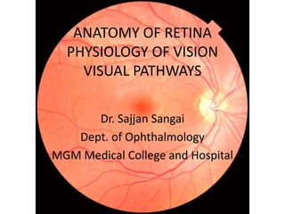
ANATOMY OF RETINA
PHYSIOLOGY OF VISION
VISUAL PATHWAYS
Dr. Sajjan Sangai
Dept. of Ophthalmology
MGM Medical College and Hospital
Embryology
• Structures that are derived from the optic
vesicle
Sensory layer of retina (Pars optica retinae)
Epithelial linings of cilliary body/iris (Pars
retinae cilliaris/ Pars retinae iridis)
Topography of Retina
• Diaphanous tissue: Purplish / red due to visual
pigment (rhodopsin).
• Most highly developed tissue of the eye.
• Extends from the optic disc to ora serrata.
• Surface area: 266 mm2
• Thickest near optic disc :0.56 mm
• Thinner towards periphery: 0.18 mm - Equator
0.10 mm - Ora
Retinal Equator
• Retinal Equator – Line where 4 vortex veins
exit , retina posterior to this is known as
posterior retina.
Retina
Optic Disc
Macula lutea
Peripheral Retina
1. Optic Disc
• Colour :Pale pink
• 1.5 mm diameter, well defined circular area
• At the optic disc- All retinal layers terminate
except nerve fibres , which pass through the
lamina cribrosa to run into optic nerve.
• Physiological Cup: Depression seen in it ,
central retinal vessels emerge through the
centre of this cup.
2. Macula Lutea/ Area Centralis
• Yellow spot – due to presence of carotenoid
pigment Xanthophyll.
• Dark area : 5.5 mm in diameter
• Situated in posterior pole of the eyeball ,
temporal to optic disc.
• Corresponds to 15 degrees of the visual field ,
accurate diurnal vision and colour
discrimination are primary functions.
Fovea
• Approximate centre of area centralis
• Posterior pole of globe
• 3-4mm temporal to the centre of optic disc.
• 0.8mm below horizontal meridian.
• Diameter-1.85mm – 5 degrees of visual field
• Avg. thickness- 0.25 mm
• Central concave indentation – Foveola is
produced.
• Downward sloping border which meets the
floor of foveal pit is known as CLIVUS.
• UMBO: Tiny depression in very centre of the
foveola ( Visible foveolar reflex on Direct
Ophthalmoscope).
Foveola
• Diameter: 0.35mm
• Thickness:0.13mm
• Highest visual acuity in retina corresponds to
only 1 degree of visual fields due to
I. Sole presence of cone photoreceptors.
II. Avascular nature.
• Foveal Avascular Zone: Located inside fovea
but outside foveola (clinically can be rendered
on FFA).
3. Peripheral Retina
• Increases the field of vision
• Divided into 4 regions
I. Near periphery-1.5mm around area centralis
II. Mid periphery- 3mm wide zone around near
periphery
III. Far periphery- from optic disc- 9-10 mm on
temporal side and 16 mm on nasal side in
the horizontal meridian(asymmetry due to
optic disc on nasal side)
IV. Ora serrata
Ora serrata
• Serrated peripheral margins where the retina
ends and cilliary body starts.
• 6mm nasally- 7mm temporally from limbus.
• 6-8 mm from limbus
• 25mm from optic nerve on nasal side
Ora serrata
Microscopic Architecture of Retina
• Contains 3 types of cells and their synapses
arranged in following layers: (Sclerad to
Vitread) are as follows:
Retinal Pigment Epithelium
Photoreceptor cell layer
External limiting membrane
Outer nuclear layer
Outer Plexiform layer
Inner nuclear layer
Inner Plexiform layer
Ganglion cell layer
Nerve fibre layer
Internal limiting membrane
Retinal Layers
Layer Degree of neurons
Pigment epithelium Neuroepithelial layer
Photoreceptor layer Neuron I (rod and cones)
Ext. limiting membrane (Percipient Elements)
Outer nuclear layer
Outer plexiform layer
Inner nuclear layer Neuron II Cerebral Layer
Inner plexiform layer Conductive and
associative elements
( Bipolar cells, Horizontal
cells, Amacrine cells ,
Centrifugal bipolars,
Ganglion cell layer Neuron III Muller fibres,
Nerve fibre layer conductive elements Astrocytes.)
Internal limiting membrane
I. Retinal Pigment Epithelium
• Fine mottling due to unequal distribution of
pigmentation within individual cells, gives a
granular appearance.
• Single layer- 5 million cells , firmly attached to its
basement lamina, lamina vitrea of brusch’s
membrane.
• Cobble stone appearance ( 4.2 million – 6.1
million)
• Area centralis- 12-18 µm width, 10-14 µm height.
• Ora- 60 µm in width
Functions:
• Photoreceptor
renewal and recycling
of vitamin A.
• Maintain integrity of
subretinal space.
• Transport of nutrients
and metabolites.
• Mechanical support
to the processes of
photoreceptor.
II. Photoreceptor layer of Rods and Cones
• 77.9 -107.3 million ( avg.92 Million) Rods
• 4.08 – 5.29 million ( avg. 4.6 million ) Cones
• Arranged as mosaics with variation.
• Cone Density and Distribution:
Maximally at Fovea ( avg. 1,99,000 cones/mm2)
with increasing eccentricity from fovea , density
of cones decrease rapidly.
• Rod density and distribution:
Avg. Horizontal diameter of rod free area at
fovea is 0.35 mm
1.25 degrees of visual fields
Present in large number (1,60,000 / mm2) in a
ring shaped zone 5-6 mm from fovea.
Visual Pigments
Rods
• Scotopic vision
• Greatest sensitivity
for blue – green light (
493 nm)
• Visual Purple/
Rhodopsin.
Cones
• Trichromatic pigments
• Short-Blue- 440 nm
• Medium- Green-
540nm
• Long- Orange- 577 nm
• Structure of Photoreceptor (in relation to
various Layers):
Part of photoreceptor Layer
Cell body and Nucleus Outer Nuclear Layer
Cell process Outer Plexiform layer
Inner and outer segments Layer of rods and cones
Rod Cells
• 40-60 µm
• Cylindrical , highly
retractile ,
transversely striated
and contains visual
purple.
• Has rod spherules.
Cone Cells
• 40-80 µm, Largest at
fovea and shortest at
periphery.
• Contains iodopsin.
• Has cone pedicles/
cone foot.
III. External Limiting Membrane
• From optic disc to ora serrata and becomes
continuous with the basal lamina between the
pigmented and non pigmented portions of
cilliary epithelium.
• Functions:
1. Selective barrier for nutrients.
2. Stabilization of position of the transducing
portion of photoreceptor.
IV. Outer Nuclear Layer
• Contains: soma and nuclei of photoreceptor cells
• Width varies.
• Nasal to disc: 45 µm – 8-9 rows of nuclei.
• Temporal to disc- 22 µm – 4 rows of nuclei.
• Fovea- 50 µm – 10 rows
• Different nuclei can be differentiated because
nuclei stains with Mallory stain:
Rod nuclei forms the bulk of this multi-layered
outer nuclear layer except in foveal region
Rods Nuclei Orange
Cones Nuclei Red
V. Outer Plexiform Layer
• Synapses between rod spherules and cone
pedicles with dendrites of bipolar cells and
processes of horizontal cells
• Marks the junction of end organs of vision
and 2nd order neurons in retina.
• Thickest at macula 51 µm consists of
predominantly of oblique fibres that have
deviated from fovea also known as HENLE’S
LAYER.
Outer 2/3rd Layer:
Inner fibres ( Axons) of
photoreceptors.
Inner 1/3rd Layer:
Dendrites of bipolar and
horizontal cells as well as
Müller cells processes.
VI. Inner Nuclear Layer
• Very thin layer.
• Disappears at fovea
• Consists of :
a) Bipolar cells
b) Horizontal cells
c) Amacrine cells
d) Soma of Müller cells
e) Capillaries of central retinal arteries
Outermost Layer- Horizontal Cell Nuclei
Outer Intermediate Layer-
Bipolar Cell nuclei
Inner Intermediate Layer-
Muller nuclei
Innermost Layer-
Amacrine and inter plexiform cell nuclei
•Contribute to vertical
communication within
the retinal layer.
•Carry out paracrine
functions.
•As principal glial cells,
act as a supportive
framework and a
nutritive function.
•Synapses with the
processes of Amacrine
cells and cell bodies of the
diffuse ganglion cells.
•Neuronal
interconnections
between
photoreceptors and
bipolar cells .
Horizontal
cells
Bipolar
cells
Amacrine
cells
Müller
cells
• Horizontal Cell:
Flat cells having numerous horizontal
associative and neuronal interconnections
between photoreceptor and bipolar cells in
outer plexiform layer.
Unlike bipolar cells information is relayed
radially through the retina , horizontal form a
network of fibres that integrate the activity of
photoreceptor cell horizontally.
• Highest at fovea,
number decreases
towards periphery
but processes branch
extensively from
centre to ora.
• Bipolar cells:
2 Degree neuron in visual circuitry.
35,676,000 bipolar cells
Oriented radially
- Perikarya- Inner nuclear layer
- Processes- Outer/ inner plexiform layer
 9 µm at fovea, 5 µm at periphery.
Types of Bipolar Cells
Rod Bipolar cells
Invaginating midget
Invaginating diffuse
Flat midget
Flat Diffuse
On centre blue cone bipolar cell
Off centre blue cone bipolar cell
Giant bi-stratified bipolar cells
Giant Diffuse Invaginating
Bipolar Cells
Type Connections Peculiarity
1. Rod Bipolar Cells
20%, Large soma profuse
dendrites
Arborize only with rod
spherules
Axons of these bipolar cells
have synapses with soma up
to 4 ganglion cells
2. Midget Bipolar cells
Small
Make connections only in
triads of cone pedicle
Invaginating- Deeply
invaginate cone pedicle
Flat- Makes superficial contact
with cone pedicle
Axons synapses with SINGLE
ganglion cell.
3. Diffuse- Makes contact with cone
pedicles only
Not with their triads
Axons synapse with number of
ganglion cells of all types.
4. Blue cone bipolar cells Innervate more than one cone
pedicle
5. Giant Bipolar cells Distinguished by extent of
their dendritic spread
VII. Inner Plexiform Layer
• Synapses between:
Axons of Bipolar
cells
( 2nd order neurons)
Ganglion cells
( 3rd order neurons)
• Amacrine cells also mediate interactions
within the layer and the interplexiform cells
receive input from the Amacrine cells
• Also contains processes of Muller cells ,
abundant microvasculature , occasional
displaced nucleus of a ganglion / Amacrine
cells.
• Layer is absent at foveola
• Thicker: 18-36 µm
• More synapses per unit area >2 million /mm2.
VIII. Ganglion Cell Layer
• Mainly composed of cell bodies 3rd order cells.
• Others: Processes of Müller cells, other
neuroglia, branches of retinal vessels are also
present.
• Layer structure:
Layers Situation
Single layer Peripheral retina
2 layers Temporal side of optic disc
6-8 layers Edge of foveola
At foveola and optic nerve head, ganglion cell
layer is absent.
1.2 million
ganglion cells
present in retina
Each produce a
single axon
Converge and
exit from the eye
as OPTIC NERVE
• Can be classified according to size, degree of
arborisation, spread of their dendrites,
pattern of synaptic connections with Amacrine
and bipolar cells.
• 2 major types:
• Project to magnocellular layer of
lateral geniculate body and exhibit
non opponent responses
M cells
(PARASOL)
• P1 – Midget cells-contribute 90% of
total ganglion cell layer @ foveola
• P2- Small Bi- StratifiedP cells
IX. Nerve Fibre Layer
Unmyelinated axons of ganglion cells
Converge at optic nerve head
Pass through lamina cribrosa
Become ensheathed by myelin
posterior to lamina cribrosa
• Contents:
1. Axons of ganglion cells- Centripetal nerve
fibre.
2. Centrifugal nerve fibre( Thicker than
centripetal).
3. Processes of Müller cells which interweave
with axons of ganglion cells .
4. Neuroglia cells present in nerve fibre.
5. Retinal vessels: Do not project on surface of
retina , rich bed of superficial capillary
network.
• 0.6- 2 µm non myelinated fibres.
• Contains microtubules, mitochondria, smooth
endoplasmic reticulum.
• Arrangement of nerve fibre in Retina:
• 1. Nasal Half: Directly to optic disc as superior
and inferior radiating fibres (Srf & Irf).
• 2. Macular region: Pass straight in temporal
part of disc as papillomacular bundle.(PMB)
• 3. Temporal retina: Arch above and below the
macular/ papillomacular bundle as superior /
inferior arcuate fibres. (Saf & Iaf).
• Arrangement in optic nerve:
Fibres from
peripheral part
Deep in retina
Occupy most
peripheral part
of optic disc
Fibres closer to
optic disc
Superficially in
retina and occupy
more central (deep)
portion of disc.
X. Internal Limiting Membrane
• Pas positive true basement membrane.
• Forms interface between retina and vitreous.
• Consists of 4 elements:
1. Collagen fibrils
2. Proteoglycans
3. Basement membrane
4. Plasma membrane
5. Plasma membrane of Müller cells and other glial
cells.
Retinal Layers Foveola Fovea Parafoveolar
region
Perifoveolar
region
RPE + +( Dark Area) + +
Photoreceptor
layer
C++++ C+++ C++R+ C++R+
ELM + + + +
ONL + + + +
OPL - + Thickest +
INL - + Thickest +
IPL - + + +
Ganglion cell
layer
- Multi-layered Thickest Multi-layered
Nerve fibre
layer
- + + +
Internal Limiting
membrane
+ (Thin) + + +
Blood Supply
• Outer four layers – Choriocapillaries
• Inner six layers- Central retina Artery
• Retina is supplied by Central Retinal Artery
Enters optic nerve on lower surface 15-20 mm
behind the globe.
Retinal arteries are end arteries and have no
anastomosis at ora serrata.
Arteries are distinguished from veins by being
brighter red and narrower.
Veins have purplish tint and are duller and of
wide calibre.
Choroidal vessels: Broader, ribbon like.
 Without any central streak
 Anastomose freely
 Easily visible in myopes and albinos.
Physiology of Vision
Initiation of vision (Photo
transduction)
Processing and
transmission of visual
sensation
Visual Perception
• Phototransduction:
Retina
Light falling upon
the retina causes
photochemical
changes
Photochemical changes
1.Rhodopsin
Bleaching
2. Rhodopsin
regeneration
3. Visual cycle
Electrical changes
Generation of
receptor potential
Rhodopsin Bleaching:
 Rhodopsin- the visual pigment present in rods
for scotopic vision.
 Maximum absorption spectrum- 500 nm
Rhodopsin
Protein- Opsin
Carotenoid- Retinine(
Vit. A aldehyde / 11
Cis Retinal)
11 Cis
Retinal
Light
All Trans
Retinal
This process is known as photodecomposition and
rhodopsin is said to be bleached.
Rhodopsin
Bathorhodopsin
Lumirhodopsin
Metarhodopsin
All trans retinal
11 Cis retinal
Rhodopsin regenration:
All trans retinal +
Vitamin A ( retinal) from
blood
11 Cis Retinal + Opsin in
rod outer segment Rhodopsin
Visual Cycle: Equilibrium between the
photodecomposition and regeneration of
visual pigments is referred to as visual cycle.
All trans
retinal
11 Cis
retinal
Rhodopsin
Excitation of nerve
OpsinOpsin
Magnocellular, Parvocellular and
Koniocellular pathways
P cells/
Parvocellular
Smaller, thinner axons
of smaller calibre
Colour sensitive with
High Spatial
resolution
M cells
/Magnocelluar
Large cells, thicker,
larger axons , faster
conducting
Transmits high
temporal motion
related information of
low spatial frequency
unrelated to colour.
Visual Perception
Stimulation
Sensation
1.Light sense
2. Form Sense
3. Sense of contrast
4. Colour sense
• Light Sense:
Light falling upon retina is gradually reduced in
intensity , there comes a point when it is no
longer perceived. This is known as Light
minimum.
• Measured when eye is dark adapted for 20-30
minutes.
• Light minimum for fovea is considerably
higher than for the Para central and peripheral
parts.
• Form sense:
Cones play major role and most acute at fovea
where it is most closely set and highly
differentiated.
Visual acuity is measured in a variety of ways
1. Recognition- Snellen’s Chart, Landolt C chart .
2. Resolution- Acuity grating
3. Localisation- Vernier grating.
• Sense of Contrast:
• Ability to perceive slight change
in luminance between regions
which are not separated by
definite borders.
• Measurement of contrast
sensitivity:
1. Pelli- Robson’s Contrast
sensitivity chart.
2. Cambridge low-contrast
gratings.
3. Arden gratings.
4. Functional acuity contrast test.
• Colour Sense: Ability of the eye to
discriminate between colours excited by light
of different wavelengths.
Cones
Short
Stimulated by
blue light
(440nm)
Medium
Stimulated by
green light
(540 nm)
Long
Stimulated by
red light (577
nm)
White colour can be formed from combination
of these colours in suitable proportions hence
normal colour vision is trichromatic.
Theories of colour vision:
a) Young- Helmholtz theory
b) Opponent colour theory of Herring
Visual Pathway
Neural
epithelium of
rods and
cones( end
organs)
Bipolar cells
in inner
nuclear layer
with its axons
in inner
reticular layer
Ganglion cells
in retina
Optic nerve,
Chiasm to
lateral
geniculate
body
Optic
radiations to
Visual cortex
• Fibres from peripheral regions in retina forms
2 distinct groups corresponding to nasal and
temporal half of retina.
Fibres from
temporal half
Optic
Chiasma
Optic tract of
same side
Fibres from
nasal half
Optic
Chiasma
Optic tract of
opposite side
Anatomy of retina
Anatomy of retina

Contenu connexe

Tendances (20)

RETINA - anatomy & physiology
RETINA - anatomy & physiologyRETINA - anatomy & physiology
RETINA - anatomy & physiology
 
Uvea anatomy
Uvea anatomyUvea anatomy
Uvea anatomy
 
Anatomy of the eyelids
Anatomy of the eyelidsAnatomy of the eyelids
Anatomy of the eyelids
 
eyelid anatomy slideshare
eyelid anatomy slideshareeyelid anatomy slideshare
eyelid anatomy slideshare
 
Aphakia
AphakiaAphakia
Aphakia
 
Central Retinal Vein Occlsion (CRVO)
Central Retinal Vein Occlsion (CRVO)Central Retinal Vein Occlsion (CRVO)
Central Retinal Vein Occlsion (CRVO)
 
Vitreous humour
Vitreous humourVitreous humour
Vitreous humour
 
Gonioscopy presentation
Gonioscopy presentationGonioscopy presentation
Gonioscopy presentation
 
Vitreous
VitreousVitreous
Vitreous
 
EXTRA OCULAR MUSCLES PHYSIOLOGY
EXTRA OCULAR MUSCLES PHYSIOLOGYEXTRA OCULAR MUSCLES PHYSIOLOGY
EXTRA OCULAR MUSCLES PHYSIOLOGY
 
Tear film test
Tear film testTear film test
Tear film test
 
Crystalline Lens
Crystalline LensCrystalline Lens
Crystalline Lens
 
Limbus
LimbusLimbus
Limbus
 
anatomy of retina
 anatomy of retina anatomy of retina
anatomy of retina
 
Optic disc evaluation
Optic disc evaluationOptic disc evaluation
Optic disc evaluation
 
Aqueous humor outflow
Aqueous humor outflowAqueous humor outflow
Aqueous humor outflow
 
anatomy of retina
anatomy of retinaanatomy of retina
anatomy of retina
 
Anatomy and physiology of cornea
Anatomy and physiology of corneaAnatomy and physiology of cornea
Anatomy and physiology of cornea
 
Direct ophthalmoscopy
Direct ophthalmoscopyDirect ophthalmoscopy
Direct ophthalmoscopy
 
Coloboma
ColobomaColoboma
Coloboma
 

En vedette

OCT - few basic facts
OCT - few basic factsOCT - few basic facts
OCT - few basic factsArindam Pande
 
OCT – Introduction and Macular disorders
OCT – Introduction and Macular disordersOCT – Introduction and Macular disorders
OCT – Introduction and Macular disordersSajjan Sangai
 
İnterpretation of optic coherence tomography images
İnterpretation of optic coherence tomography imagesİnterpretation of optic coherence tomography images
İnterpretation of optic coherence tomography imagesSinan çalışkan
 
Optical Coherence Tomography
Optical Coherence TomographyOptical Coherence Tomography
Optical Coherence TomographyManoj Aryal
 
Optical coherence tomography
Optical coherence tomographyOptical coherence tomography
Optical coherence tomographySinan çalışkan
 
The basics of retinal oct ophso.net
The basics of retinal oct ophso.netThe basics of retinal oct ophso.net
The basics of retinal oct ophso.netkebaplik
 
Spectralis oct normal anatomy & systematic interpretation.
Spectralis oct normal anatomy & systematic interpretation.Spectralis oct normal anatomy & systematic interpretation.
Spectralis oct normal anatomy & systematic interpretation.oxfordshireloc
 
Optical Coherence Tomography - principle and uses in ophthalmology
Optical Coherence Tomography - principle and uses in ophthalmologyOptical Coherence Tomography - principle and uses in ophthalmology
Optical Coherence Tomography - principle and uses in ophthalmologytapan_jakkal
 

En vedette (8)

OCT - few basic facts
OCT - few basic factsOCT - few basic facts
OCT - few basic facts
 
OCT – Introduction and Macular disorders
OCT – Introduction and Macular disordersOCT – Introduction and Macular disorders
OCT – Introduction and Macular disorders
 
İnterpretation of optic coherence tomography images
İnterpretation of optic coherence tomography imagesİnterpretation of optic coherence tomography images
İnterpretation of optic coherence tomography images
 
Optical Coherence Tomography
Optical Coherence TomographyOptical Coherence Tomography
Optical Coherence Tomography
 
Optical coherence tomography
Optical coherence tomographyOptical coherence tomography
Optical coherence tomography
 
The basics of retinal oct ophso.net
The basics of retinal oct ophso.netThe basics of retinal oct ophso.net
The basics of retinal oct ophso.net
 
Spectralis oct normal anatomy & systematic interpretation.
Spectralis oct normal anatomy & systematic interpretation.Spectralis oct normal anatomy & systematic interpretation.
Spectralis oct normal anatomy & systematic interpretation.
 
Optical Coherence Tomography - principle and uses in ophthalmology
Optical Coherence Tomography - principle and uses in ophthalmologyOptical Coherence Tomography - principle and uses in ophthalmology
Optical Coherence Tomography - principle and uses in ophthalmology
 

Similaire à Anatomy of retina

Similaire à Anatomy of retina (20)

ANATOMY OF RETINA - DR.RUTHRA.pptx
ANATOMY OF RETINA - DR.RUTHRA.pptxANATOMY OF RETINA - DR.RUTHRA.pptx
ANATOMY OF RETINA - DR.RUTHRA.pptx
 
Anatomy and physiology of retina
Anatomy and physiology of retinaAnatomy and physiology of retina
Anatomy and physiology of retina
 
Anatomy and Physiology of retina
Anatomy and Physiology of retinaAnatomy and Physiology of retina
Anatomy and Physiology of retina
 
Retina
RetinaRetina
Retina
 
Anatomy of retina
Anatomy of retinaAnatomy of retina
Anatomy of retina
 
Anatomy of Retina by Robin Singh ( BMCO )
Anatomy of Retina by Robin Singh ( BMCO )Anatomy of Retina by Robin Singh ( BMCO )
Anatomy of Retina by Robin Singh ( BMCO )
 
Anatomy of the retina
Anatomy of the retinaAnatomy of the retina
Anatomy of the retina
 
Retina 1
Retina 1Retina 1
Retina 1
 
Retina and layers
Retina and layersRetina and layers
Retina and layers
 
Suraj chhetri retina
Suraj chhetri retinaSuraj chhetri retina
Suraj chhetri retina
 
Retina : an overview
Retina : an overviewRetina : an overview
Retina : an overview
 
ANATOMY OF RETINA.pptx
ANATOMY OF RETINA.pptxANATOMY OF RETINA.pptx
ANATOMY OF RETINA.pptx
 
Anatomy of Retina
Anatomy of RetinaAnatomy of Retina
Anatomy of Retina
 
Retina
RetinaRetina
Retina
 
ANATOMY OF RETINA.pptx
ANATOMY OF RETINA.pptxANATOMY OF RETINA.pptx
ANATOMY OF RETINA.pptx
 
Anatomy of Retina and vitreous
Anatomy of Retina and vitreousAnatomy of Retina and vitreous
Anatomy of Retina and vitreous
 
Anatomy and Applied aspects of Uvea
Anatomy and Applied aspects of UveaAnatomy and Applied aspects of Uvea
Anatomy and Applied aspects of Uvea
 
Retina-Anat.pdf
Retina-Anat.pdfRetina-Anat.pdf
Retina-Anat.pdf
 
Anatomy of Retina
Anatomy of RetinaAnatomy of Retina
Anatomy of Retina
 
Anatomy of retina.pptx
Anatomy of retina.pptxAnatomy of retina.pptx
Anatomy of retina.pptx
 

Dernier

Call Girls Hebbal Just Call 7001305949 Top Class Call Girl Service Available
Call Girls Hebbal Just Call 7001305949 Top Class Call Girl Service AvailableCall Girls Hebbal Just Call 7001305949 Top Class Call Girl Service Available
Call Girls Hebbal Just Call 7001305949 Top Class Call Girl Service Availablenarwatsonia7
 
Call Girls Service Chennai Jiya 7001305949 Independent Escort Service Chennai
Call Girls Service Chennai Jiya 7001305949 Independent Escort Service ChennaiCall Girls Service Chennai Jiya 7001305949 Independent Escort Service Chennai
Call Girls Service Chennai Jiya 7001305949 Independent Escort Service ChennaiNehru place Escorts
 
Noida Sector 135 Call Girls ( 9873940964 ) Book Hot And Sexy Girls In A Few C...
Noida Sector 135 Call Girls ( 9873940964 ) Book Hot And Sexy Girls In A Few C...Noida Sector 135 Call Girls ( 9873940964 ) Book Hot And Sexy Girls In A Few C...
Noida Sector 135 Call Girls ( 9873940964 ) Book Hot And Sexy Girls In A Few C...rajnisinghkjn
 
Kolkata Call Girls Services 9907093804 @24x7 High Class Babes Here Call Now
Kolkata Call Girls Services 9907093804 @24x7 High Class Babes Here Call NowKolkata Call Girls Services 9907093804 @24x7 High Class Babes Here Call Now
Kolkata Call Girls Services 9907093804 @24x7 High Class Babes Here Call NowNehru place Escorts
 
Russian Call Girls Chickpet - 7001305949 Booking and charges genuine rate for...
Russian Call Girls Chickpet - 7001305949 Booking and charges genuine rate for...Russian Call Girls Chickpet - 7001305949 Booking and charges genuine rate for...
Russian Call Girls Chickpet - 7001305949 Booking and charges genuine rate for...narwatsonia7
 
High Profile Call Girls Jaipur Vani 8445551418 Independent Escort Service Jaipur
High Profile Call Girls Jaipur Vani 8445551418 Independent Escort Service JaipurHigh Profile Call Girls Jaipur Vani 8445551418 Independent Escort Service Jaipur
High Profile Call Girls Jaipur Vani 8445551418 Independent Escort Service Jaipurparulsinha
 
Call Girls Kanakapura Road Just Call 7001305949 Top Class Call Girl Service A...
Call Girls Kanakapura Road Just Call 7001305949 Top Class Call Girl Service A...Call Girls Kanakapura Road Just Call 7001305949 Top Class Call Girl Service A...
Call Girls Kanakapura Road Just Call 7001305949 Top Class Call Girl Service A...narwatsonia7
 
See the 2,456 pharmacies on the National E-Pharmacy Platform
See the 2,456 pharmacies on the National E-Pharmacy PlatformSee the 2,456 pharmacies on the National E-Pharmacy Platform
See the 2,456 pharmacies on the National E-Pharmacy PlatformKweku Zurek
 
Housewife Call Girls Bangalore - Call 7001305949 Rs-3500 with A/C Room Cash o...
Housewife Call Girls Bangalore - Call 7001305949 Rs-3500 with A/C Room Cash o...Housewife Call Girls Bangalore - Call 7001305949 Rs-3500 with A/C Room Cash o...
Housewife Call Girls Bangalore - Call 7001305949 Rs-3500 with A/C Room Cash o...narwatsonia7
 
Book Call Girls in Kasavanahalli - 7001305949 with real photos and phone numbers
Book Call Girls in Kasavanahalli - 7001305949 with real photos and phone numbersBook Call Girls in Kasavanahalli - 7001305949 with real photos and phone numbers
Book Call Girls in Kasavanahalli - 7001305949 with real photos and phone numbersnarwatsonia7
 
College Call Girls Vyasarpadi Whatsapp 7001305949 Independent Escort Service
College Call Girls Vyasarpadi Whatsapp 7001305949 Independent Escort ServiceCollege Call Girls Vyasarpadi Whatsapp 7001305949 Independent Escort Service
College Call Girls Vyasarpadi Whatsapp 7001305949 Independent Escort ServiceNehru place Escorts
 
Call Girls In Andheri East Call 9920874524 Book Hot And Sexy Girls
Call Girls In Andheri East Call 9920874524 Book Hot And Sexy GirlsCall Girls In Andheri East Call 9920874524 Book Hot And Sexy Girls
Call Girls In Andheri East Call 9920874524 Book Hot And Sexy Girlsnehamumbai
 
call girls in munirka DELHI 🔝 >༒9540349809 🔝 genuine Escort Service 🔝✔️✔️
call girls in munirka  DELHI 🔝 >༒9540349809 🔝 genuine Escort Service 🔝✔️✔️call girls in munirka  DELHI 🔝 >༒9540349809 🔝 genuine Escort Service 🔝✔️✔️
call girls in munirka DELHI 🔝 >༒9540349809 🔝 genuine Escort Service 🔝✔️✔️saminamagar
 
Pharmaceutical Marketting: Unit-5, Pricing
Pharmaceutical Marketting: Unit-5, PricingPharmaceutical Marketting: Unit-5, Pricing
Pharmaceutical Marketting: Unit-5, PricingArunagarwal328757
 
Call Girls Service Noida Maya 9711199012 Independent Escort Service Noida
Call Girls Service Noida Maya 9711199012 Independent Escort Service NoidaCall Girls Service Noida Maya 9711199012 Independent Escort Service Noida
Call Girls Service Noida Maya 9711199012 Independent Escort Service NoidaPooja Gupta
 
Glomerular Filtration rate and its determinants.pptx
Glomerular Filtration rate and its determinants.pptxGlomerular Filtration rate and its determinants.pptx
Glomerular Filtration rate and its determinants.pptxDr.Nusrat Tariq
 
Russian Call Girl Brookfield - 7001305949 Escorts Service 50% Off with Cash O...
Russian Call Girl Brookfield - 7001305949 Escorts Service 50% Off with Cash O...Russian Call Girl Brookfield - 7001305949 Escorts Service 50% Off with Cash O...
Russian Call Girl Brookfield - 7001305949 Escorts Service 50% Off with Cash O...narwatsonia7
 
Call Girls Whitefield Just Call 7001305949 Top Class Call Girl Service Available
Call Girls Whitefield Just Call 7001305949 Top Class Call Girl Service AvailableCall Girls Whitefield Just Call 7001305949 Top Class Call Girl Service Available
Call Girls Whitefield Just Call 7001305949 Top Class Call Girl Service Availablenarwatsonia7
 
Call Girls Jayanagar Just Call 7001305949 Top Class Call Girl Service Available
Call Girls Jayanagar Just Call 7001305949 Top Class Call Girl Service AvailableCall Girls Jayanagar Just Call 7001305949 Top Class Call Girl Service Available
Call Girls Jayanagar Just Call 7001305949 Top Class Call Girl Service Availablenarwatsonia7
 
Call Girls Jp Nagar Just Call 7001305949 Top Class Call Girl Service Available
Call Girls Jp Nagar Just Call 7001305949 Top Class Call Girl Service AvailableCall Girls Jp Nagar Just Call 7001305949 Top Class Call Girl Service Available
Call Girls Jp Nagar Just Call 7001305949 Top Class Call Girl Service Availablenarwatsonia7
 

Dernier (20)

Call Girls Hebbal Just Call 7001305949 Top Class Call Girl Service Available
Call Girls Hebbal Just Call 7001305949 Top Class Call Girl Service AvailableCall Girls Hebbal Just Call 7001305949 Top Class Call Girl Service Available
Call Girls Hebbal Just Call 7001305949 Top Class Call Girl Service Available
 
Call Girls Service Chennai Jiya 7001305949 Independent Escort Service Chennai
Call Girls Service Chennai Jiya 7001305949 Independent Escort Service ChennaiCall Girls Service Chennai Jiya 7001305949 Independent Escort Service Chennai
Call Girls Service Chennai Jiya 7001305949 Independent Escort Service Chennai
 
Noida Sector 135 Call Girls ( 9873940964 ) Book Hot And Sexy Girls In A Few C...
Noida Sector 135 Call Girls ( 9873940964 ) Book Hot And Sexy Girls In A Few C...Noida Sector 135 Call Girls ( 9873940964 ) Book Hot And Sexy Girls In A Few C...
Noida Sector 135 Call Girls ( 9873940964 ) Book Hot And Sexy Girls In A Few C...
 
Kolkata Call Girls Services 9907093804 @24x7 High Class Babes Here Call Now
Kolkata Call Girls Services 9907093804 @24x7 High Class Babes Here Call NowKolkata Call Girls Services 9907093804 @24x7 High Class Babes Here Call Now
Kolkata Call Girls Services 9907093804 @24x7 High Class Babes Here Call Now
 
Russian Call Girls Chickpet - 7001305949 Booking and charges genuine rate for...
Russian Call Girls Chickpet - 7001305949 Booking and charges genuine rate for...Russian Call Girls Chickpet - 7001305949 Booking and charges genuine rate for...
Russian Call Girls Chickpet - 7001305949 Booking and charges genuine rate for...
 
High Profile Call Girls Jaipur Vani 8445551418 Independent Escort Service Jaipur
High Profile Call Girls Jaipur Vani 8445551418 Independent Escort Service JaipurHigh Profile Call Girls Jaipur Vani 8445551418 Independent Escort Service Jaipur
High Profile Call Girls Jaipur Vani 8445551418 Independent Escort Service Jaipur
 
Call Girls Kanakapura Road Just Call 7001305949 Top Class Call Girl Service A...
Call Girls Kanakapura Road Just Call 7001305949 Top Class Call Girl Service A...Call Girls Kanakapura Road Just Call 7001305949 Top Class Call Girl Service A...
Call Girls Kanakapura Road Just Call 7001305949 Top Class Call Girl Service A...
 
See the 2,456 pharmacies on the National E-Pharmacy Platform
See the 2,456 pharmacies on the National E-Pharmacy PlatformSee the 2,456 pharmacies on the National E-Pharmacy Platform
See the 2,456 pharmacies on the National E-Pharmacy Platform
 
Housewife Call Girls Bangalore - Call 7001305949 Rs-3500 with A/C Room Cash o...
Housewife Call Girls Bangalore - Call 7001305949 Rs-3500 with A/C Room Cash o...Housewife Call Girls Bangalore - Call 7001305949 Rs-3500 with A/C Room Cash o...
Housewife Call Girls Bangalore - Call 7001305949 Rs-3500 with A/C Room Cash o...
 
Book Call Girls in Kasavanahalli - 7001305949 with real photos and phone numbers
Book Call Girls in Kasavanahalli - 7001305949 with real photos and phone numbersBook Call Girls in Kasavanahalli - 7001305949 with real photos and phone numbers
Book Call Girls in Kasavanahalli - 7001305949 with real photos and phone numbers
 
College Call Girls Vyasarpadi Whatsapp 7001305949 Independent Escort Service
College Call Girls Vyasarpadi Whatsapp 7001305949 Independent Escort ServiceCollege Call Girls Vyasarpadi Whatsapp 7001305949 Independent Escort Service
College Call Girls Vyasarpadi Whatsapp 7001305949 Independent Escort Service
 
Call Girls In Andheri East Call 9920874524 Book Hot And Sexy Girls
Call Girls In Andheri East Call 9920874524 Book Hot And Sexy GirlsCall Girls In Andheri East Call 9920874524 Book Hot And Sexy Girls
Call Girls In Andheri East Call 9920874524 Book Hot And Sexy Girls
 
call girls in munirka DELHI 🔝 >༒9540349809 🔝 genuine Escort Service 🔝✔️✔️
call girls in munirka  DELHI 🔝 >༒9540349809 🔝 genuine Escort Service 🔝✔️✔️call girls in munirka  DELHI 🔝 >༒9540349809 🔝 genuine Escort Service 🔝✔️✔️
call girls in munirka DELHI 🔝 >༒9540349809 🔝 genuine Escort Service 🔝✔️✔️
 
Pharmaceutical Marketting: Unit-5, Pricing
Pharmaceutical Marketting: Unit-5, PricingPharmaceutical Marketting: Unit-5, Pricing
Pharmaceutical Marketting: Unit-5, Pricing
 
Call Girls Service Noida Maya 9711199012 Independent Escort Service Noida
Call Girls Service Noida Maya 9711199012 Independent Escort Service NoidaCall Girls Service Noida Maya 9711199012 Independent Escort Service Noida
Call Girls Service Noida Maya 9711199012 Independent Escort Service Noida
 
Glomerular Filtration rate and its determinants.pptx
Glomerular Filtration rate and its determinants.pptxGlomerular Filtration rate and its determinants.pptx
Glomerular Filtration rate and its determinants.pptx
 
Russian Call Girl Brookfield - 7001305949 Escorts Service 50% Off with Cash O...
Russian Call Girl Brookfield - 7001305949 Escorts Service 50% Off with Cash O...Russian Call Girl Brookfield - 7001305949 Escorts Service 50% Off with Cash O...
Russian Call Girl Brookfield - 7001305949 Escorts Service 50% Off with Cash O...
 
Call Girls Whitefield Just Call 7001305949 Top Class Call Girl Service Available
Call Girls Whitefield Just Call 7001305949 Top Class Call Girl Service AvailableCall Girls Whitefield Just Call 7001305949 Top Class Call Girl Service Available
Call Girls Whitefield Just Call 7001305949 Top Class Call Girl Service Available
 
Call Girls Jayanagar Just Call 7001305949 Top Class Call Girl Service Available
Call Girls Jayanagar Just Call 7001305949 Top Class Call Girl Service AvailableCall Girls Jayanagar Just Call 7001305949 Top Class Call Girl Service Available
Call Girls Jayanagar Just Call 7001305949 Top Class Call Girl Service Available
 
Call Girls Jp Nagar Just Call 7001305949 Top Class Call Girl Service Available
Call Girls Jp Nagar Just Call 7001305949 Top Class Call Girl Service AvailableCall Girls Jp Nagar Just Call 7001305949 Top Class Call Girl Service Available
Call Girls Jp Nagar Just Call 7001305949 Top Class Call Girl Service Available
 

Anatomy of retina

  • 1. ANATOMY OF RETINA PHYSIOLOGY OF VISION VISUAL PATHWAYS Dr. Sajjan Sangai Dept. of Ophthalmology MGM Medical College and Hospital
  • 2. Embryology • Structures that are derived from the optic vesicle Sensory layer of retina (Pars optica retinae) Epithelial linings of cilliary body/iris (Pars retinae cilliaris/ Pars retinae iridis)
  • 3. Topography of Retina • Diaphanous tissue: Purplish / red due to visual pigment (rhodopsin). • Most highly developed tissue of the eye. • Extends from the optic disc to ora serrata. • Surface area: 266 mm2 • Thickest near optic disc :0.56 mm • Thinner towards periphery: 0.18 mm - Equator 0.10 mm - Ora
  • 4. Retinal Equator • Retinal Equator – Line where 4 vortex veins exit , retina posterior to this is known as posterior retina.
  • 7. • Colour :Pale pink • 1.5 mm diameter, well defined circular area • At the optic disc- All retinal layers terminate except nerve fibres , which pass through the lamina cribrosa to run into optic nerve. • Physiological Cup: Depression seen in it , central retinal vessels emerge through the centre of this cup.
  • 8. 2. Macula Lutea/ Area Centralis • Yellow spot – due to presence of carotenoid pigment Xanthophyll. • Dark area : 5.5 mm in diameter • Situated in posterior pole of the eyeball , temporal to optic disc. • Corresponds to 15 degrees of the visual field , accurate diurnal vision and colour discrimination are primary functions.
  • 9.
  • 10. Fovea • Approximate centre of area centralis • Posterior pole of globe • 3-4mm temporal to the centre of optic disc. • 0.8mm below horizontal meridian. • Diameter-1.85mm – 5 degrees of visual field • Avg. thickness- 0.25 mm
  • 11. • Central concave indentation – Foveola is produced. • Downward sloping border which meets the floor of foveal pit is known as CLIVUS. • UMBO: Tiny depression in very centre of the foveola ( Visible foveolar reflex on Direct Ophthalmoscope).
  • 12. Foveola • Diameter: 0.35mm • Thickness:0.13mm • Highest visual acuity in retina corresponds to only 1 degree of visual fields due to I. Sole presence of cone photoreceptors. II. Avascular nature. • Foveal Avascular Zone: Located inside fovea but outside foveola (clinically can be rendered on FFA).
  • 13. 3. Peripheral Retina • Increases the field of vision • Divided into 4 regions I. Near periphery-1.5mm around area centralis II. Mid periphery- 3mm wide zone around near periphery III. Far periphery- from optic disc- 9-10 mm on temporal side and 16 mm on nasal side in the horizontal meridian(asymmetry due to optic disc on nasal side) IV. Ora serrata
  • 14. Ora serrata • Serrated peripheral margins where the retina ends and cilliary body starts. • 6mm nasally- 7mm temporally from limbus. • 6-8 mm from limbus • 25mm from optic nerve on nasal side
  • 16. Microscopic Architecture of Retina • Contains 3 types of cells and their synapses arranged in following layers: (Sclerad to Vitread) are as follows:
  • 17. Retinal Pigment Epithelium Photoreceptor cell layer External limiting membrane Outer nuclear layer Outer Plexiform layer Inner nuclear layer Inner Plexiform layer Ganglion cell layer Nerve fibre layer Internal limiting membrane
  • 19. Layer Degree of neurons Pigment epithelium Neuroepithelial layer Photoreceptor layer Neuron I (rod and cones) Ext. limiting membrane (Percipient Elements) Outer nuclear layer Outer plexiform layer Inner nuclear layer Neuron II Cerebral Layer Inner plexiform layer Conductive and associative elements ( Bipolar cells, Horizontal cells, Amacrine cells , Centrifugal bipolars, Ganglion cell layer Neuron III Muller fibres, Nerve fibre layer conductive elements Astrocytes.) Internal limiting membrane
  • 20. I. Retinal Pigment Epithelium • Fine mottling due to unequal distribution of pigmentation within individual cells, gives a granular appearance. • Single layer- 5 million cells , firmly attached to its basement lamina, lamina vitrea of brusch’s membrane. • Cobble stone appearance ( 4.2 million – 6.1 million) • Area centralis- 12-18 µm width, 10-14 µm height. • Ora- 60 µm in width
  • 21. Functions: • Photoreceptor renewal and recycling of vitamin A. • Maintain integrity of subretinal space. • Transport of nutrients and metabolites. • Mechanical support to the processes of photoreceptor.
  • 22. II. Photoreceptor layer of Rods and Cones • 77.9 -107.3 million ( avg.92 Million) Rods • 4.08 – 5.29 million ( avg. 4.6 million ) Cones • Arranged as mosaics with variation. • Cone Density and Distribution: Maximally at Fovea ( avg. 1,99,000 cones/mm2) with increasing eccentricity from fovea , density of cones decrease rapidly.
  • 23. • Rod density and distribution: Avg. Horizontal diameter of rod free area at fovea is 0.35 mm 1.25 degrees of visual fields Present in large number (1,60,000 / mm2) in a ring shaped zone 5-6 mm from fovea.
  • 24. Visual Pigments Rods • Scotopic vision • Greatest sensitivity for blue – green light ( 493 nm) • Visual Purple/ Rhodopsin. Cones • Trichromatic pigments • Short-Blue- 440 nm • Medium- Green- 540nm • Long- Orange- 577 nm
  • 25. • Structure of Photoreceptor (in relation to various Layers): Part of photoreceptor Layer Cell body and Nucleus Outer Nuclear Layer Cell process Outer Plexiform layer Inner and outer segments Layer of rods and cones
  • 26. Rod Cells • 40-60 µm • Cylindrical , highly retractile , transversely striated and contains visual purple. • Has rod spherules.
  • 27. Cone Cells • 40-80 µm, Largest at fovea and shortest at periphery. • Contains iodopsin. • Has cone pedicles/ cone foot.
  • 28. III. External Limiting Membrane • From optic disc to ora serrata and becomes continuous with the basal lamina between the pigmented and non pigmented portions of cilliary epithelium. • Functions: 1. Selective barrier for nutrients. 2. Stabilization of position of the transducing portion of photoreceptor.
  • 30. • Contains: soma and nuclei of photoreceptor cells • Width varies. • Nasal to disc: 45 µm – 8-9 rows of nuclei. • Temporal to disc- 22 µm – 4 rows of nuclei. • Fovea- 50 µm – 10 rows • Different nuclei can be differentiated because nuclei stains with Mallory stain: Rod nuclei forms the bulk of this multi-layered outer nuclear layer except in foveal region Rods Nuclei Orange Cones Nuclei Red
  • 32. • Synapses between rod spherules and cone pedicles with dendrites of bipolar cells and processes of horizontal cells • Marks the junction of end organs of vision and 2nd order neurons in retina. • Thickest at macula 51 µm consists of predominantly of oblique fibres that have deviated from fovea also known as HENLE’S LAYER.
  • 33. Outer 2/3rd Layer: Inner fibres ( Axons) of photoreceptors. Inner 1/3rd Layer: Dendrites of bipolar and horizontal cells as well as Müller cells processes.
  • 34. VI. Inner Nuclear Layer • Very thin layer. • Disappears at fovea • Consists of : a) Bipolar cells b) Horizontal cells c) Amacrine cells d) Soma of Müller cells e) Capillaries of central retinal arteries
  • 35. Outermost Layer- Horizontal Cell Nuclei Outer Intermediate Layer- Bipolar Cell nuclei Inner Intermediate Layer- Muller nuclei Innermost Layer- Amacrine and inter plexiform cell nuclei
  • 36. •Contribute to vertical communication within the retinal layer. •Carry out paracrine functions. •As principal glial cells, act as a supportive framework and a nutritive function. •Synapses with the processes of Amacrine cells and cell bodies of the diffuse ganglion cells. •Neuronal interconnections between photoreceptors and bipolar cells . Horizontal cells Bipolar cells Amacrine cells Müller cells
  • 37. • Horizontal Cell: Flat cells having numerous horizontal associative and neuronal interconnections between photoreceptor and bipolar cells in outer plexiform layer. Unlike bipolar cells information is relayed radially through the retina , horizontal form a network of fibres that integrate the activity of photoreceptor cell horizontally.
  • 38. • Highest at fovea, number decreases towards periphery but processes branch extensively from centre to ora.
  • 39. • Bipolar cells: 2 Degree neuron in visual circuitry. 35,676,000 bipolar cells Oriented radially - Perikarya- Inner nuclear layer - Processes- Outer/ inner plexiform layer  9 µm at fovea, 5 µm at periphery.
  • 40. Types of Bipolar Cells Rod Bipolar cells Invaginating midget Invaginating diffuse Flat midget Flat Diffuse On centre blue cone bipolar cell Off centre blue cone bipolar cell Giant bi-stratified bipolar cells Giant Diffuse Invaginating
  • 41. Bipolar Cells Type Connections Peculiarity 1. Rod Bipolar Cells 20%, Large soma profuse dendrites Arborize only with rod spherules Axons of these bipolar cells have synapses with soma up to 4 ganglion cells 2. Midget Bipolar cells Small Make connections only in triads of cone pedicle Invaginating- Deeply invaginate cone pedicle Flat- Makes superficial contact with cone pedicle Axons synapses with SINGLE ganglion cell. 3. Diffuse- Makes contact with cone pedicles only Not with their triads Axons synapse with number of ganglion cells of all types. 4. Blue cone bipolar cells Innervate more than one cone pedicle 5. Giant Bipolar cells Distinguished by extent of their dendritic spread
  • 43. • Synapses between: Axons of Bipolar cells ( 2nd order neurons) Ganglion cells ( 3rd order neurons)
  • 44. • Amacrine cells also mediate interactions within the layer and the interplexiform cells receive input from the Amacrine cells • Also contains processes of Muller cells , abundant microvasculature , occasional displaced nucleus of a ganglion / Amacrine cells. • Layer is absent at foveola • Thicker: 18-36 µm • More synapses per unit area >2 million /mm2.
  • 45. VIII. Ganglion Cell Layer • Mainly composed of cell bodies 3rd order cells. • Others: Processes of Müller cells, other neuroglia, branches of retinal vessels are also present.
  • 46. • Layer structure: Layers Situation Single layer Peripheral retina 2 layers Temporal side of optic disc 6-8 layers Edge of foveola At foveola and optic nerve head, ganglion cell layer is absent.
  • 47. 1.2 million ganglion cells present in retina Each produce a single axon Converge and exit from the eye as OPTIC NERVE
  • 48. • Can be classified according to size, degree of arborisation, spread of their dendrites, pattern of synaptic connections with Amacrine and bipolar cells. • 2 major types: • Project to magnocellular layer of lateral geniculate body and exhibit non opponent responses M cells (PARASOL) • P1 – Midget cells-contribute 90% of total ganglion cell layer @ foveola • P2- Small Bi- StratifiedP cells
  • 49. IX. Nerve Fibre Layer Unmyelinated axons of ganglion cells Converge at optic nerve head Pass through lamina cribrosa Become ensheathed by myelin posterior to lamina cribrosa
  • 50. • Contents: 1. Axons of ganglion cells- Centripetal nerve fibre. 2. Centrifugal nerve fibre( Thicker than centripetal). 3. Processes of Müller cells which interweave with axons of ganglion cells . 4. Neuroglia cells present in nerve fibre. 5. Retinal vessels: Do not project on surface of retina , rich bed of superficial capillary network.
  • 51. • 0.6- 2 µm non myelinated fibres. • Contains microtubules, mitochondria, smooth endoplasmic reticulum.
  • 52. • Arrangement of nerve fibre in Retina: • 1. Nasal Half: Directly to optic disc as superior and inferior radiating fibres (Srf & Irf). • 2. Macular region: Pass straight in temporal part of disc as papillomacular bundle.(PMB) • 3. Temporal retina: Arch above and below the macular/ papillomacular bundle as superior / inferior arcuate fibres. (Saf & Iaf).
  • 53. • Arrangement in optic nerve: Fibres from peripheral part Deep in retina Occupy most peripheral part of optic disc Fibres closer to optic disc Superficially in retina and occupy more central (deep) portion of disc.
  • 54. X. Internal Limiting Membrane • Pas positive true basement membrane. • Forms interface between retina and vitreous. • Consists of 4 elements: 1. Collagen fibrils 2. Proteoglycans 3. Basement membrane 4. Plasma membrane 5. Plasma membrane of Müller cells and other glial cells.
  • 55. Retinal Layers Foveola Fovea Parafoveolar region Perifoveolar region RPE + +( Dark Area) + + Photoreceptor layer C++++ C+++ C++R+ C++R+ ELM + + + + ONL + + + + OPL - + Thickest + INL - + Thickest + IPL - + + + Ganglion cell layer - Multi-layered Thickest Multi-layered Nerve fibre layer - + + + Internal Limiting membrane + (Thin) + + +
  • 56. Blood Supply • Outer four layers – Choriocapillaries • Inner six layers- Central retina Artery • Retina is supplied by Central Retinal Artery Enters optic nerve on lower surface 15-20 mm behind the globe. Retinal arteries are end arteries and have no anastomosis at ora serrata.
  • 57. Arteries are distinguished from veins by being brighter red and narrower. Veins have purplish tint and are duller and of wide calibre. Choroidal vessels: Broader, ribbon like.  Without any central streak  Anastomose freely  Easily visible in myopes and albinos.
  • 58.
  • 59. Physiology of Vision Initiation of vision (Photo transduction) Processing and transmission of visual sensation Visual Perception
  • 60. • Phototransduction: Retina Light falling upon the retina causes photochemical changes Photochemical changes 1.Rhodopsin Bleaching 2. Rhodopsin regeneration 3. Visual cycle Electrical changes Generation of receptor potential
  • 61. Rhodopsin Bleaching:  Rhodopsin- the visual pigment present in rods for scotopic vision.  Maximum absorption spectrum- 500 nm Rhodopsin Protein- Opsin Carotenoid- Retinine( Vit. A aldehyde / 11 Cis Retinal)
  • 62. 11 Cis Retinal Light All Trans Retinal This process is known as photodecomposition and rhodopsin is said to be bleached.
  • 64. Rhodopsin regenration: All trans retinal + Vitamin A ( retinal) from blood 11 Cis Retinal + Opsin in rod outer segment Rhodopsin
  • 65. Visual Cycle: Equilibrium between the photodecomposition and regeneration of visual pigments is referred to as visual cycle. All trans retinal 11 Cis retinal Rhodopsin Excitation of nerve OpsinOpsin
  • 66. Magnocellular, Parvocellular and Koniocellular pathways P cells/ Parvocellular Smaller, thinner axons of smaller calibre Colour sensitive with High Spatial resolution M cells /Magnocelluar Large cells, thicker, larger axons , faster conducting Transmits high temporal motion related information of low spatial frequency unrelated to colour.
  • 67. Visual Perception Stimulation Sensation 1.Light sense 2. Form Sense 3. Sense of contrast 4. Colour sense
  • 68. • Light Sense: Light falling upon retina is gradually reduced in intensity , there comes a point when it is no longer perceived. This is known as Light minimum. • Measured when eye is dark adapted for 20-30 minutes. • Light minimum for fovea is considerably higher than for the Para central and peripheral parts.
  • 69. • Form sense: Cones play major role and most acute at fovea where it is most closely set and highly differentiated. Visual acuity is measured in a variety of ways 1. Recognition- Snellen’s Chart, Landolt C chart . 2. Resolution- Acuity grating 3. Localisation- Vernier grating.
  • 70. • Sense of Contrast: • Ability to perceive slight change in luminance between regions which are not separated by definite borders. • Measurement of contrast sensitivity: 1. Pelli- Robson’s Contrast sensitivity chart. 2. Cambridge low-contrast gratings. 3. Arden gratings. 4. Functional acuity contrast test.
  • 71. • Colour Sense: Ability of the eye to discriminate between colours excited by light of different wavelengths. Cones Short Stimulated by blue light (440nm) Medium Stimulated by green light (540 nm) Long Stimulated by red light (577 nm)
  • 72. White colour can be formed from combination of these colours in suitable proportions hence normal colour vision is trichromatic. Theories of colour vision: a) Young- Helmholtz theory b) Opponent colour theory of Herring
  • 73. Visual Pathway Neural epithelium of rods and cones( end organs) Bipolar cells in inner nuclear layer with its axons in inner reticular layer Ganglion cells in retina Optic nerve, Chiasm to lateral geniculate body Optic radiations to Visual cortex
  • 74.
  • 75. • Fibres from peripheral regions in retina forms 2 distinct groups corresponding to nasal and temporal half of retina. Fibres from temporal half Optic Chiasma Optic tract of same side Fibres from nasal half Optic Chiasma Optic tract of opposite side